• 文献检索
  • 文档翻译
  • 深度研究
  • 学术资讯
  • Suppr Zotero 插件Zotero 插件
  • 邀请有礼
  • 套餐&价格
  • 历史记录
应用&插件
Suppr Zotero 插件Zotero 插件浏览器插件Mac 客户端Windows 客户端微信小程序
定价
高级版会员购买积分包购买API积分包
服务
文献检索文档翻译深度研究API 文档MCP 服务
关于我们
关于 Suppr公司介绍联系我们用户协议隐私条款
关注我们

Suppr 超能文献

核心技术专利:CN118964589B侵权必究
粤ICP备2023148730 号-1Suppr @ 2026

文献检索

告别复杂PubMed语法,用中文像聊天一样搜索,搜遍4000万医学文献。AI智能推荐,让科研检索更轻松。

立即免费搜索

文件翻译

保留排版,准确专业,支持PDF/Word/PPT等文件格式,支持 12+语言互译。

免费翻译文档

深度研究

AI帮你快速写综述,25分钟生成高质量综述,智能提取关键信息,辅助科研写作。

立即免费体验

利用对比增强超声血流速度成像技术在体测定兔主动脉血流动力学壁切应力。

Determining Haemodynamic Wall Shear Stress in the Rabbit Aorta In Vivo Using Contrast-Enhanced Ultrasound Image Velocimetry.

机构信息

Department of Bioengineering, Imperial College London, Prince Consort Road, London, SW7 2AZ, UK.

出版信息

Ann Biomed Eng. 2020 Jun;48(6):1728-1739. doi: 10.1007/s10439-020-02484-2. Epub 2020 Mar 4.

DOI:10.1007/s10439-020-02484-2
PMID:32130594
原文链接:https://pmc.ncbi.nlm.nih.gov/articles/PMC7280334/
Abstract

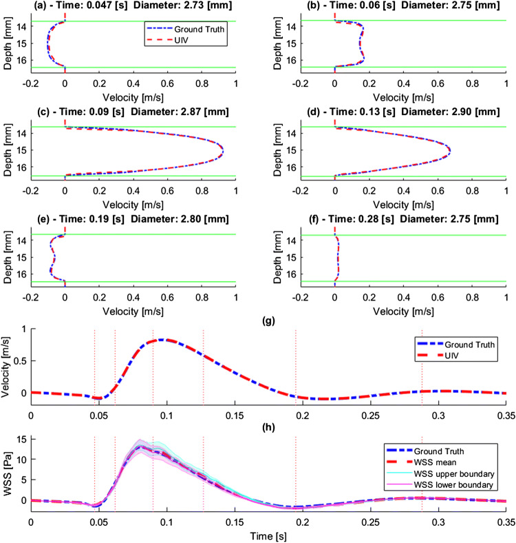
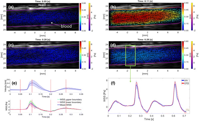
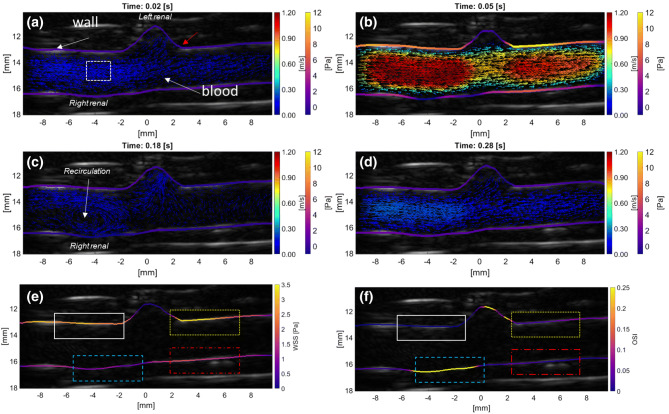
Abnormal blood flow and wall shear stress (WSS) can cause and be caused by cardiovascular disease. To date, however, no standard method has been established for mapping WSS in vivo. Here we demonstrate wide-field assessment of WSS in the rabbit abdominal aorta using contrast-enhanced ultrasound image velocimetry (UIV). Flow and WSS measurements were made independent of beam angle, curvature or branching. Measurements were validated in an in silico model of the rabbit thoracic aorta with moving walls and pulsatile flow. Mean errors over a cardiac cycle for velocity and WSS were 0.34 and 1.69%, respectively. In vivo time average WSS in a straight segment of the suprarenal aorta correlated highly with simulations (PC = 0.99) with a mean deviation of 0.29 Pa or 5.16%. To assess fundamental plausibility of the measurement, UIV WSS was compared to an analytic approximation derived from the Poiseuille equation; the discrepancy was 17%. Mapping of WSS was also demonstrated in regions of arterial branching. High time average WSS (TAWSS = 3.4 Pa) and oscillatory flow (OSI = 0.3) were observed near the origin of conduit arteries. In conclusion, we have demonstrated that contrast-enhanced UIV is capable of measuring spatiotemporal variation in flow velocity, arterial wall location and hence WSS in vivo with high accuracy over a large field of view.

摘要

异常的血流和壁切应力(WSS)可导致心血管疾病,并可由心血管疾病引起。然而,迄今为止,尚未建立用于体内映射 WSS 的标准方法。在这里,我们使用对比增强超声图像速度(UIV)演示了在兔腹主动脉中进行 WSS 的宽视场评估。流量和 WSS 的测量与波束角度、曲率或分支无关。在具有移动壁和脉动流的兔胸主动脉的计算机模拟中验证了测量值。心动周期内速度和 WSS 的平均误差分别为 0.34%和 1.69%。在肾上段主动脉的直段内进行的体内平均壁切应力与模拟高度相关(PC=0.99),平均偏差为 0.29 Pa 或 5.16%。为了评估测量的基本合理性,将 UIV WSS 与从泊肃叶方程得出的解析近似值进行了比较;差异为 17%。在动脉分支区域也进行了 WSS 映射。在导管动脉起源附近观察到高时间平均壁切应力(TAWSS=3.4 Pa)和振荡流(OSI=0.3)。总之,我们已经证明,对比增强 UIV 能够以高精度在大视场范围内测量体内血流速度、动脉壁位置以及因此的 WSS 的时空变化。

https://cdn.ncbi.nlm.nih.gov/pmc/blobs/b9e1/7280334/df69670dc044/10439_2020_2484_Fig7_HTML.jpg
https://cdn.ncbi.nlm.nih.gov/pmc/blobs/b9e1/7280334/988a1d0b1796/10439_2020_2484_Fig1_HTML.jpg
https://cdn.ncbi.nlm.nih.gov/pmc/blobs/b9e1/7280334/8ab265b44434/10439_2020_2484_Fig2_HTML.jpg
https://cdn.ncbi.nlm.nih.gov/pmc/blobs/b9e1/7280334/f46017074055/10439_2020_2484_Fig3_HTML.jpg
https://cdn.ncbi.nlm.nih.gov/pmc/blobs/b9e1/7280334/6ac71dd2db89/10439_2020_2484_Fig4_HTML.jpg
https://cdn.ncbi.nlm.nih.gov/pmc/blobs/b9e1/7280334/41936c83c21f/10439_2020_2484_Fig5_HTML.jpg
https://cdn.ncbi.nlm.nih.gov/pmc/blobs/b9e1/7280334/c62e3190c4e7/10439_2020_2484_Fig6_HTML.jpg
https://cdn.ncbi.nlm.nih.gov/pmc/blobs/b9e1/7280334/df69670dc044/10439_2020_2484_Fig7_HTML.jpg

相似文献

1
Determining Haemodynamic Wall Shear Stress in the Rabbit Aorta In Vivo Using Contrast-Enhanced Ultrasound Image Velocimetry.利用对比增强超声血流速度成像技术在体测定兔主动脉血流动力学壁切应力。
Ann Biomed Eng. 2020 Jun;48(6):1728-1739. doi: 10.1007/s10439-020-02484-2. Epub 2020 Mar 4.
2
Contrast Agent-Free Assessment of Blood Flow and Wall Shear Stress in the Rabbit Aorta using Ultrasound Image Velocimetry.使用超声影像速度描绘技术对兔主动脉血流和壁面切应力进行对比剂自由评估。
Ultrasound Med Biol. 2022 Mar;48(3):437-449. doi: 10.1016/j.ultrasmedbio.2021.10.010. Epub 2021 Dec 5.
3
Measurement of Wall Shear Stress Exerted by Flowing Blood in the Human Carotid Artery: Ultrasound Doppler Velocimetry and Echo Particle Image Velocimetry.人体颈动脉中流动血液所施加的壁面剪应力测量:超声多普勒测速法与回声粒子图像测速法。
Ultrasound Med Biol. 2018 Jul;44(7):1392-1401. doi: 10.1016/j.ultrasmedbio.2018.02.013. Epub 2018 Apr 17.
4
Echo Particle Image Velocimetry for Estimation of Carotid Artery Wall Shear Stress: Repeatability, Reproducibility and Comparison with Phase-Contrast Magnetic Resonance Imaging.用于估计颈动脉壁切应力的回波粒子图像测速技术:重复性、再现性以及与相位对比磁共振成像的比较
Ultrasound Med Biol. 2017 Aug;43(8):1618-1627. doi: 10.1016/j.ultrasmedbio.2017.03.020. Epub 2017 May 10.
5
Effects of arterial blood flow on walls of the abdominal aorta: distributions of wall shear stress and oscillatory shear index determined by phase-contrast magnetic resonance imaging.动脉血流对腹主动脉壁的影响:通过相位对比磁共振成像确定的壁面剪应力和振荡剪切指数分布
Heart Vessels. 2016 Jul;31(7):1168-75. doi: 10.1007/s00380-015-0758-x. Epub 2015 Oct 19.
6
Flow and wall shear stress characterization after endovascular aneurysm repair and endovascular aneurysm sealing in an infrarenal aneurysm model.肾下腹主动脉瘤模型中血管内动脉瘤修复术和血管内动脉瘤封堵术后的血流及壁面剪应力特征
J Vasc Surg. 2017 Dec;66(6):1844-1853. doi: 10.1016/j.jvs.2016.10.077. Epub 2017 Mar 9.
7
Spatio-Temporal Flow and Wall Shear Stress Mapping Based on Incoherent Ensemble-Correlation of Ultrafast Contrast Enhanced Ultrasound Images.基于超快对比增强超声图像非相干总体相关性的时空流与壁面切应力映射
Ultrasound Med Biol. 2018 Jan;44(1):134-152. doi: 10.1016/j.ultrasmedbio.2017.08.930. Epub 2017 Oct 13.
8
Hemodynamic effects on atherosclerosis-prone coronary artery: wall shear stress/rate distribution and impedance phase angle in coronary and aortic circulation.对易患动脉粥样硬化冠状动脉的血流动力学影响:冠状动脉和主动脉循环中的壁面剪应力/速率分布及阻抗相角
Yonsei Med J. 2001 Aug;42(4):375-83. doi: 10.3349/ymj.2001.42.4.375.
9
Two-dimensional velocity measurements in a pulsatile flow model of the normal abdominal aorta simulating different hemodynamic conditions.在模拟不同血流动力学条件的正常腹主动脉搏动血流模型中进行二维速度测量。
J Biomech. 1993 Oct;26(10):1237-47. doi: 10.1016/0021-9290(93)90071-l.
10
Numerical simulation of steady flow fields in a model of abdominal aorta with its peripheral branches.腹主动脉及其外周分支模型中稳定流场的数值模拟。
J Biomech. 2002 Aug;35(8):1115-22. doi: 10.1016/s0021-9290(02)00044-1.

引用本文的文献

1
Enhanced ultrasound particle image velocimetry (E-uPIV) enables fast flow mapping of microvasculature.增强型超声粒子图像测速技术(E-uPIV)能够对微血管进行快速血流成像。
Commun Eng. 2025 May 14;4(1):88. doi: 10.1038/s44172-025-00423-4.
2
An In vivo Pilot Study to Estimate the Swelling of the Aneurysm Wall Rabbit Model Generated with Pulsed Fluid Against the Aneurysm Wall.一项体内初步研究,旨在评估用脉冲流体冲击动脉瘤壁所生成的兔动脉瘤壁模型的肿胀情况。
Ann Biomed Eng. 2025 Feb;53(2):406-419. doi: 10.1007/s10439-024-03633-7. Epub 2024 Oct 22.
3
A Pseudo-Spectral Method for Wall Shear Stress Estimation from Doppler Ultrasound Imaging in Coronary Arteries.

本文引用的文献

1
4-D Echo-Particle Image Velocimetry in a Left Ventricular Phantom.左心室模型中的 4-D 超声粒子图像测速
Ultrasound Med Biol. 2020 Mar;46(3):805-817. doi: 10.1016/j.ultrasmedbio.2019.11.020. Epub 2020 Jan 8.
2
3-D Flow Reconstruction Using Divergence-Free Interpolation of Multiple 2-D Contrast-Enhanced Ultrasound Particle Imaging Velocimetry Measurements.使用多幅二维对比增强超声粒子成像测速测量值的无散插值进行 3-D 流重建。
Ultrasound Med Biol. 2019 Mar;45(3):795-810. doi: 10.1016/j.ultrasmedbio.2018.10.031. Epub 2019 Jan 4.
3
3-D Velocity and Volume Flow Measurement In Vivo Using Speckle Decorrelation and 2-D High-Frame-Rate Contrast-Enhanced Ultrasound.
一种基于冠状动脉多普勒超声成像估计壁面剪应力的伪谱方法。
Cardiovasc Eng Technol. 2024 Dec;15(6):647-666. doi: 10.1007/s13239-024-00741-2. Epub 2024 Aug 5.
4
mechanical characterization of arterial wall using an inverse analysis procedure: application on an animal model of intracranial aneurysm.使用逆向分析程序对动脉壁进行力学特性分析:在颅内动脉瘤动物模型上的应用
R Soc Open Sci. 2024 Apr 17;11(4):231936. doi: 10.1098/rsos.231936. eCollection 2024 Apr.
5
Dynamic Coronary Blood Flow Velocity and Wall Shear Stress Estimation Using Ultrasound in an Ex Vivo Porcine Heart.应用超声技术于离体猪心模型测量冠状动脉血流速度和壁面切应力
Cardiovasc Eng Technol. 2024 Feb;15(1):65-76. doi: 10.1007/s13239-023-00697-9. Epub 2023 Nov 14.
6
Contrast Agent-Free Assessment of Blood Flow and Wall Shear Stress in the Rabbit Aorta using Ultrasound Image Velocimetry.使用超声影像速度描绘技术对兔主动脉血流和壁面切应力进行对比剂自由评估。
Ultrasound Med Biol. 2022 Mar;48(3):437-449. doi: 10.1016/j.ultrasmedbio.2021.10.010. Epub 2021 Dec 5.
7
Fructose plus High-Salt Diet in Early Life Results in Salt-Sensitive Cardiovascular Changes in Mature Male Sprague Dawley Rats.生命早期果糖加高盐饮食导致成熟雄性 Sprague Dawley 大鼠的盐敏感性心血管变化。
Nutrients. 2021 Sep 8;13(9):3129. doi: 10.3390/nu13093129.
8
Investigation of Wall Shear Stress in Cardiovascular Research and in Clinical Practice-From Bench to Bedside.血管壁切应力在心血管研究和临床实践中的应用——从基础到临床。
Int J Mol Sci. 2021 May 26;22(11):5635. doi: 10.3390/ijms22115635.
9
The Progress of Advanced Ultrasonography in Assessing Aortic Stiffness and the Application Discrepancy between Humans and Rodents.先进超声技术在评估主动脉僵硬度方面的进展以及人与啮齿动物之间的应用差异。
Diagnostics (Basel). 2021 Mar 6;11(3):454. doi: 10.3390/diagnostics11030454.
利用散斑相关和二维高速帧频对比增强超声技术在体测量三维速度和容积流量。
IEEE Trans Ultrason Ferroelectr Freq Control. 2018 Dec;65(12):2233-2244. doi: 10.1109/TUFFC.2018.2850535. Epub 2018 Jun 27.
4
High-Frame-Rate Contrast-Enhanced Ultrasound for Velocimetry in the Human Abdominal Aorta.高帧率对比增强超声在人体腹主动脉速度测量中的应用。
IEEE Trans Ultrason Ferroelectr Freq Control. 2018 Dec;65(12):2245-2254. doi: 10.1109/TUFFC.2018.2846416. Epub 2018 Jun 11.
5
Adaptive Spatiotemporal SVD Clutter Filtering for Ultrafast Doppler Imaging Using Similarity of Spatial Singular Vectors.基于空域奇异向量相似性的超快多普勒成像自适应时空 SVD 杂波滤波
IEEE Trans Med Imaging. 2018 Jul;37(7):1574-1586. doi: 10.1109/TMI.2018.2789499.
6
Measurement of Wall Shear Stress Exerted by Flowing Blood in the Human Carotid Artery: Ultrasound Doppler Velocimetry and Echo Particle Image Velocimetry.人体颈动脉中流动血液所施加的壁面剪应力测量:超声多普勒测速法与回声粒子图像测速法。
Ultrasound Med Biol. 2018 Jul;44(7):1392-1401. doi: 10.1016/j.ultrasmedbio.2018.02.013. Epub 2018 Apr 17.
7
Spatio-Temporal Flow and Wall Shear Stress Mapping Based on Incoherent Ensemble-Correlation of Ultrafast Contrast Enhanced Ultrasound Images.基于超快对比增强超声图像非相干总体相关性的时空流与壁面切应力映射
Ultrasound Med Biol. 2018 Jan;44(1):134-152. doi: 10.1016/j.ultrasmedbio.2017.08.930. Epub 2017 Oct 13.
8
Echo Particle Image Velocimetry for Estimation of Carotid Artery Wall Shear Stress: Repeatability, Reproducibility and Comparison with Phase-Contrast Magnetic Resonance Imaging.用于估计颈动脉壁切应力的回波粒子图像测速技术:重复性、再现性以及与相位对比磁共振成像的比较
Ultrasound Med Biol. 2017 Aug;43(8):1618-1627. doi: 10.1016/j.ultrasmedbio.2017.03.020. Epub 2017 May 10.
9
Wall Shear Rate Measurement: Validation of a New Method Through Multiphysics Simulations.壁面切变率测量:一种新方法的多物理场模拟验证。
IEEE Trans Ultrason Ferroelectr Freq Control. 2017 Jan;64(1):66-77. doi: 10.1109/TUFFC.2016.2608442.
10
4D ultrafast ultrasound flow imaging: in vivo quantification of arterial volumetric flow rate in a single heartbeat.4D超快超声血流成像:单心跳期间动脉容积流速的体内定量分析
Phys Med Biol. 2016 Dec 7;61(23):L48-L61. doi: 10.1088/0031-9155/61/23/L48. Epub 2016 Nov 3.