Turku Bioscience Centre, University of Turku and Åbo Akademi University, FI-20520 Turku, Finland.
MRC-Laboratory for Molecular Cell Biology, University College London, London WC1E 6BT, U.K.
Nano Lett. 2020 Apr 8;20(4):2230-2245. doi: 10.1021/acs.nanolett.9b04083. Epub 2020 Mar 20.
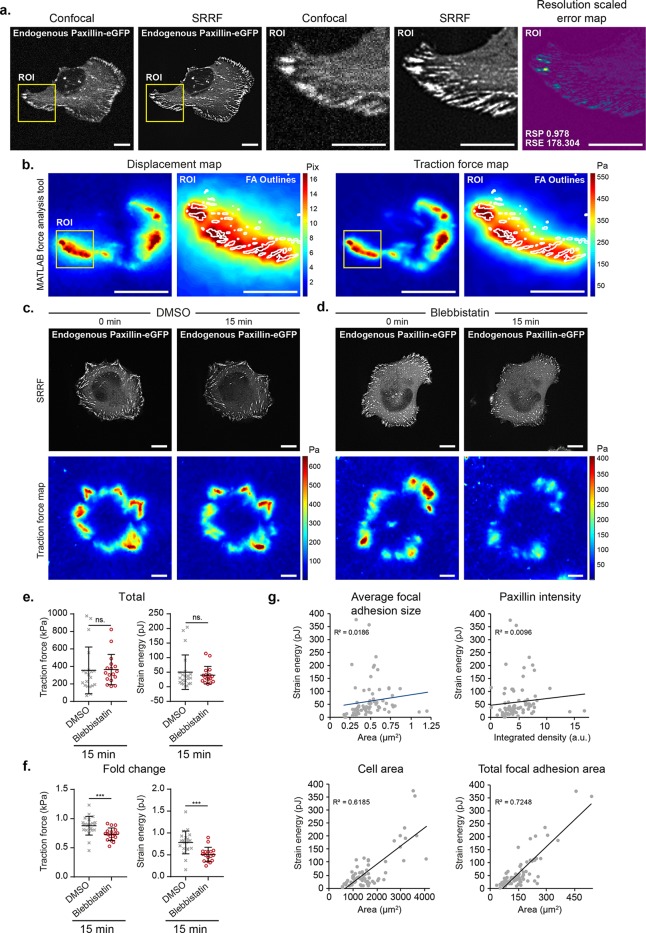
Cellular mechanics play a crucial role in tissue homeostasis and are often misregulated in disease. Traction force microscopy is one of the key methods that has enabled researchers to study fundamental aspects of mechanobiology; however, traction force microscopy is limited by poor resolution. Here, we propose a simplified protocol and imaging strategy that enhances the output of traction force microscopy by increasing i) achievable bead density and ii) the accuracy of bead tracking. Our approach relies on super-resolution microscopy, enabled by fluorescence fluctuation analysis. Our pipeline can be used on spinning-disk confocal or widefield microscopes and is compatible with available analysis software. In addition, we demonstrate that our workflow can be used to gain biologically relevant information and is suitable for fast long-term live measurement of traction forces even in light-sensitive cells. Finally, using fluctuation-based traction force microscopy, we observe that filopodia align to the force field generated by focal adhesions.
细胞力学在组织动态平衡中起着关键作用,在疾病中往往失调。牵引力显微镜是使研究人员能够研究机械生物学基本方面的关键方法之一;然而,牵引力显微镜受到分辨率差的限制。在这里,我们提出了一种简化的方案和成像策略,通过增加 i)可实现的珠密度和 ii)珠跟踪的准确性来提高牵引力显微镜的输出。我们的方法依赖于超分辨率显微镜,这得益于荧光波动分析。我们的流水线可用于旋转盘共聚焦或宽场显微镜,并且与可用的分析软件兼容。此外,我们证明我们的工作流程可以用于获取与生物学相关的信息,并且适用于快速长期的牵引力活测量,即使在对光敏感的细胞中也是如此。最后,使用基于波动的牵引力显微镜,我们观察到丝状伪足与由焦点黏附产生的力场对齐。