Department of Medical Physics and Bioengineering, University Hospitals Bristol NHS Foundation Trust, Bristol, BS28HW, UK.
Umea Functional Brain Imaging Center, Umea University, 901 87, Umea, Sweden.
Med Phys. 2020 Jun;47(6):2380-2391. doi: 10.1002/mp.14127. Epub 2020 Mar 31.
Many methods are available to segment structural magnetic resonance (MR) images of the brain into different tissue types. These have generally been developed for research purposes but there is some clinical use in the diagnosis of neurodegenerative diseases such as dementia. The potential exists for computed tomography (CT) segmentation to be used in place of MRI segmentation, but this will require a method to verify the accuracy of CT processing, particularly if algorithms developed for MR are used, as MR has notably greater tissue contrast.
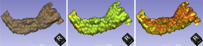
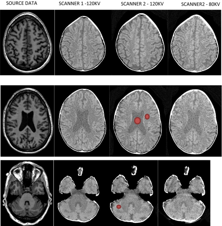
To investigate these issues we have created a three-dimensional (3D) printed brain with realistic Hounsfield unit (HU) values based on tissue maps segmented directly from an individual T1 MRI scan of a normal subject. Several T1 MRI scans of normal subjects from the ADNI database were segmented using SPM12 and used to create stereolithography files of different tissues for 3D printing. The attenuation properties of several material blends were investigated, and three suitable formulations were used to print an object expected to have realistic geometry and attenuation properties. A skull was simulated by coating the object with plaster of Paris impregnated bandages. Using two CT scanners, the realism of the phantom was assessed by the measurement of HU values, SPM12 segmentation and comparison with the source data used to create the phantom.
Realistic relative HU values were measured although a subtraction of 60 was required to obtain equivalence with the expected values (gray matter 32.9-35.8 phantom, 29.9-34.2 literature). Segmentation of images acquired at different kVps/mAs showed excellent agreement with the source data (Dice Similarity Coefficient 0.79 for gray matter). The performance of two scanners with two segmentation methods was compared, with the scanners found to have similar performance and with one segmentation method clearly superior to the other.
The ability to use 3D printing to create a realistic (in terms of geometry and attenuation properties) head phantom has been demonstrated and used in an initial assessment of CT segmentation accuracy using freely available software developed for MRI.
有许多方法可将大脑的结构磁共振(MR)图像分割成不同的组织类型。这些方法通常是为研究目的而开发的,但在痴呆等神经退行性疾病的诊断中也有一些临床应用。CT 分割有可能替代 MRI 分割,但这需要一种方法来验证 CT 处理的准确性,特别是如果使用为 MRI 开发的算法,因为 MR 具有明显更高的组织对比度。
为了研究这些问题,我们根据从正常受试者的 T1MR 扫描中直接分割的组织图谱,创建了一个具有真实亨氏单位(HU)值的三维(3D)打印脑。使用 SPM12 对 ADNI 数据库中的几个正常受试者的 T1MRI 扫描进行分割,并用于为 3D 打印创建不同组织的立体光刻文件。研究了几种材料混合物的衰减特性,并使用三种合适的配方打印了一个预期具有真实几何形状和衰减特性的物体。通过用石膏绷带浸渍的绷带涂层物体来模拟颅骨。使用两台 CT 扫描仪,通过测量 HU 值、SPM12 分割以及与用于创建幻影的源数据进行比较,评估了幻影的逼真度。
尽管需要减去 60 才能获得与预期值(灰质 32.9-35.8 幻影,29.9-34.2 文献)等效的逼真相对 HU 值。在不同 kVp/mAs 下采集的图像的分割与源数据显示出极好的一致性(灰质的骰子相似性系数为 0.79)。比较了两台扫描仪和两种分割方法的性能,发现扫描仪的性能相似,一种分割方法明显优于另一种。
已经证明了使用 3D 打印创建逼真(在几何形状和衰减特性方面)头部幻影的能力,并在使用为 MRI 开发的免费可用软件初步评估 CT 分割准确性方面进行了应用。