• 文献检索
  • 文档翻译
  • 深度研究
  • 学术资讯
  • Suppr Zotero 插件Zotero 插件
  • 邀请有礼
  • 套餐&价格
  • 历史记录
应用&插件
Suppr Zotero 插件Zotero 插件浏览器插件Mac 客户端Windows 客户端微信小程序
定价
高级版会员购买积分包购买API积分包
服务
文献检索文档翻译深度研究API 文档MCP 服务
关于我们
关于 Suppr公司介绍联系我们用户协议隐私条款
关注我们

Suppr 超能文献

核心技术专利:CN118964589B侵权必究
粤ICP备2023148730 号-1Suppr @ 2026

文献检索

告别复杂PubMed语法,用中文像聊天一样搜索,搜遍4000万医学文献。AI智能推荐,让科研检索更轻松。

立即免费搜索

文件翻译

保留排版,准确专业,支持PDF/Word/PPT等文件格式,支持 12+语言互译。

免费翻译文档

深度研究

AI帮你快速写综述,25分钟生成高质量综述,智能提取关键信息,辅助科研写作。

立即免费体验

通过人工智能搜索存档组织病理学图像实现全癌诊断共识。

Pan-cancer diagnostic consensus through searching archival histopathology images using artificial intelligence.

作者信息

Kalra Shivam, Tizhoosh H R, Shah Sultaan, Choi Charles, Damaskinos Savvas, Safarpoor Amir, Shafiei Sobhan, Babaie Morteza, Diamandis Phedias, Campbell Clinton J V, Pantanowitz Liron

机构信息

Huron Digital Pathology, St. Jacobs, ON Canada.

2Kimia Lab, University of Waterloo, Waterloo, ON Canada.

出版信息

NPJ Digit Med. 2020 Mar 10;3:31. doi: 10.1038/s41746-020-0238-2. eCollection 2020.

DOI:10.1038/s41746-020-0238-2
PMID:32195366
原文链接:https://pmc.ncbi.nlm.nih.gov/articles/PMC7064517/
Abstract

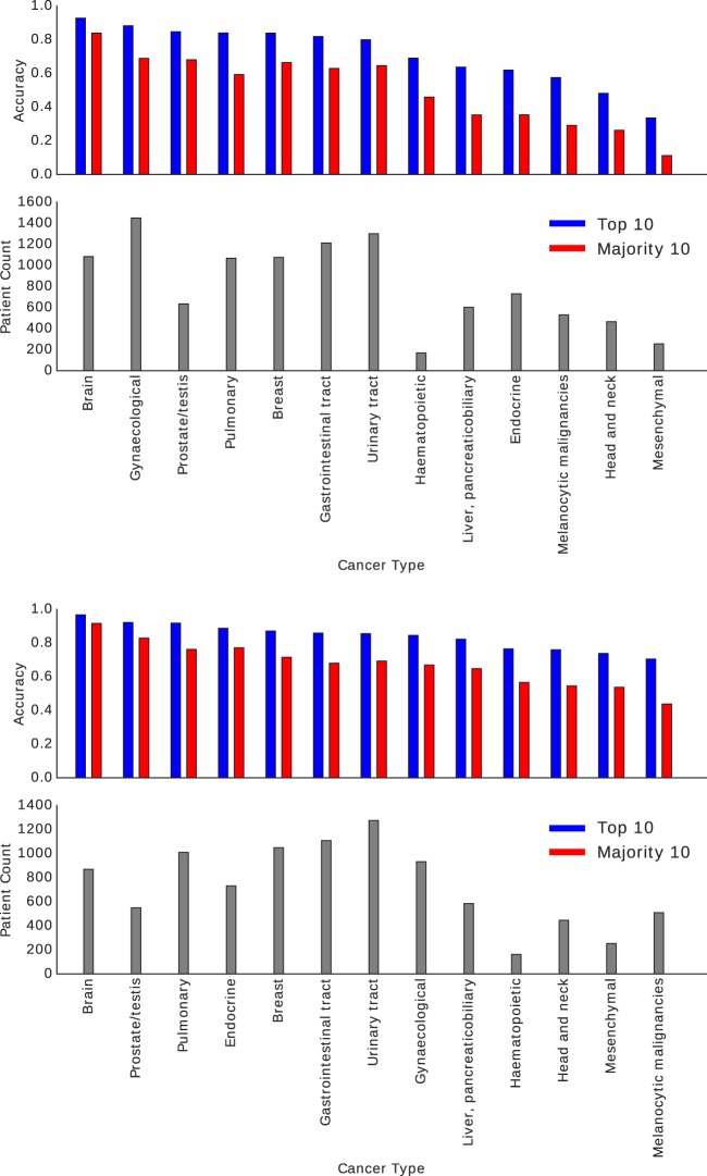
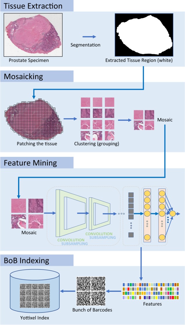
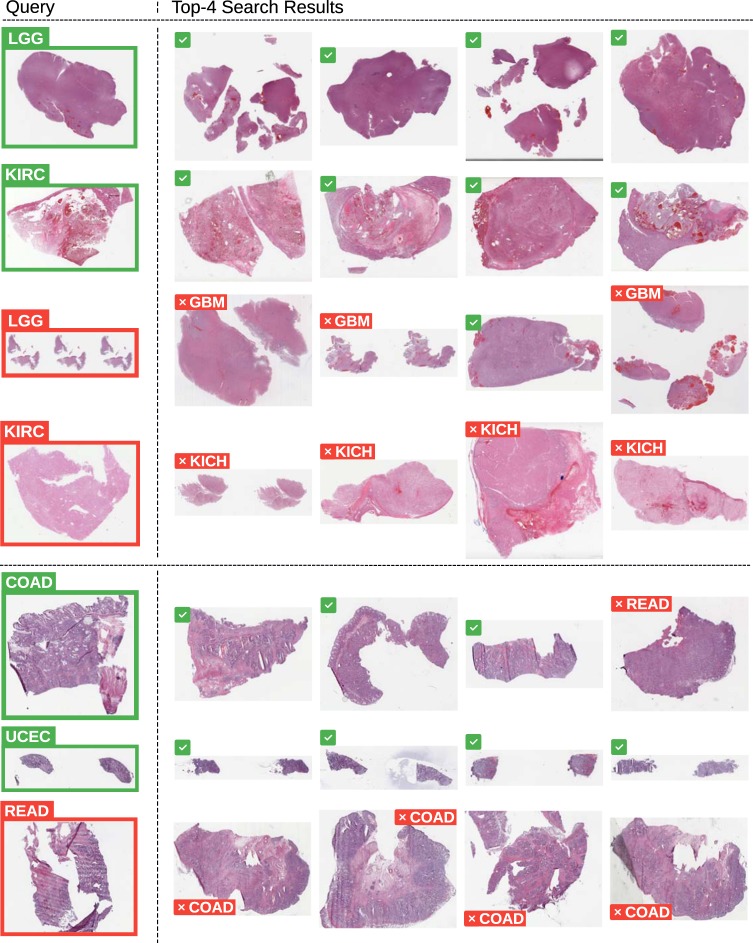
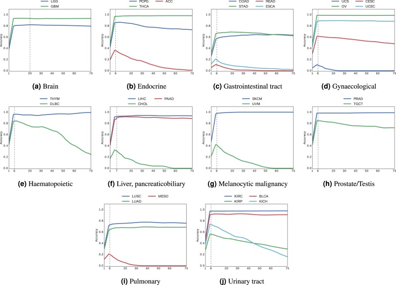
The emergence of digital pathology has opened new horizons for histopathology. Artificial intelligence (AI) algorithms are able to operate on digitized slides to assist pathologists with different tasks. Whereas AI-involving classification and segmentation methods have obvious benefits for image analysis, image search represents a fundamental shift in computational pathology. Matching the pathology of new patients with already diagnosed and curated cases offers pathologists a new approach to improve diagnostic accuracy through visual inspection of similar cases and computational majority vote for consensus building. In this study, we report the results from searching the largest public repository (The Cancer Genome Atlas, TCGA) of whole-slide images from almost 11,000 patients. We successfully indexed and searched almost 30,000 high-resolution digitized slides constituting 16 terabytes of data comprised of 20 million 1000 × 1000 pixels image patches. The TCGA image database covers 25 anatomic sites and contains 32 cancer subtypes. High-performance storage and GPU power were employed for experimentation. The results were assessed with conservative "majority voting" to build consensus for subtype diagnosis through vertical search and demonstrated high accuracy values for both frozen section slides (e.g., bladder urothelial carcinoma 93%, kidney renal clear cell carcinoma 97%, and ovarian serous cystadenocarcinoma 99%) and permanent histopathology slides (e.g., prostate adenocarcinoma 98%, skin cutaneous melanoma 99%, and thymoma 100%). The key finding of this validation study was that computational consensus appears to be possible for rendering diagnoses if a sufficiently large number of searchable cases are available for each cancer subtype.

摘要

数字病理学的出现为组织病理学开辟了新的视野。人工智能(AI)算法能够对数字化切片进行操作,以协助病理学家完成不同的任务。虽然涉及AI的分类和分割方法在图像分析方面具有明显优势,但图像搜索代表了计算病理学的一个根本性转变。将新患者的病理学与已诊断和整理的病例进行匹配,为病理学家提供了一种新方法,通过目视检查相似病例并进行计算多数投票以达成共识,从而提高诊断准确性。在本研究中,我们报告了在近11000名患者的全切片图像的最大公共存储库(癌症基因组图谱,TCGA)中进行搜索的结果。我们成功地对近30000张高分辨率数字化切片进行了索引和搜索,这些切片构成了16太字节的数据,由2000万个1000×1000像素的图像块组成。TCGA图像数据库涵盖25个解剖部位,包含32种癌症亚型。实验采用了高性能存储和GPU算力。通过垂直搜索,用保守的“多数投票”对结果进行评估,以达成亚型诊断的共识,结果显示冰冻切片(如膀胱尿路上皮癌93%、肾透明细胞癌97%、卵巢浆液性囊腺癌99%)和永久性组织病理学切片(如前列腺腺癌98%、皮肤黑色素瘤99%、胸腺瘤100%)的诊断准确率都很高。这项验证研究的关键发现是,如果每个癌症亚型有足够数量的可搜索病例,那么通过计算达成诊断共识似乎是可行的。

https://cdn.ncbi.nlm.nih.gov/pmc/blobs/f5d2/7064517/bfecd3a89cae/41746_2020_238_Fig9_HTML.jpg
https://cdn.ncbi.nlm.nih.gov/pmc/blobs/f5d2/7064517/a1216708b3b3/41746_2020_238_Fig1_HTML.jpg
https://cdn.ncbi.nlm.nih.gov/pmc/blobs/f5d2/7064517/b1ba09cfb428/41746_2020_238_Fig2_HTML.jpg
https://cdn.ncbi.nlm.nih.gov/pmc/blobs/f5d2/7064517/2a0093fe1731/41746_2020_238_Fig3_HTML.jpg
https://cdn.ncbi.nlm.nih.gov/pmc/blobs/f5d2/7064517/b25cfb11d6b5/41746_2020_238_Fig4_HTML.jpg
https://cdn.ncbi.nlm.nih.gov/pmc/blobs/f5d2/7064517/2494ef28dc7b/41746_2020_238_Fig5_HTML.jpg
https://cdn.ncbi.nlm.nih.gov/pmc/blobs/f5d2/7064517/c5927cab4264/41746_2020_238_Fig6_HTML.jpg
https://cdn.ncbi.nlm.nih.gov/pmc/blobs/f5d2/7064517/ae55270540a5/41746_2020_238_Fig7_HTML.jpg
https://cdn.ncbi.nlm.nih.gov/pmc/blobs/f5d2/7064517/0b4d676d208e/41746_2020_238_Fig8_HTML.jpg
https://cdn.ncbi.nlm.nih.gov/pmc/blobs/f5d2/7064517/bfecd3a89cae/41746_2020_238_Fig9_HTML.jpg

相似文献

1
Pan-cancer diagnostic consensus through searching archival histopathology images using artificial intelligence.通过人工智能搜索存档组织病理学图像实现全癌诊断共识。
NPJ Digit Med. 2020 Mar 10;3:31. doi: 10.1038/s41746-020-0238-2. eCollection 2020.
2
A Fully Automated Artificial Intelligence System to Assist Pathologists' Diagnosis to Predict Histologically High-grade Urothelial Carcinoma from Digitized Urine Cytology Slides Using Deep Learning.一种全自动人工智能系统,利用深度学习从数字化尿液细胞学载玻片辅助病理学家诊断预测组织学高级别尿路上皮癌。
Eur Urol Oncol. 2024 Apr;7(2):258-265. doi: 10.1016/j.euo.2023.11.009. Epub 2023 Dec 7.
3
Recognition of Cutaneous Melanoma on Digitized Histopathological Slides Artificial Intelligence Algorithm.数字化组织病理切片上皮肤黑色素瘤的识别 人工智能算法
Front Oncol. 2020 Aug 20;10:1559. doi: 10.3389/fonc.2020.01559. eCollection 2020.
4
Automated diagnosis of 7 canine skin tumors using machine learning on H&E-stained whole slide images.基于 H&E 染色全切片图像的机器学习对 7 例犬皮肤肿瘤的自动诊断
Vet Pathol. 2023 Nov;60(6):865-875. doi: 10.1177/03009858231189205. Epub 2023 Jul 29.
5
AI (artificial intelligence) in histopathology--from image analysis to automated diagnosis.组织病理学中的人工智能——从图像分析到自动诊断。
Folia Histochem Cytobiol. 2009 Jan;47(3):355-61. doi: 10.2478/v10042-009-0087-y.
6
AI Deployment on GBM Diagnosis: A Novel Approach to Analyze Histopathological Images Using Image Feature-Based Analysis.人工智能在胶质母细胞瘤诊断中的应用:一种基于图像特征分析的组织病理学图像分析新方法。
Cancers (Basel). 2023 Oct 19;15(20):5063. doi: 10.3390/cancers15205063.
7
An artificial intelligence algorithm for prostate cancer diagnosis in whole slide images of core needle biopsies: a blinded clinical validation and deployment study.一种用于经皮穿刺活检全切片图像中前列腺癌诊断的人工智能算法:一项盲法临床验证与应用研究。
Lancet Digit Health. 2020 Aug;2(8):e407-e416. doi: 10.1016/S2589-7500(20)30159-X.
8
Whole slide imaging equivalency and efficiency study: experience at a large academic center.全 slides 成像等效性和效率研究:大型学术中心的经验。
Mod Pathol. 2019 Jul;32(7):916-928. doi: 10.1038/s41379-019-0205-0. Epub 2019 Feb 18.
9
Semi-supervised training of deep convolutional neural networks with heterogeneous data and few local annotations: An experiment on prostate histopathology image classification.基于异构数据和少量局部标注的深度卷积神经网络的半监督学习:前列腺组织病理学图像分类实验。
Med Image Anal. 2021 Oct;73:102165. doi: 10.1016/j.media.2021.102165. Epub 2021 Jul 14.
10
Evaluation of panoramic digital images using Panoptiq for frozen section diagnosis.使用Panoptiq对全景数字图像进行评估以用于冰冻切片诊断。
J Pathol Inform. 2016 May 4;7:26. doi: 10.4103/2153-3539.181770. eCollection 2016.

引用本文的文献

1
Enhancing image retrieval through optimal barcode representation.通过优化条形码表示增强图像检索。
Sci Rep. 2025 Aug 7;15(1):28847. doi: 10.1038/s41598-025-14576-x.
2
Histology image analysis of 13 healthy tissues reveals molecular-histological correlations.对13种健康组织的组织学图像分析揭示了分子与组织学之间的相关性。
Sci Rep. 2025 Jul 23;15(1):26812. doi: 10.1038/s41598-025-11853-7.
3
Applications of artificial intelligence in the analysis of histopathology images of gliomas: a review.人工智能在胶质瘤组织病理学图像分析中的应用:综述

本文引用的文献

1
Yottixel - An Image Search Engine for Large Archives of Histopathology Whole Slide Images.Yottixel——一个用于大型组织病理学全切片图像档案的图像搜索引擎。
Med Image Anal. 2020 Oct;65:101757. doi: 10.1016/j.media.2020.101757. Epub 2020 Jun 24.
2
Clinical-grade computational pathology using weakly supervised deep learning on whole slide images.基于全切片图像的弱监督深度学习的临床级计算病理学。
Nat Med. 2019 Aug;25(8):1301-1309. doi: 10.1038/s41591-019-0508-1. Epub 2019 Jul 15.
3
Digital pathology and artificial intelligence.数字病理学与人工智能。
Npj Imaging. 2024 Jul 1;2(1):16. doi: 10.1038/s44303-024-00020-8.
4
Pathomics Image Analysis of Tumor Infiltrating Lymphocytes (TILs) in Colon Cancer.结肠癌中肿瘤浸润淋巴细胞(TILs)的病理组学图像分析
Res Sq. 2025 Apr 1:rs.3.rs-6173056. doi: 10.21203/rs.3.rs-6173056/v1.
5
Foundation Models for Histopathology-Fanfare or Flair.组织病理学基础模型——是大张旗鼓还是虚张声势。
Mayo Clin Proc Digit Health. 2024 Mar 5;2(1):165-174. doi: 10.1016/j.mcpdig.2024.02.003. eCollection 2024 Mar.
6
Digital pathology and artificial intelligence in renal cell carcinoma focusing on feature extraction: a literature review.聚焦特征提取的肾细胞癌数字病理学与人工智能:文献综述
Front Oncol. 2025 Jan 24;15:1516264. doi: 10.3389/fonc.2025.1516264. eCollection 2025.
7
Using digital pathology to standardize and automate histological evaluations of environmental samples.利用数字病理学实现环境样本组织学评估的标准化和自动化。
Environ Toxicol Chem. 2025 Feb 1;44(2):306-317. doi: 10.1093/etojnl/vgae038.
8
Validation of histopathology foundation models through whole slide image retrieval.通过全切片图像检索验证组织病理学基础模型。
Sci Rep. 2025 Feb 1;15(1):3990. doi: 10.1038/s41598-025-88545-9.
9
Clinical Validation of Deep Learning for Segmentation of Multiple Dental Features in Periapical Radiographs.深度学习用于根尖片多牙特征分割的临床验证
Bioengineering (Basel). 2024 Oct 5;11(10):1001. doi: 10.3390/bioengineering11101001.
10
Automatic feature segmentation in dental panoramic radiographs.口腔全景 X 光片的自动特征分割。
Sci Prog. 2024 Oct-Dec;107(4):368504241286659. doi: 10.1177/00368504241286659.
Lancet Oncol. 2019 May;20(5):e253-e261. doi: 10.1016/S1470-2045(19)30154-8.
4
A Fast and Refined Cancer Regions Segmentation Framework in Whole-slide Breast Pathological Images.一种用于全玻片乳腺病理图像的快速精细化癌症区域分割框架。
Sci Rep. 2019 Jan 29;9(1):882. doi: 10.1038/s41598-018-37492-9.
5
Digital pathology: Review of current opportunities and challenges for oral pathologists.数字病理学:口腔病理学家当前机遇与挑战的综述。
J Oral Pathol Med. 2019 Apr;48(4):263-269. doi: 10.1111/jop.12825. Epub 2019 Feb 8.
6
Artificial Intelligence and Digital Pathology: Challenges and Opportunities.人工智能与数字病理学:挑战与机遇
J Pathol Inform. 2018 Nov 14;9:38. doi: 10.4103/jpi.jpi_53_18. eCollection 2018.
7
Machine Learning Methods for Histopathological Image Analysis.用于组织病理学图像分析的机器学习方法
Comput Struct Biotechnol J. 2018 Feb 9;16:34-42. doi: 10.1016/j.csbj.2018.01.001. eCollection 2018.
8
Review of diagnostic error in anatomical pathology and the role and value of second opinions in error prevention.解剖病理学中诊断错误的回顾,以及第二意见在错误预防中的作用和价值。
J Clin Pathol. 2018 Nov;71(11):995-1000. doi: 10.1136/jclinpath-2018-205226. Epub 2018 Aug 1.
9
Representing Medical Images With Encoded Local Projections.用编码局部投影表示医学图像。
IEEE Trans Biomed Eng. 2018 Oct;65(10):2267-2277. doi: 10.1109/TBME.2018.2791567. Epub 2018 Jan 10.
10
Deep Learning in Microscopy Image Analysis: A Survey.深度学习在显微镜图像分析中的应用:综述。
IEEE Trans Neural Netw Learn Syst. 2018 Oct;29(10):4550-4568. doi: 10.1109/TNNLS.2017.2766168. Epub 2017 Nov 22.