• 文献检索
  • 文档翻译
  • 深度研究
  • 学术资讯
  • Suppr Zotero 插件Zotero 插件
  • 邀请有礼
  • 套餐&价格
  • 历史记录
应用&插件
Suppr Zotero 插件Zotero 插件浏览器插件Mac 客户端Windows 客户端微信小程序
定价
高级版会员购买积分包购买API积分包
服务
文献检索文档翻译深度研究API 文档MCP 服务
关于我们
关于 Suppr公司介绍联系我们用户协议隐私条款
关注我们

Suppr 超能文献

核心技术专利:CN118964589B侵权必究
粤ICP备2023148730 号-1Suppr @ 2026

文献检索

告别复杂PubMed语法,用中文像聊天一样搜索,搜遍4000万医学文献。AI智能推荐,让科研检索更轻松。

立即免费搜索

文件翻译

保留排版,准确专业,支持PDF/Word/PPT等文件格式,支持 12+语言互译。

免费翻译文档

深度研究

AI帮你快速写综述,25分钟生成高质量综述,智能提取关键信息,辅助科研写作。

立即免费体验

通过无监督模糊聚类对CT图像上的全肿瘤负荷进行组织特异性和可解释的子分割。

Tissue-specific and interpretable sub-segmentation of whole tumour burden on CT images by unsupervised fuzzy clustering.

作者信息

Rundo Leonardo, Beer Lucian, Ursprung Stephan, Martin-Gonzalez Paula, Markowetz Florian, Brenton James D, Crispin-Ortuzar Mireia, Sala Evis, Woitek Ramona

机构信息

Department of Radiology, University of Cambridge, Cambridge CB2 0QQ, UK; Cancer Research UK Cambridge Centre, University of Cambridge, Cambridge CB2 0RE, UK.

Department of Radiology, University of Cambridge, Cambridge CB2 0QQ, UK; Cancer Research UK Cambridge Centre, University of Cambridge, Cambridge CB2 0RE, UK; Department of Biomedical Imaging and Image-guided Therapy, Medical University Vienna, Vienna 1090, Austria.

出版信息

Comput Biol Med. 2020 May;120:103751. doi: 10.1016/j.compbiomed.2020.103751. Epub 2020 Apr 10.

DOI:10.1016/j.compbiomed.2020.103751
PMID:32421652
原文链接:https://pmc.ncbi.nlm.nih.gov/articles/PMC7248575/
Abstract

BACKGROUND

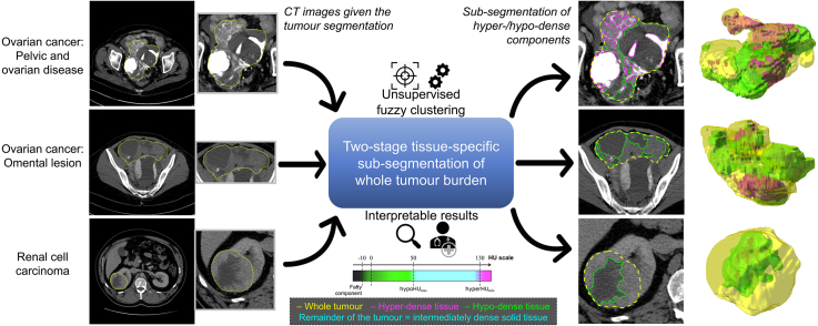
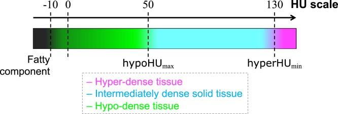
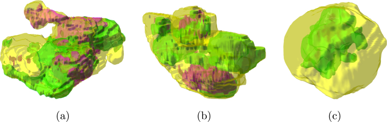
Cancer typically exhibits genotypic and phenotypic heterogeneity, which can have prognostic significance and influence therapy response. Computed Tomography (CT)-based radiomic approaches calculate quantitative features of tumour heterogeneity at a mesoscopic level, regardless of macroscopic areas of hypo-dense (i.e., cystic/necrotic), hyper-dense (i.e., calcified), or intermediately dense (i.e., soft tissue) portions.

METHOD

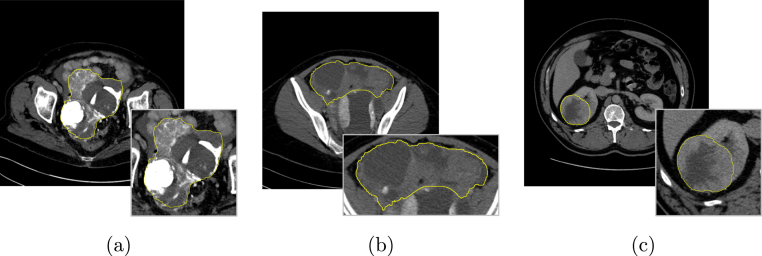
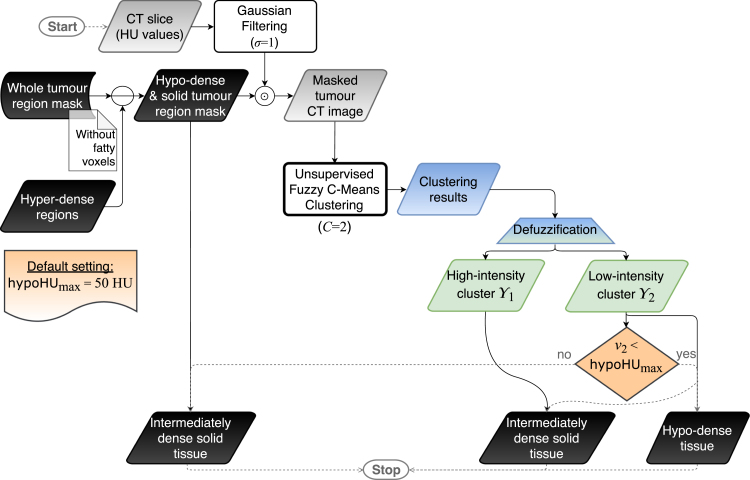
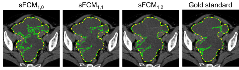
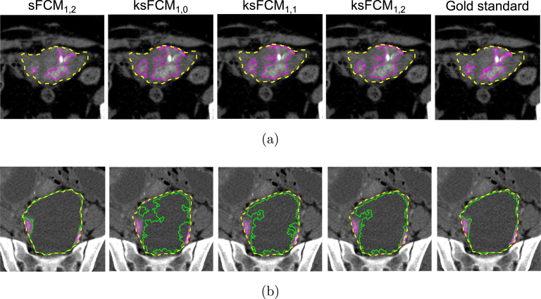
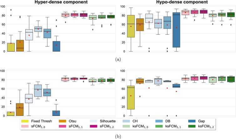
With the goal of achieving the automated sub-segmentation of these three tissue types, we present here a two-stage computational framework based on unsupervised Fuzzy C-Means Clustering (FCM) techniques. No existing approach has specifically addressed this task so far. Our tissue-specific image sub-segmentation was tested on ovarian cancer (pelvic/ovarian and omental disease) and renal cell carcinoma CT datasets using both overlap-based and distance-based metrics for evaluation.

RESULTS

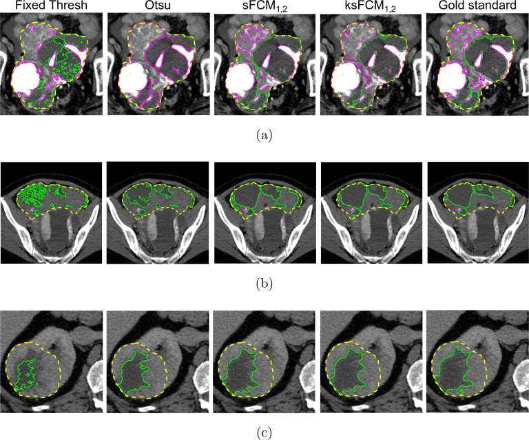
On all tested sub-segmentation tasks, our two-stage segmentation approach outperformed conventional segmentation techniques: fixed multi-thresholding, the Otsu method, and automatic cluster number selection heuristics for the K-means clustering algorithm. In addition, experiments showed that the integration of the spatial information into the FCM algorithm generally achieves more accurate segmentation results, whilst the kernelised FCM versions are not beneficial. The best spatial FCM configuration achieved average Dice similarity coefficient values starting from 81.94±4.76 and 83.43±3.81 for hyper-dense and hypo-dense components, respectively, for the investigated sub-segmentation tasks.

CONCLUSIONS

The proposed intelligent framework could be readily integrated into clinical research environments and provides robust tools for future radiomic biomarker validation.

摘要

背景

癌症通常表现出基因和表型异质性,这可能具有预后意义并影响治疗反应。基于计算机断层扫描(CT)的放射组学方法可在介观水平计算肿瘤异质性的定量特征,而无需考虑低密度(即囊性/坏死性)、高密度(即钙化)或中等密度(即软组织)部分的宏观区域。

方法

为了实现这三种组织类型的自动子分割,我们在此提出一种基于无监督模糊C均值聚类(FCM)技术的两阶段计算框架。到目前为止,尚无现有方法专门解决此任务。我们的组织特异性图像子分割在卵巢癌(盆腔/卵巢和网膜疾病)和肾细胞癌CT数据集上进行了测试,使用基于重叠和基于距离的指标进行评估。

结果

在所有测试的子分割任务中,我们的两阶段分割方法优于传统分割技术:固定多阈值分割、大津法以及K均值聚类算法的自动聚类数选择启发式方法。此外,实验表明,将空间信息集成到FCM算法中通常可获得更准确的分割结果,而核化的FCM版本并无益处。对于所研究的子分割任务,最佳空间FCM配置分别为高密度和低密度成分实现的平均骰子相似系数值,起始值分别为81.94±4.76和83.43±3.81。

结论

所提出的智能框架可轻松集成到临床研究环境中,并为未来放射组学生物标志物验证提供强大工具。

https://cdn.ncbi.nlm.nih.gov/pmc/blobs/cc3e/7248575/23cd81051f4c/gr11.jpg
https://cdn.ncbi.nlm.nih.gov/pmc/blobs/cc3e/7248575/889a674ed4f8/fx1.jpg
https://cdn.ncbi.nlm.nih.gov/pmc/blobs/cc3e/7248575/331dc4957795/gr1.jpg
https://cdn.ncbi.nlm.nih.gov/pmc/blobs/cc3e/7248575/38b3a21c0dda/gr2.jpg
https://cdn.ncbi.nlm.nih.gov/pmc/blobs/cc3e/7248575/e694e683a421/gr3.jpg
https://cdn.ncbi.nlm.nih.gov/pmc/blobs/cc3e/7248575/c3b0c63a61c8/gr4.jpg
https://cdn.ncbi.nlm.nih.gov/pmc/blobs/cc3e/7248575/35bc4e351de3/gr5.jpg
https://cdn.ncbi.nlm.nih.gov/pmc/blobs/cc3e/7248575/0be7d0258443/gr6.jpg
https://cdn.ncbi.nlm.nih.gov/pmc/blobs/cc3e/7248575/517ec12d4f55/gr7.jpg
https://cdn.ncbi.nlm.nih.gov/pmc/blobs/cc3e/7248575/9e29c35e7004/gr8.jpg
https://cdn.ncbi.nlm.nih.gov/pmc/blobs/cc3e/7248575/e486ac7092c4/gr9.jpg
https://cdn.ncbi.nlm.nih.gov/pmc/blobs/cc3e/7248575/2481368fdb19/gr10.jpg
https://cdn.ncbi.nlm.nih.gov/pmc/blobs/cc3e/7248575/23cd81051f4c/gr11.jpg

相似文献

1
Tissue-specific and interpretable sub-segmentation of whole tumour burden on CT images by unsupervised fuzzy clustering.通过无监督模糊聚类对CT图像上的全肿瘤负荷进行组织特异性和可解释的子分割。
Comput Biol Med. 2020 May;120:103751. doi: 10.1016/j.compbiomed.2020.103751. Epub 2020 Apr 10.
2
Learning fuzzy clustering for SPECT/CT segmentation via convolutional neural networks.通过卷积神经网络学习用于SPECT/CT分割的模糊聚类
Med Phys. 2021 Jul;48(7):3860-3877. doi: 10.1002/mp.14903. Epub 2021 May 28.
3
A novel fuzzy C-means algorithm for unsupervised heterogeneous tumor quantification in PET.一种用于 PET 中无监督异质肿瘤定量的新型模糊 C 均值算法。
Med Phys. 2010 Mar;37(3):1309-24. doi: 10.1118/1.3301610.
4
Brain tissue segmentation using fuzzy clustering techniques.使用模糊聚类技术进行脑组织分割。
Technol Health Care. 2015;23(5):571-80. doi: 10.3233/THC-151012.
5
Automated fibroglandular tissue segmentation and volumetric density estimation in breast MRI using an atlas-aided fuzzy C-means method.基于图谱辅助模糊 C 均值法的乳腺 MRI 中纤维腺体组织自动分割及容积密度估测
Med Phys. 2013 Dec;40(12):122302. doi: 10.1118/1.4829496.
6
Brain tumor segmentation approach based on the extreme learning machine and significantly fast and robust fuzzy C-means clustering algorithms running on Raspberry Pi hardware.基于极端学习机和显著快速稳健的模糊 C 均值聚类算法在 Raspberry Pi 硬件上运行的脑肿瘤分割方法。
Med Hypotheses. 2020 Mar;136:109507. doi: 10.1016/j.mehy.2019.109507. Epub 2019 Nov 18.
7
A segmentation of brain MRI images utilizing intensity and contextual information by Markov random field.利用马尔可夫随机场的强度和上下文信息对脑 MRI 图像进行分割。
Comput Assist Surg (Abingdon). 2017 Dec;22(sup1):200-211. doi: 10.1080/24699322.2017.1389398. Epub 2017 Oct 26.
8
Dynamically learned PSO based neighborhood influenced fuzzy c-means for pre-treatment and post-treatment organ segmentation from CT images.基于动态学习粒子群优化算法的邻域影响模糊 c 均值算法用于 CT 图像中治疗前和治疗后器官分割
Comput Methods Programs Biomed. 2021 Apr;202:105971. doi: 10.1016/j.cmpb.2021.105971. Epub 2021 Feb 4.
9
Suspicious Lesion Segmentation on Brain, Mammograms and Breast MR Images Using New Optimized Spatial Feature Based Super-Pixel Fuzzy C-Means Clustering.基于新型优化空间特征的超像素模糊 C 均值聚类的脑、乳腺 X 线摄影术及乳腺磁共振图像可疑病变分割。
J Digit Imaging. 2019 Apr;32(2):322-335. doi: 10.1007/s10278-018-0149-9.
10
Efficient model-informed co-segmentation of tumors on PET/CT driven by clustering and classification information.基于聚类和分类信息驱动的 PET/CT 上肿瘤的高效模型引导的共分割。
Comput Biol Med. 2024 Sep;180:108980. doi: 10.1016/j.compbiomed.2024.108980. Epub 2024 Aug 12.

引用本文的文献

1
Evolving and Novel Applications of Artificial Intelligence in Cancer Imaging.人工智能在癌症成像中的不断发展与新应用
Cancers (Basel). 2025 Apr 30;17(9):1510. doi: 10.3390/cancers17091510.
2
Fuzzy C-means clustering algorithm applied in computed tomography images of patients with intracranial hemorrhage.模糊C均值聚类算法在颅内出血患者计算机断层扫描图像中的应用。
Front Neuroinform. 2024 Oct 23;18:1440304. doi: 10.3389/fninf.2024.1440304. eCollection 2024.
3
PET/CT-based 3D multi-class semantic segmentation of ovarian cancer and the stability of the extracted radiomics features.

本文引用的文献

1
A novel framework for MR image segmentation and quantification by using MedGA.利用 MedGA 实现磁共振图像分割和定量分析的新框架
Comput Methods Programs Biomed. 2019 Jul;176:159-172. doi: 10.1016/j.cmpb.2019.04.016. Epub 2019 Apr 17.
2
A Road Map for Translational Research on Artificial Intelligence in Medical Imaging: From the 2018 National Institutes of Health/RSNA/ACR/The Academy Workshop.人工智能在医学影像转化研究路线图:来自 2018 年美国国立卫生研究院/北美放射学会/美国放射学院/美国学院的研讨会。
J Am Coll Radiol. 2019 Sep;16(9 Pt A):1179-1189. doi: 10.1016/j.jacr.2019.04.014. Epub 2019 May 28.
3
Radiological images and machine learning: Trends, perspectives, and prospects.
基于PET/CT的卵巢癌三维多类别语义分割及提取的影像组学特征的稳定性
Phys Eng Sci Med. 2024 Dec;47(4):1739-1749. doi: 10.1007/s13246-024-01485-y. Epub 2024 Sep 23.
4
AI-based automated segmentation for ovarian/adnexal masses and their internal components on ultrasound imaging.基于人工智能的超声成像中卵巢/附件肿块及其内部成分的自动分割
J Med Imaging (Bellingham). 2024 Jul;11(4):044505. doi: 10.1117/1.JMI.11.4.044505. Epub 2024 Aug 6.
5
Integrated radiogenomics models predict response to neoadjuvant chemotherapy in high grade serous ovarian cancer.整合放射基因组学模型预测高级别浆液性卵巢癌对新辅助化疗的反应。
Nat Commun. 2023 Oct 24;14(1):6756. doi: 10.1038/s41467-023-41820-7.
6
CT-based deep learning segmentation of ovarian cancer and the stability of the extracted radiomics features.基于CT的卵巢癌深度学习分割及提取的影像组学特征的稳定性
Quant Imaging Med Surg. 2023 Aug 1;13(8):5218-5229. doi: 10.21037/qims-22-1135. Epub 2023 Jun 13.
7
Predictive Factors of Local Recurrence after Colorectal Cancer Liver Metastases Thermal Ablation.结直肠癌肝转移热消融术后局部复发的预测因素
J Imaging. 2023 Mar 10;9(3):66. doi: 10.3390/jimaging9030066.
8
Fully Automated Segmentation Models of Supratentorial Meningiomas Assisted by Inclusion of Normal Brain Images.纳入正常脑图像辅助的幕上脑膜瘤全自动分割模型
J Imaging. 2022 Dec 15;8(12):327. doi: 10.3390/jimaging8120327.
9
Artificial intelligence and machine learning in cancer imaging.癌症成像中的人工智能与机器学习
Commun Med (Lond). 2022 Oct 27;2:133. doi: 10.1038/s43856-022-00199-0. eCollection 2022.
10
Neural-Symbolic Ensemble Learning for early-stage prediction of critical state of Covid-19 patients.基于神经符号集成学习的新冠肺炎患者危急状态早期预测。
Med Biol Eng Comput. 2022 Dec;60(12):3461-3474. doi: 10.1007/s11517-022-02674-1. Epub 2022 Oct 6.
放射影像学与机器学习:趋势、视角与展望。
Comput Biol Med. 2019 May;108:354-370. doi: 10.1016/j.compbiomed.2019.02.017. Epub 2019 Feb 27.
4
Explainable artificial intelligence for breast cancer: A visual case-based reasoning approach.可解释人工智能在乳腺癌中的应用:一种基于案例的可视化推理方法。
Artif Intell Med. 2019 Mar;94:42-53. doi: 10.1016/j.artmed.2019.01.001. Epub 2019 Jan 14.
5
Constrained-CNN losses for weakly supervised segmentation.约束卷积神经网络损失的弱监督分割。
Med Image Anal. 2019 May;54:88-99. doi: 10.1016/j.media.2019.02.009. Epub 2019 Feb 13.
6
How clinical imaging can assess cancer biology.临床影像学如何评估癌症生物学。
Insights Imaging. 2019 Mar 4;10(1):28. doi: 10.1186/s13244-019-0703-0.
7
A mathematical-descriptor of tumor-mesoscopic-structure from computed-tomography images annotates prognostic- and molecular-phenotypes of epithelial ovarian cancer.从计算机断层扫描图像中提取的肿瘤介观结构的数学描述符可标注上皮性卵巢癌的预后和分子表型。
Nat Commun. 2019 Feb 15;10(1):764. doi: 10.1038/s41467-019-08718-9.
8
Diagnostic Imaging for Solid Renal Tumors: A Pictorial Review.实性肾肿瘤的诊断性影像学:图文综述
Kidney Cancer. 2018 Aug 1;2(2):79-93. doi: 10.3233/KCA-180028.
9
Association between CT-texture-derived tumor heterogeneity, outcomes, and BRCA mutation status in patients with high-grade serous ovarian cancer.高级别浆液性卵巢癌患者 CT 纹理特征肿瘤异质性与结局及 BRCA 基因突变状态的相关性研究。
Abdom Radiol (NY). 2019 Jun;44(6):2040-2047. doi: 10.1007/s00261-018-1840-5.
10
Enhanced detection of circulating tumor DNA by fragment size analysis.通过片段大小分析增强循环肿瘤 DNA 的检测。
Sci Transl Med. 2018 Nov 7;10(466). doi: 10.1126/scitranslmed.aat4921.