Kang Sohee
Department of Dentistry, Yeungnam University Hospital, Daegu, Korea.
Yeungnam Univ J Med. 2020 Jul;37(3):217-225. doi: 10.12701/yujm.2020.00248. Epub 2020 May 22.
To provide a long-term bacterial seal through the formation of reparative dentin bridge, calcium silicate-based pulp capping materials have been used at sites of pulpal exposure. The aim of this study was to evaluate the mineralization-inducing potentials of calcium silicate-based pulp capping materials (ProRoot MTA [PR], Biodentine [BD], and TheraCal LC [TC]) in human dental pulp cells (HDPCs).
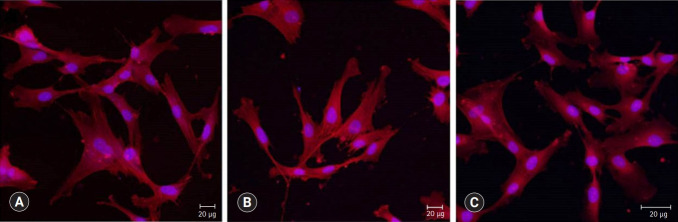
Specimens of test materials were placed in deionized water for various incubation times to measure the pH variation and the concentration of calcium released. The morphology of HDPCs cultured on the specimens was examined using a confocal laser scanning microscope (CLSM). Alizarin red S staining and alkaline phosphatase assays were used to evaluate mineralization-inducing potentials of the capping materials.
BD showed the highest calcium release in all test periods, followed by PR and TC. (p<0.05). All experimental groups showed high alkalinity after 1 day, except at 14 days. BD showed the highest cell viability compared with PR and TC after 1 and 3 days, while TC showed the lowest value (p<0.05). The CLSM analysis showed that cells were well adhered and expressed actin filaments for all pulp capping materials. Mineralization by PR and BD groups was higher than that by TC group based on alizarin red S staining. BD showed significantly higher alkaline phosphatase activity than PR and TC, while TC showed the lowest value (p<0.05).
Within the limitations of the in vitro study, BD had higher mineralization-inducing potential than PR and TC.
为了通过形成修复性牙本质桥来提供长期的细菌封闭,基于硅酸钙的牙髓盖髓材料已被用于牙髓暴露部位。本研究的目的是评估基于硅酸钙的牙髓盖髓材料(ProRoot MTA [PR]、Biodentine [BD] 和 TheraCal LC [TC])在人牙髓细胞(HDPCs)中的矿化诱导潜力。
将测试材料标本置于去离子水中不同孵育时间,以测量pH值变化和钙释放浓度。使用共聚焦激光扫描显微镜(CLSM)检查在标本上培养的HDPCs的形态。茜素红S染色和碱性磷酸酶测定用于评估盖髓材料的矿化诱导潜力。
在所有测试期间,BD的钙释放量最高,其次是PR和TC。(p<0.05)。除14天外,所有实验组在1天后均显示高碱度。在1天和3天后,与PR和TC相比,BD显示出最高的细胞活力,而TC显示出最低值(p<0.05)。CLSM分析表明,所有牙髓盖髓材料的细胞均良好粘附并表达肌动蛋白丝。基于茜素红S染色,PR和BD组的矿化程度高于TC组。BD显示出比PR和TC显著更高的碱性磷酸酶活性,而TC显示出最低值(p<0.05)。
在体外研究的局限性内,BD比PR和TC具有更高的矿化诱导潜力。