Department of Dental Laboratory Science and Engineering, Hana Sciences Hall B #374, Korea University, 145, Anam-ro, Seongbuk-gu, Seoul, Republic of Korea, 02841.
Institute of Health Science Research, Hana Sciences Hall B #473, Korea University, 145, Anam-ro, Seongbuk-gu, Seoul, Republic of Korea, 02841.
BMC Oral Health. 2020 Jun 1;20(1):157. doi: 10.1186/s12903-020-01150-2.
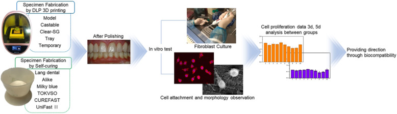
Three-dimensional (3D) printing is widely used in the fabrication of dental prostheses; however, the influence of dental materials used for 3D printing on temporary restoration of fibroblasts in tissues is unclear. Thus, the influence of different dental materials on fibroblasts were investigated.
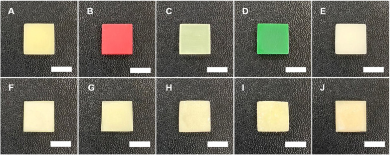
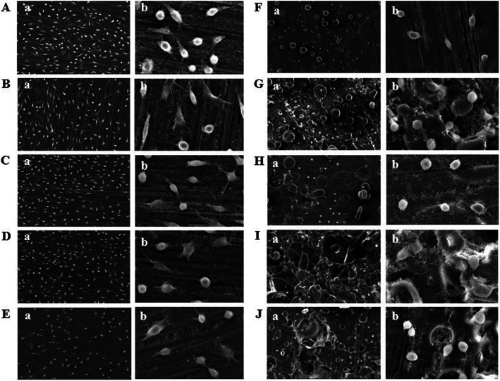
Digital light processing (DLP) type 3D printing was used. Specimens in the control group were fabricated by mixing liquid and powder self-curing resin restoration materials. The temporary resin materials used were Model, Castable, Clear-SG, Tray, and Temporary, and the self-curing resin materials used were Lang dental, Alike, Milky blue, TOKVSO CUREFAST, and UniFast III. Fibroblast cells were cultured on each specimen and subsequently post-treated for analysis. Morphology of the adhered cells were observed using a confocal laser scanning microscope (CLSM) and a scanning electron microscope (SEM).
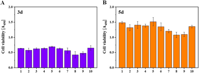
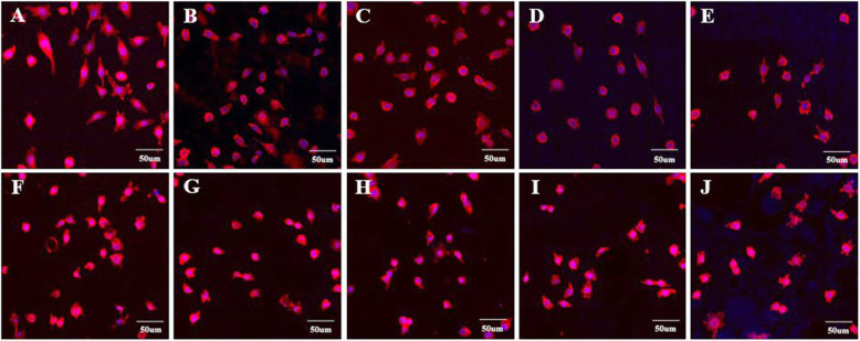
CLSM and SEM cell imaging revealed that the 3D printed material group presented better cell adhesion with well-distributed filopodia compared to that in the conventional resin material group. Cell proliferation was significantly higher in the 3D printing materials.
Superior cytocompatibility of the specimens fabricated through 3D printing and polishing process was demonstrated with the proof of better cell adhesion and higher cell proliferation.
三维(3D)打印技术广泛应用于牙科修复体的制作中;然而,用于 3D 打印的牙科材料对组织中成纤维细胞的临时修复的影响尚不清楚。因此,研究了不同牙科材料对成纤维细胞的影响。
使用数字光处理(DLP)型 3D 打印。对照组标本由混合液体和粉末自固化树脂修复材料制成。使用的临时树脂材料有 Model、Castable、Clear-SG、Tray 和 Temporary,自固化树脂材料有 Lang dental、Alike、Milky blue、TOKVSO CUREFAST 和 UniFast III。将成纤维细胞培养在每个标本上,然后进行后处理分析。使用共聚焦激光扫描显微镜(CLSM)和扫描电子显微镜(SEM)观察细胞的形态。
CLSM 和 SEM 细胞成像显示,与传统树脂材料组相比,3D 打印材料组的细胞黏附性更好,丝状伪足分布更均匀。3D 打印材料的细胞增殖显著更高。
通过 3D 打印和抛光工艺制作的标本具有更好的细胞黏附性和更高的细胞增殖能力,证明其具有更好的细胞相容性。