• 文献检索
  • 文档翻译
  • 深度研究
  • 学术资讯
  • Suppr Zotero 插件Zotero 插件
  • 邀请有礼
  • 套餐&价格
  • 历史记录
应用&插件
Suppr Zotero 插件Zotero 插件浏览器插件Mac 客户端Windows 客户端微信小程序
定价
高级版会员购买积分包购买API积分包
服务
文献检索文档翻译深度研究API 文档MCP 服务
关于我们
关于 Suppr公司介绍联系我们用户协议隐私条款
关注我们

Suppr 超能文献

核心技术专利:CN118964589B侵权必究
粤ICP备2023148730 号-1Suppr @ 2026

文献检索

告别复杂PubMed语法,用中文像聊天一样搜索,搜遍4000万医学文献。AI智能推荐,让科研检索更轻松。

立即免费搜索

文件翻译

保留排版,准确专业,支持PDF/Word/PPT等文件格式,支持 12+语言互译。

免费翻译文档

深度研究

AI帮你快速写综述,25分钟生成高质量综述,智能提取关键信息,辅助科研写作。

立即免费体验

氧化铜纳米颗粒通过铜离子介导的 P38MAPK 激活诱导血管内皮细胞氧化 DNA 损伤和细胞死亡。

Copper Oxide Nanoparticles Induce Oxidative DNA Damage and Cell Death via Copper Ion-Mediated P38 MAPK Activation in Vascular Endothelial Cells.

机构信息

College of Pharmacy, Chongqing Medical University, Chongqing, People's Republic of China.

Institute of Life Sciences, Chongqing Medical University, Chongqing, People's Republic of China.

出版信息

Int J Nanomedicine. 2020 May 8;15:3291-3302. doi: 10.2147/IJN.S241157. eCollection 2020.

DOI:10.2147/IJN.S241157
PMID:32494130
原文链接:https://pmc.ncbi.nlm.nih.gov/articles/PMC7229313/
Abstract

BACKGROUND

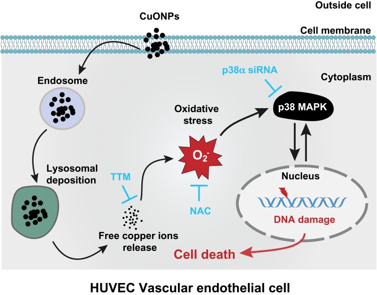
Inhaled nanoparticles can cross pulmonary air-blood barrier into circulation and cause vascular endothelial injury and progression of cardiovascular disease. However, the molecular mechanism underlying the vascular toxicity of copper oxide nanoparticles (CuONPs) remains unclear. We have recently demonstrated that the release of copper ions and the accumulation of superoxide anions contributed to CuONPs-induced cell death in human umbilical vein endothelial cells (HUVECs). Herein, we further demonstrate the mechanism underlying copper ions-induced cell death in HUVECs.

METHODS AND RESULTS

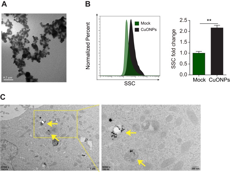
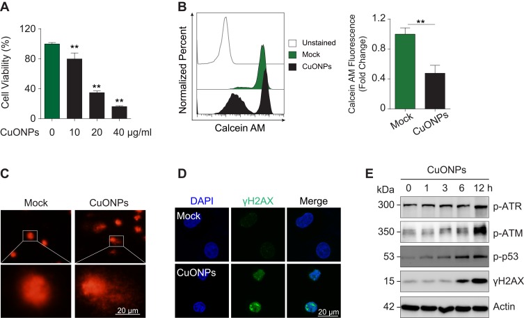
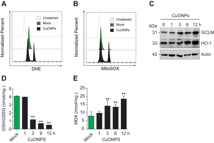
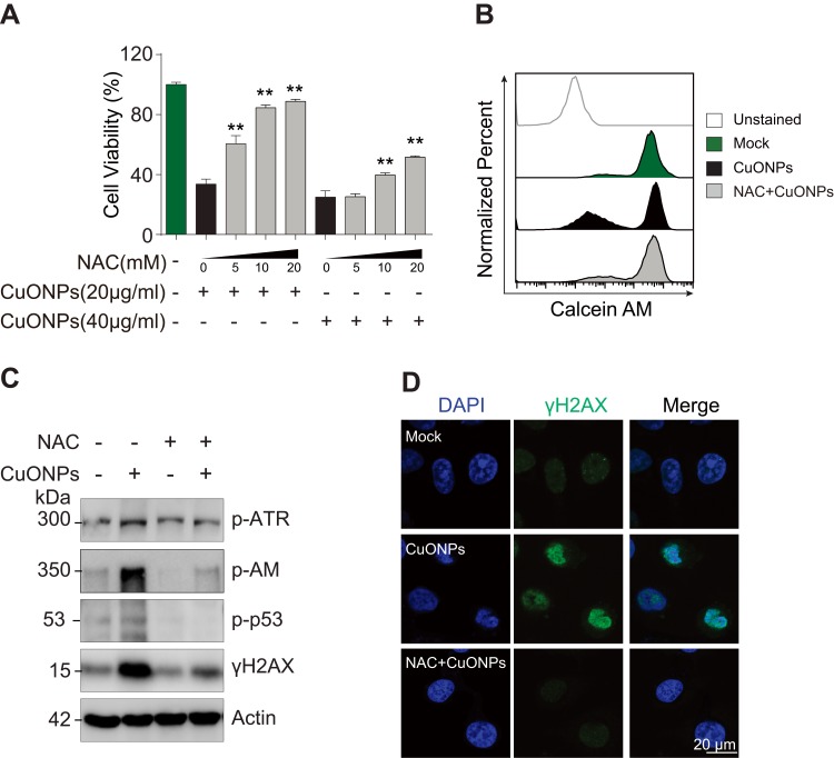
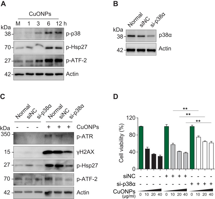
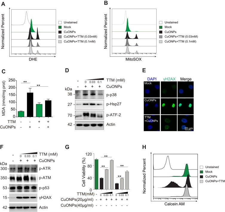
CuONPs were suspended in culture medium and vigorously vortexed for several seconds before exposure. After treatment with CuONPs, HUVECs were collected, and cell function assays were conducted to elucidate cellular processes including cell viability, oxidative stress, DNA damage and cell signaling pathways. We demonstrated that CuONPs uptake induced DNA damage in HUVECs as evidenced by γH2AX foci formation and increased phosphorylation levels of ATR, ATM, p53 and H2AX. Meanwhile, we showed that CuONPs exposure induced oxidative stress, indicated by the increase of cellular levels of superoxide anions, the upregulation of protein levels of heme oxygenase-1 (HO-1) and glutamate-cysteine ligase modifier subunit (GCLM), the elevation of the levels of malondialdehyde (MDA), but the reduction of glutathione to glutathione disulfide ratio. We also found that antioxidant N-acetyl-L-cysteine (NAC) could ameliorate CuONPs-induced oxidative stress and cell death. Interestingly, we demonstrated that p38 mitogen-activated protein kinase (MAPK) signaling pathway was activated in CuONPs-treated HUVECs, while p38α MAPK knockdown by siRNA significantly rescued HUVECs from CuONPs-induced DNA damage and cell death. Importantly, we showed that copper ions chelator tetrathiomolybdate (TTM) could alleviate CuONPs-induced oxidative stress, DNA damage, p38 MAPK pathway activation and cell death in HUVECs.

CONCLUSION

We demonstrated that CuONPs induced oxidative DNA damage and cell death via copper ions-mediated p38 MAPK activation in HUVECs, suggesting that the release of copper ions was the upstream activator for CuONPs-induced vascular endothelial toxicity, and the copper ions chelator TTM can alleviate CuONPs-associated cardiovascular disease.

摘要

背景

吸入的纳米颗粒可以穿过肺气血屏障进入循环系统,导致血管内皮损伤,并促进心血管疾病的发生和发展。然而,氧化铜纳米颗粒(CuONPs)引起血管毒性的分子机制尚不清楚。我们最近的研究表明,铜离子的释放和超氧阴离子的积累导致了 CuONPs 诱导的人脐静脉内皮细胞(HUVEC)死亡。在此,我们进一步研究了铜离子诱导 HUVEC 死亡的机制。

方法和结果

CuONPs 在暴露前先悬浮于培养基中,剧烈涡旋数秒。用 CuONPs 处理 HUVECs 后,收集细胞,进行细胞功能测定,以阐明包括细胞活力、氧化应激、DNA 损伤和细胞信号通路在内的细胞过程。我们证明,CuONPs 的摄取导致 HUVECs 的 DNA 损伤,表现为 γH2AX 焦点形成和 ATR、ATM、p53 和 H2AX 的磷酸化水平增加。同时,我们表明,CuONPs 暴露诱导氧化应激,表现为细胞中超氧阴离子水平增加,血红素加氧酶-1(HO-1)和谷氨酸-半胱氨酸连接酶修饰亚基(GCLM)蛋白水平上调,丙二醛(MDA)水平升高,而谷胱甘肽/谷胱甘肽二硫化物比值降低。我们还发现抗氧化剂 N-乙酰-L-半胱氨酸(NAC)可以改善 CuONPs 诱导的氧化应激和细胞死亡。有趣的是,我们证明 p38 丝裂原活化蛋白激酶(MAPK)信号通路在 CuONPs 处理的 HUVECs 中被激活,而 siRNA 敲低 p38α MAPK 可显著挽救 HUVECs 免受 CuONPs 诱导的 DNA 损伤和细胞死亡。重要的是,我们表明铜离子螯合剂四硫钼酸盐(TTM)可以减轻 CuONPs 诱导的氧化应激、DNA 损伤、p38 MAPK 通路激活和 HUVECs 中的细胞死亡。

结论

我们证明,CuONPs 通过铜离子介导的 p38 MAPK 激活诱导 HUVECs 发生氧化 DNA 损伤和细胞死亡,提示铜离子的释放是 CuONPs 诱导血管内皮毒性的上游激活剂,铜离子螯合剂 TTM 可以减轻 CuONPs 相关的心血管疾病。

https://cdn.ncbi.nlm.nih.gov/pmc/blobs/b3f8/7229313/d588304c9e24/IJN-15-3291-g0007.jpg
https://cdn.ncbi.nlm.nih.gov/pmc/blobs/b3f8/7229313/9368dd80b313/IJN-15-3291-g0001.jpg
https://cdn.ncbi.nlm.nih.gov/pmc/blobs/b3f8/7229313/0356369bb949/IJN-15-3291-g0002.jpg
https://cdn.ncbi.nlm.nih.gov/pmc/blobs/b3f8/7229313/82180ded80e8/IJN-15-3291-g0003.jpg
https://cdn.ncbi.nlm.nih.gov/pmc/blobs/b3f8/7229313/d1b6b6fca213/IJN-15-3291-g0004.jpg
https://cdn.ncbi.nlm.nih.gov/pmc/blobs/b3f8/7229313/0a12aa24c944/IJN-15-3291-g0005.jpg
https://cdn.ncbi.nlm.nih.gov/pmc/blobs/b3f8/7229313/b60bb3012e1d/IJN-15-3291-g0006.jpg
https://cdn.ncbi.nlm.nih.gov/pmc/blobs/b3f8/7229313/d588304c9e24/IJN-15-3291-g0007.jpg

相似文献

1
Copper Oxide Nanoparticles Induce Oxidative DNA Damage and Cell Death via Copper Ion-Mediated P38 MAPK Activation in Vascular Endothelial Cells.氧化铜纳米颗粒通过铜离子介导的 P38MAPK 激活诱导血管内皮细胞氧化 DNA 损伤和细胞死亡。
Int J Nanomedicine. 2020 May 8;15:3291-3302. doi: 10.2147/IJN.S241157. eCollection 2020.
2
The NADPH oxidase 4 protects vascular endothelial cells from copper oxide nanoparticles-induced oxidative stress and cell death.NADPH 氧化酶 4 可保护血管内皮细胞免受氧化铜纳米颗粒诱导的氧化应激和细胞死亡。
Life Sci. 2020 Jul 1;252:117571. doi: 10.1016/j.lfs.2020.117571. Epub 2020 Mar 19.
3
Disruption of the superoxide anions-mitophagy regulation axis mediates copper oxide nanoparticles-induced vascular endothelial cell death.破坏超氧阴离子-自噬调节轴介导氧化铜纳米颗粒诱导的血管内皮细胞死亡。
Free Radic Biol Med. 2018 Dec;129:268-278. doi: 10.1016/j.freeradbiomed.2018.09.032. Epub 2018 Sep 21.
4
Lysosomal deposition of copper oxide nanoparticles triggers HUVEC cells death.铜氧化物纳米粒子在溶酶体中的沉积引发了人脐静脉内皮细胞的死亡。
Biomaterials. 2018 Apr;161:228-239. doi: 10.1016/j.biomaterials.2018.01.048. Epub 2018 Feb 3.
5
Willow bark extract increases antioxidant enzymes and reduces oxidative stress through activation of Nrf2 in vascular endothelial cells and Caenorhabditis elegans.柳树皮提取物通过激活血管内皮细胞和秀丽隐杆线虫中的Nrf2来增加抗氧化酶并降低氧化应激。
Free Radic Biol Med. 2013 Dec;65:1506-1515. doi: 10.1016/j.freeradbiomed.2012.12.006. Epub 2012 Dec 28.
6
Exogenous sodium hydrosulfide protects against high glucose‑induced injury and inflammation in human umbilical vein endothelial cells by inhibiting necroptosis via the p38 MAPK signaling pathway.外源性硫氢化钠通过抑制 p38MAPK 信号通路抑制坏死性凋亡对高糖诱导的人脐静脉内皮细胞损伤和炎症起保护作用。
Mol Med Rep. 2021 Jan;23(1). doi: 10.3892/mmr.2020.11706. Epub 2020 Nov 20.
7
Downregulation of cathepsin C alleviates endothelial cell dysfunction by suppressing p38 MAPK/NF-κB pathway in preeclampsia.下调组织蛋白酶 C 通过抑制子痫前期中 p38MAPK/NF-κB 通路减轻内皮细胞功能障碍。
Bioengineered. 2022 Feb;13(2):3019-3028. doi: 10.1080/21655979.2021.2023994.
8
Cytotoxicity and genotoxicity of copper oxide nanoparticles in human skin keratinocytes cells.氧化铜纳米粒子对人皮肤角质细胞的细胞毒性和遗传毒性。
Int J Toxicol. 2013 Jul;32(4):296-307. doi: 10.1177/1091581813487563. Epub 2013 May 10.
9
Dysregulated expression of ACTN4 contributes to endothelial cell injury via the activation of the p38-MAPK/p53 apoptosis pathway in preeclampsia.ACTN4 表达失调通过激活子痫前期内皮细胞中 p38-MAPK/p53 凋亡通路导致内皮细胞损伤。
J Physiol Biochem. 2019 Nov;75(4):475-487. doi: 10.1007/s13105-019-00700-9. Epub 2019 Aug 10.
10
Epigallocatechin-3-gallate protects HUVECs from PM2.5-induced oxidative stress injury by activating critical antioxidant pathways.表没食子儿茶素-3-没食子酸酯通过激活关键抗氧化途径保护人脐静脉内皮细胞免受细颗粒物2.5诱导的氧化应激损伤。
Molecules. 2015 Apr 14;20(4):6626-39. doi: 10.3390/molecules20046626.

引用本文的文献

1
Endothelial activation in thromboangiitis obliterans: mechanisms and therapeutic horizons.血栓闭塞性脉管炎中的内皮细胞激活:机制与治疗前景
Front Immunol. 2025 Aug 27;16:1668203. doi: 10.3389/fimmu.2025.1668203. eCollection 2025.
2
Recent advances in copper sulfide nanoparticles for cancer diagnosis and therapy.用于癌症诊断与治疗的硫化铜纳米颗粒的最新进展
Mater Today Bio. 2025 Aug 13;34:102197. doi: 10.1016/j.mtbio.2025.102197. eCollection 2025 Oct.
3
Rapid Size-Dependent Impact of Cu and CuO Nanoparticles on Lentil Seeds and Leaves Using Biospeckle Optical Coherence Tomography.

本文引用的文献

1
Cu selective chelators relieve copper-induced oxidative stress .铜选择性螯合剂可减轻铜诱导的氧化应激。
Chem Sci. 2018 Oct 2;9(41):7916-7930. doi: 10.1039/c8sc04041a. eCollection 2018 Nov 7.
2
Autophagy-dependent release of zinc ions is critical for acute lung injury triggered by zinc oxide nanoparticles.自噬依赖性锌离子释放对于氧化锌纳米颗粒引发的急性肺损伤至关重要。
Nanotoxicology. 2018 Nov;12(9):1068-1091. doi: 10.1080/17435390.2018.1513094. Epub 2018 Oct 14.
3
Transcriptional responses and mechanisms of copper nanoparticle toxicology on zebrafish embryos.
使用生物散斑光学相干断层扫描技术研究铜和氧化铜纳米颗粒对小扁豆种子和叶片的快速尺寸依赖性影响。
Nanomaterials (Basel). 2025 Aug 8;15(16):1214. doi: 10.3390/nano15161214.
4
Targeting cuproptosis in liver cancer: Molecular mechanisms and therapeutic implications.靶向肝癌中的铜死亡:分子机制与治疗意义
Apoptosis. 2025 Aug 7. doi: 10.1007/s10495-025-02150-9.
5
Deciphering Cuproptosis in Sepsis: Mechanisms, Consequences, and Therapeutic Opportunities.解析脓毒症中的铜死亡:机制、后果及治疗机遇
J Inflamm Res. 2025 Jul 25;18:9879-9890. doi: 10.2147/JIR.S533967. eCollection 2025.
6
Copper Homeostasis and Cuproptosis As Potential Intervention Strategy in Atherosclerosis.铜稳态与铜死亡作为动脉粥样硬化潜在干预策略
J Cardiovasc Transl Res. 2025 Jul 21. doi: 10.1007/s12265-025-10661-8.
7
Ultrasmall Copper-Based Nanozyme Eye Drops for Effective Antioxidative Therapy of Ocular Surface Diseases.用于眼表疾病有效抗氧化治疗的超小铜基纳米酶眼药水
ACS Omega. 2025 Jun 16;10(25):26478-26487. doi: 10.1021/acsomega.5c00103. eCollection 2025 Jul 1.
8
A novel cuproptosis-associated LncRNA model predicting prognostic and immunotherapy response for glioma.一种预测胶质瘤预后和免疫治疗反应的新型铜死亡相关长链非编码RNA模型。
Discov Oncol. 2025 Jun 13;16(1):1089. doi: 10.1007/s12672-025-02912-6.
9
Understanding the Mechanism of Cardiotoxicity Induced by Nanomaterials: A Comprehensive Review.理解纳米材料诱导心脏毒性的机制:综述
Small Sci. 2025 Feb 20;5(5):2400498. doi: 10.1002/smsc.202400498. eCollection 2025 May.
10
A review on the green synthesis of metal (Ag, Cu, and Au) and metal oxide (ZnO, MgO, CoO, and TiO) nanoparticles using plant extracts for developing antimicrobial properties.一篇关于利用植物提取物进行金属(银、铜和金)及金属氧化物(氧化锌、氧化镁、氧化钴和二氧化钛)纳米颗粒的绿色合成以开发抗菌性能的综述。
Nanoscale Adv. 2025 Mar 7;7(9):2446-2473. doi: 10.1039/d5na00037h. eCollection 2025 Apr 29.
铜纳米颗粒对斑马鱼胚胎的毒理学转录反应和机制。
J Hazard Mater. 2018 Feb 15;344:1057-1068. doi: 10.1016/j.jhazmat.2017.11.039. Epub 2017 Dec 2.
4
Copper nanoparticles induce early fibrotic changes in the liver via TGF-β/Smad signaling and cause immunosuppressive effects in rats.铜纳米颗粒通过 TGF-β/Smad 信号通路诱导肝脏早期纤维化改变,并在大鼠中产生免疫抑制作用。
Nanotoxicology. 2018 Aug;12(6):637-651. doi: 10.1080/17435390.2018.1472313. Epub 2018 May 30.
5
Lysosomal deposition of copper oxide nanoparticles triggers HUVEC cells death.铜氧化物纳米粒子在溶酶体中的沉积引发了人脐静脉内皮细胞的死亡。
Biomaterials. 2018 Apr;161:228-239. doi: 10.1016/j.biomaterials.2018.01.048. Epub 2018 Feb 3.
6
Copper oxide nanoparticles induce collagen deposition via TGF-β1/Smad3 signaling in human airway epithelial cells.氧化铜纳米颗粒通过 TGF-β1/Smad3 信号通路诱导人呼吸道上皮细胞胶原沉积。
Nanotoxicology. 2018 Apr;12(3):239-250. doi: 10.1080/17435390.2018.1432778. Epub 2018 Jan 31.
7
Zinc oxide nanoparticles harness autophagy to induce cell death in lung epithelial cells.氧化锌纳米颗粒利用自噬作用诱导肺上皮细胞死亡。
Cell Death Dis. 2017 Jul 27;8(7):e2954. doi: 10.1038/cddis.2017.337.
8
LAMP-2 mediates oxidative stress-dependent cell death in Zn-treated lung epithelium cells.LAMP-2介导锌处理的肺上皮细胞中氧化应激依赖性细胞死亡。
Biochem Biophys Res Commun. 2017 Jun 17;488(1):177-181. doi: 10.1016/j.bbrc.2017.05.030. Epub 2017 May 5.
9
Inhaled Nanoparticles Accumulate at Sites of Vascular Disease.吸入纳米颗粒在血管疾病部位蓄积。
ACS Nano. 2017 May 23;11(5):4542-4552. doi: 10.1021/acsnano.6b08551. Epub 2017 Apr 26.
10
Flow Cytometry-Based Quantification of Cellular Au Nanoparticles.基于流式细胞术的细胞金纳米粒子定量分析
Anal Chem. 2017 Feb 21;89(4):2449-2456. doi: 10.1021/acs.analchem.6b04418. Epub 2017 Feb 6.