Institute of Psychiatry and Neurosciences of Paris, INSERM U1266, Université de Paris, 75014 Paris, France.
Brain Plasticity Unit, ESPCI Paris, CNRS, PSL Research University, 75005 Paris, France.
Proc Natl Acad Sci U S A. 2020 Jun 30;117(26):15270-15280. doi: 10.1073/pnas.1920475117. Epub 2020 Jun 15.
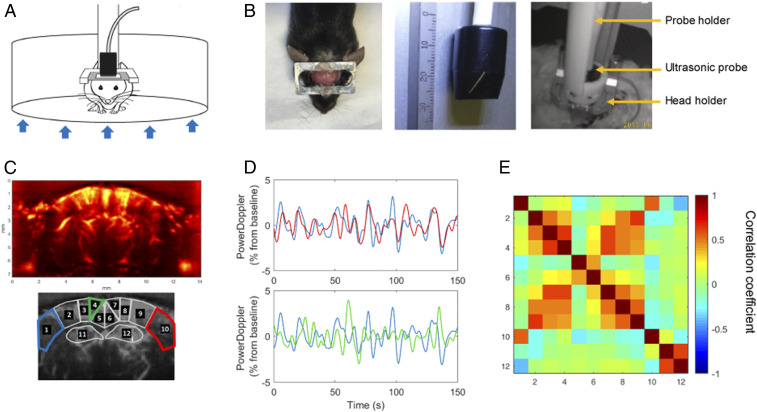
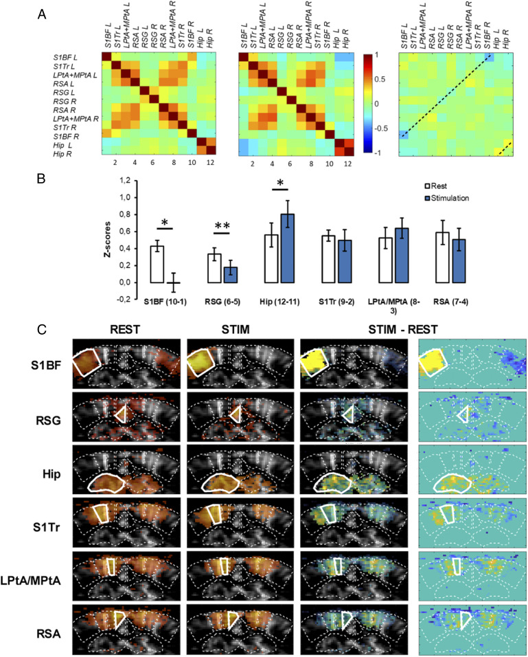
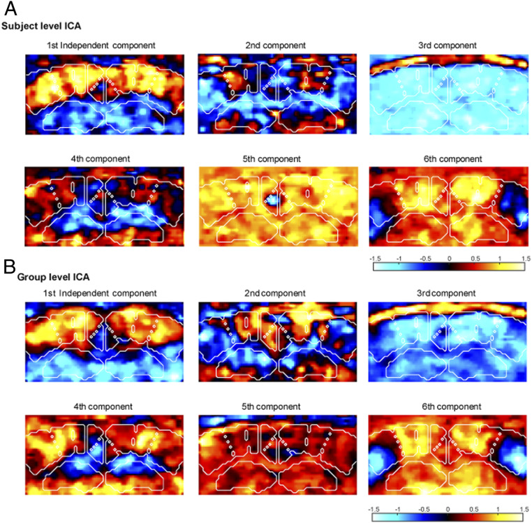
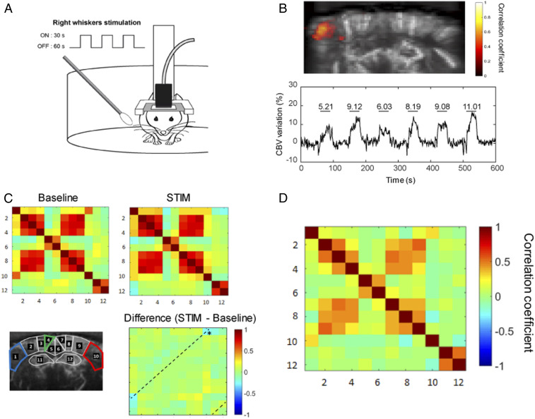
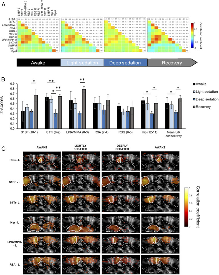
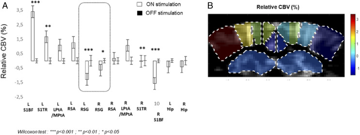
The default mode network (DMN) has been defined in functional brain imaging studies as a set of highly connected brain areas, which are active during wakeful rest and inactivated during task-based stimulation. DMN function is characteristically impaired in major neuropsychiatric diseases, emphasizing its interest for translational research. However, in the mouse, a major preclinical rodent model, there is still no functional imaging evidence supporting DMN deactivation and deconnection during high-demanding cognitive/sensory tasks. Here we have developed functional ultrasound (fUS) imaging to properly visualize both activation levels and functional connectivity patterns, in head-restrained awake and behaving mice, and investigated their modulation during a sensory-task, whisker stimulation. We identified reproducible and highly symmetric resting-state networks, with overall connectivity strength directly proportional to the wakefulness level of the animal. We show that unilateral whisker stimulation leads to the expected activation of the contralateral barrel cortex in lightly sedated mice, while interhemispheric inhibition reduces activity in the ipsilateral barrel cortex. Whisker stimulation also leads to elevated bilateral connectivity in the hippocampus. Importantly, in addition to functional changes in these major hubs of tactile information processing, whisker stimulation during genuine awake resting-state periods leads to highly specific reductions both in activation and interhemispheric correlation within the restrosplenial cortex, a major hub of the DMN. These results validate an imaging technique for the study of activation and connectivity in the lightly sedated awake mouse brain and provide evidence supporting an evolutionary preserved function of the DMN, putatively improving translational relevance of preclinical models of neuropsychiatric diseases.
默认模式网络(DMN)在功能脑成像研究中被定义为一组高度连接的脑区,这些脑区在清醒休息时活跃,而在基于任务的刺激时失活。DMN 功能在重大神经精神疾病中受损,强调了其对转化研究的兴趣。然而,在主要的临床前啮齿动物模型——老鼠中,仍然没有功能成像证据支持在高要求的认知/感觉任务期间 DMN 的去激活和去连接。在这里,我们开发了功能超声(fUS)成像,以便在头部固定的清醒和行为小鼠中正确可视化激活水平和功能连接模式,并研究了它们在感觉任务——胡须刺激期间的调制。我们确定了可重复且高度对称的静息状态网络,整体连接强度与动物的清醒水平直接成正比。我们表明,单侧胡须刺激会导致轻度镇静的小鼠对侧桶状皮层的预期激活,而半球间抑制会降低同侧桶状皮层的活性。胡须刺激还会导致海马双侧连接增加。重要的是,除了在这些触觉信息处理的主要中枢的功能变化之外,在真正清醒的静息状态期间进行胡须刺激会导致在 restrosplenial 皮层内的激活和半球间相关性的高度特异性降低,这是 DMN 的主要中枢之一。这些结果验证了一种用于研究轻度镇静清醒小鼠大脑中激活和连接的成像技术,并提供了支持 DMN 的进化保守功能的证据,推测提高了神经精神疾病临床前模型的转化相关性。