Huang Jiacheng, Tian Wuwei, Zhang Lele, Huang Qiang, Lin Shengzhang, Ding Yong, Liang Wenjie, Zheng Shusen
Department of Hepatobiliary and Pancreatic Surgery, The First Affiliated Hospital, College of Medicine, Zhejiang University, Hangzhou, China.
Collaborative Innovation Center for Diagnosis and Treatment of Infectious Diseases, The First Affiliated Hospital, College of Medicine, Zhejiang University, Hangzhou, China.
Front Oncol. 2020 Jun 26;10:887. doi: 10.3389/fonc.2020.00887. eCollection 2020.
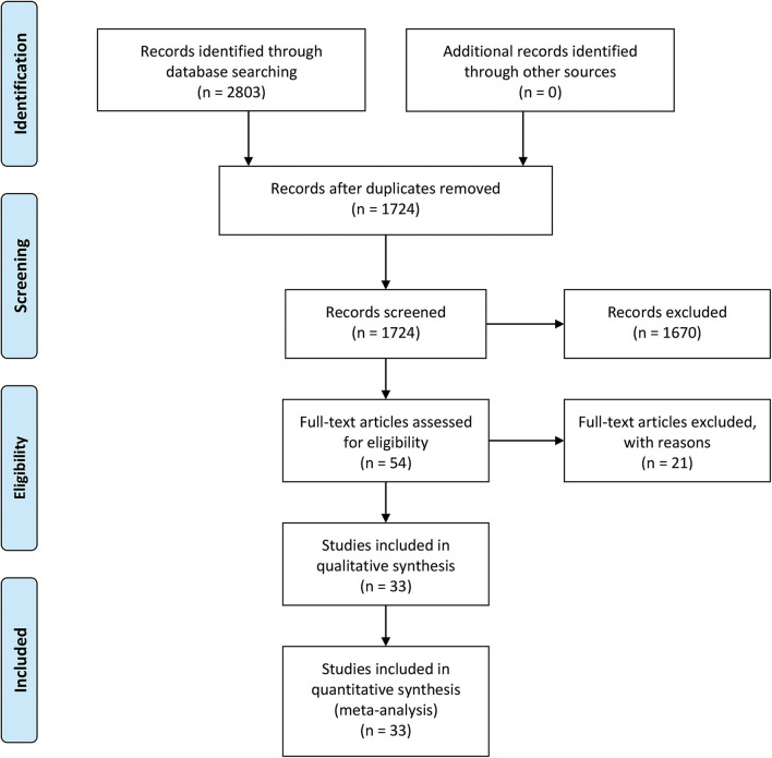
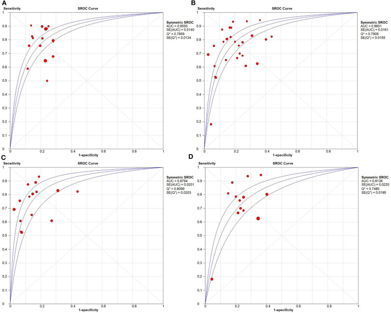
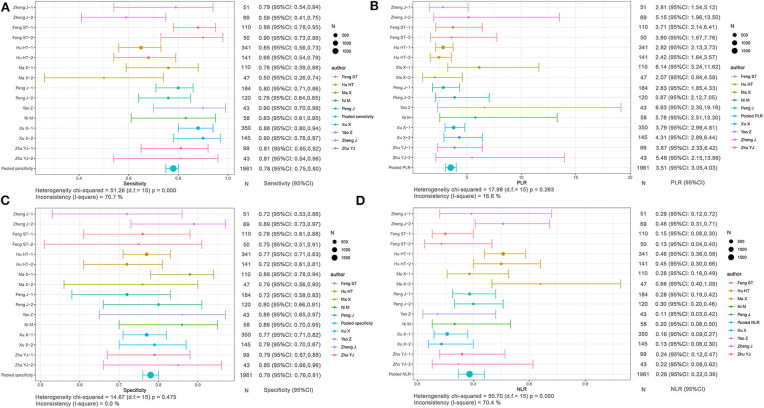
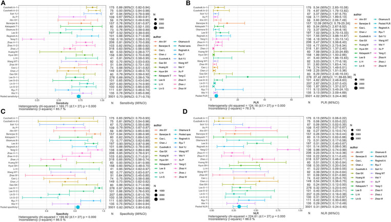
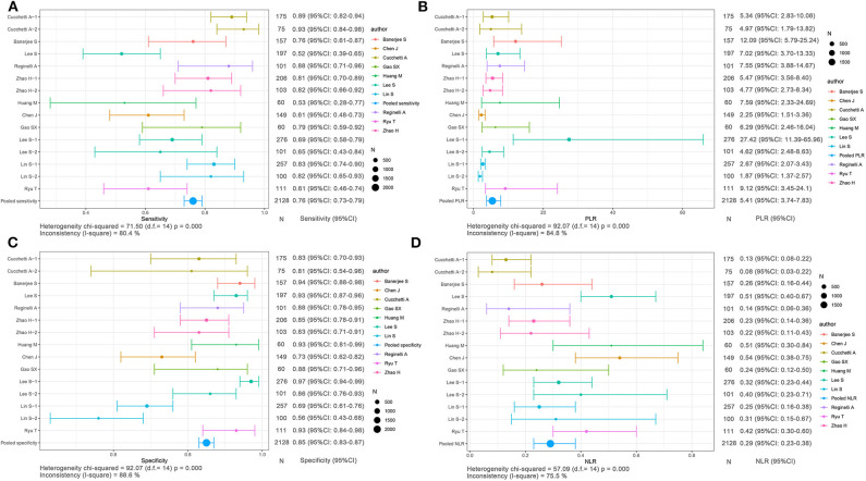
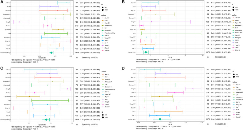
To compare the predictive power between radiomics and non-radiomics (conventional imaging and functional imaging methods) for preoperative evaluation of microvascular invasion (MVI) in hepatocellular carcinoma (HCC). Comprehensive publications were screened in PubMed, Embase, and Cochrane Library. Studies focusing on the discrimination values of imaging methods, including radiomics and non-radiomics methods, for MVI evaluation were included in our meta-analysis. Thirty-three imaging studies with 5,462 cases, focusing on preoperative evaluation of MVI status in HCC, were included. The sensitivity and specificity of MVI prediction in HCC were 0.78 [95% confidence interval (CI): 0.75-0.80; = 70.7%] and 0.78 (95% CI: 0.76-0.81; = 0.0%) for radiomics, respectively, and were 0.73 (95% CI: 0.71-0.75; = 83.7%) and 0.82 (95% CI: 0.80-0.83; = 86.5%) for non-radiomics, respectively. The areas under the receiver operation curves for radiomics and non-radiomics to predict MVI status in HCC were 0.8550 and 0.8601, respectively, showing no significant difference. The imaging method is feasible to predict the MVI state of HCC. Radiomics method based on medical image data is a promising application in clinical practice and can provide quantifiable image features. With the help of these features, highly consistent prediction performance will be achieved in anticipation.
比较放射组学与非放射组学(传统成像和功能成像方法)在肝细胞癌(HCC)微血管侵犯(MVI)术前评估中的预测能力。在PubMed、Embase和Cochrane图书馆中筛选综合出版物。纳入我们荟萃分析的研究聚焦于成像方法(包括放射组学和非放射组学方法)对MVI评估的判别值。纳入了33项影像学研究,共5462例病例,聚焦于HCC中MVI状态的术前评估。放射组学对HCC中MVI预测的敏感性和特异性分别为0.78[95%置信区间(CI):0.75 - 0.80;I² = 70.7%]和0.78(95%CI:0.76 - 0.81;I² = 0.0%),非放射组学的敏感性和特异性分别为0.73(95%CI:0.71 - 0.75;I² = 83.7%)和0.82(95%CI:0.80 - 0.83;I² = 86.5%)。放射组学和非放射组学预测HCC中MVI状态的受试者操作曲线下面积分别为0.8550和0.8601,无显著差异。该成像方法可用于预测HCC的MVI状态。基于医学图像数据的放射组学方法在临床实践中有广阔的应用前景,可提供可量化的图像特征。借助这些特征,有望实现高度一致的预测性能。