Wen Xuehua, Li Yumei, He Xiaodong, Xu Yuyun, Shu Zhenyu, Hu Xingfei, Chen Junfa, Jiang Hongyang, Gong Xiangyang
Department of Radiology, Zhejiang Provincial People's Hospital, Affiliated People's Hospital of Hangzhou Medical College, Hangzhou, China.
Institute of Artificial Intelligence and Remote Imaging, Hangzhou Medical College, Hangzhou, China.
Front Neurosci. 2020 Jul 7;14:708. doi: 10.3389/fnins.2020.00708. eCollection 2020.
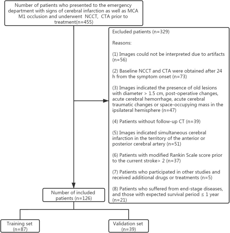
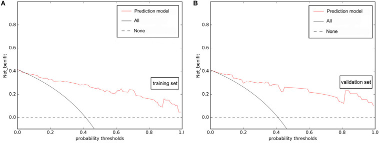
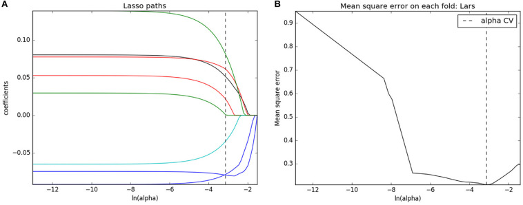
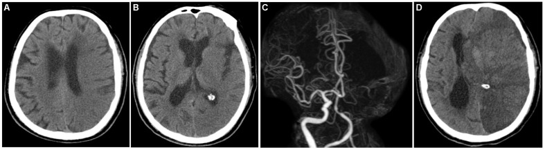
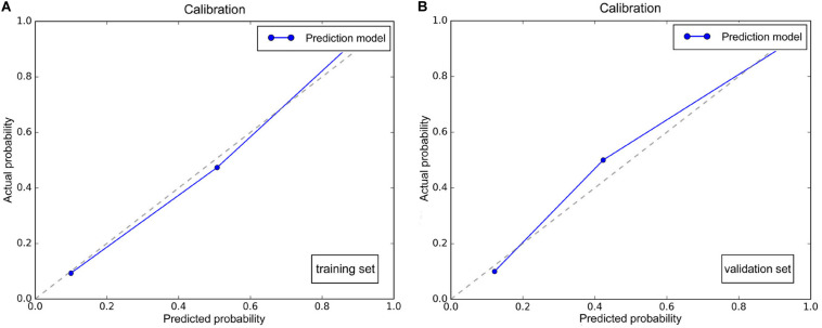
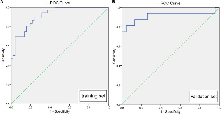
Malignant middle cerebral artery infarction (mMCAi) is a serious complication of cerebral infarction usually associated with poor patient prognosis. In this retrospective study, we analyzed clinical information as well as non-contrast computed tomography (NCCT) and computed tomography angiography (CTA) data from patients with cerebral infarction in the middle cerebral artery (MCA) territory acquired within 24 h from symptoms onset. Then, we aimed to develop a model based on the radiomics signature to predict the development of mMCAi in cerebral infarction patients. Patients were divided randomly into training ( = 87) and validation ( = 39) sets. A total of 396 texture features were extracted from each NCCT image from the 126 patients. The least absolute shrinkage and selection operator regression analysis was used to reduce the feature dimension and construct an accurate radiomics signature based on the remaining texture features. Subsequently, we developed a model based on the radiomics signature and Alberta Stroke Program Early CT Score (ASPECTS) based on NCCT to predict mMCAi. Our prediction model showed a good predictive performance with an AUC of 0.917 [95% confidence interval (CI), 0.863-0.972] and 0.913 [95% CI, 0.795-1] in the training and validation sets, respectively. Additionally, the decision curve analysis (DCA) validated the clinical efficacy of the combined risk factors of radiomics signature and ASPECTS based on NCCT in the prediction of mMCAi development in patients with acute stroke across a wide range of threshold probabilities. Our research indicates that radiomics signature can be an instrumental tool to predict the risk of mMCAi.
大脑中动脉恶性梗死(mMCAi)是脑梗死的一种严重并发症,通常与患者预后不良相关。在这项回顾性研究中,我们分析了症状发作后24小时内获得的大脑中动脉(MCA)区域脑梗死患者的临床信息以及非增强计算机断层扫描(NCCT)和计算机断层扫描血管造影(CTA)数据。然后,我们旨在开发一种基于放射组学特征的模型,以预测脑梗死患者发生mMCAi的情况。患者被随机分为训练组( = 87)和验证组( = 39)。从126例患者的每张NCCT图像中提取了总共396个纹理特征。使用最小绝对收缩和选择算子回归分析来降低特征维度,并基于剩余的纹理特征构建准确的放射组学特征。随后,我们开发了一种基于放射组学特征和基于NCCT的阿尔伯塔卒中项目早期CT评分(ASPECTS)的模型来预测mMCAi。我们的预测模型在训练组和验证组中的预测性能良好,AUC分别为0.917 [95%置信区间(CI),0.863 - 0.972]和0.913 [95% CI,0.795 - 1]。此外,决策曲线分析(DCA)验证了基于NCCT的放射组学特征和ASPECTS联合危险因素在预测广泛阈值概率范围内急性卒中患者发生mMCAi方面的临床疗效。我们的研究表明,放射组学特征可以成为预测mMCAi风险的有用工具。