Department of Oral Diagnosis and Pathology Federal University of Rio de Janeiro School of Dentistry Av. Carlos Chagas Filho 373, Prédio do CCS, Bloco K, 2° andar, Sala 56 Ilha da Cidade Universitária, Rio de Janeiro/RJ. 21.941-902
Med Oral Patol Oral Cir Bucal. 2021 May 1;26(3):e284-e291. doi: 10.4317/medoral.24168.
Pigmented lesions are uncommon in the oral mucosa, and studies investigating the incidence and types of these lesions are desired to improve the diagnostic knowledge of clinicians. The aim of this study was to analyze the distribution of oral pigmented lesions in a Brazilian population.
A retrospective descriptive cross-sectional study was performed. Oral pigmented lesions were retrieved from the files of two oral and maxillofacial pathology services from Brazil over a 45-year period (1974-2019). The clinical data and the diagnoses of each case were retrieved and included in a Microsoft Excel® database.
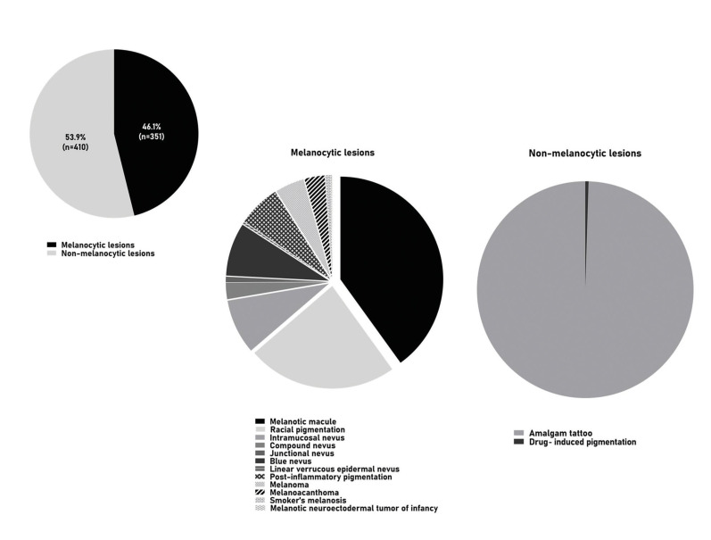
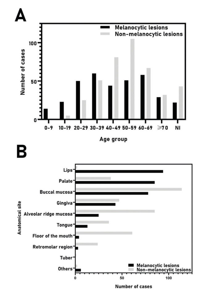
From 77.074 lesions diagnosed in this period, 761 (0.99%) represented pigmented lesions of the oral mucosa, including 351 (46.1%) melanocytic and 410 (53.9%) non-melanocytic lesions, with a higher incidence in females (73.2%) between the fourth and seventh decades of life. Amalgam tattoo (53.6%) represented the most common lesion, followed by melanotic macule (18.3%) and racial pigmentation (10.8%). Other pigmented lesions included nevus (9.9%), post-inflammatory pigmentation (3%), melanoma (2.1%), melanoacanthoma (1.4%), smoker's melanosis (0.4%), drug-induced pigmentation (0.3%), and melanotic neuroectodermal tumor of infancy (0.1%). The buccal mucosa was the most commonly affected site (25.2%), followed by the alveolar ridge (14.5%), and gingiva (11.8%).
The current findings were similar to previous studies with minor differences due methodology and characteristics of the services from where lesions were retrieved. The knowledge of these data may contribute to a better understanding of oral pigmented lesions and assist clinicians to better recognize and manage them.
口腔黏膜的色素性病变并不常见,研究这些病变的发生率和类型有助于提高临床医生的诊断知识。本研究旨在分析巴西人群口腔色素性病变的分布。
这是一项回顾性描述性的横断面研究。从巴西的两个口腔颌面病理服务机构的档案中检索了 45 年间(1974 年至 2019 年)的口腔色素性病变。检索了每个病例的临床数据和诊断,并将其纳入 Microsoft Excel®数据库。
在此期间诊断的 77074 个病变中,761 个(0.99%)为口腔黏膜的色素性病变,包括 351 个(46.1%)黑素细胞性和 410 个(53.9%)非黑素细胞性病变,女性发病率(73.2%)较高,发病年龄在第四至第七个十年。汞合金纹身(53.6%)是最常见的病变,其次是色素斑(18.3%)和种族性色素沉着(10.8%)。其他色素性病变包括痣(9.9%)、炎症后色素沉着(3%)、黑色素瘤(2.1%)、黑素棘皮瘤(1.4%)、吸烟者色素沉着(0.4%)、药物诱导的色素沉着(0.3%)和婴儿黑色素神经外胚层肿瘤(0.1%)。最常受累的部位是颊黏膜(25.2%),其次是牙槽嵴(14.5%)和牙龈(11.8%)。
由于研究方法和病变来源服务机构的特点不同,本研究结果与以往研究相似,但存在一些差异。了解这些数据可能有助于更好地理解口腔色素性病变,并帮助临床医生更好地识别和处理它们。