Siller Ina G, Epping Niklas-Maximilian, Lavrentieva Antonina, Scheper Thomas, Bahnemann Janina
Institute of Technical Chemistry, Leibniz University Hannover, Callinstraße 5, 30167 Hannover, Germany.
Materials (Basel). 2020 Sep 25;13(19):4290. doi: 10.3390/ma13194290.
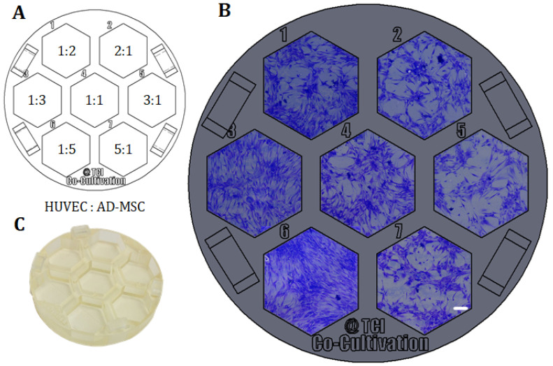
Due to the ever-increasing resolution of 3D printing technology, additive manufacturing is now even used to produce complex devices for laboratory applications. Personalized experimental devices or entire cultivation systems of almost unlimited complexity can potentially be manufactured within hours from start to finish-an enormous potential for experimental parallelization in a highly controllable environment. This study presents customized 3D-printed co-cultivation systems, which qualify for angiogenesis studies. In these systems, endothelial and mesenchymal stem cells (AD-MSC) were indirectly co-cultivated-that is, both cell types were physically separated through a rigid, 3D-printed barrier in the middle, while still sharing the same cell culture medium that allows for the exchange of signalling molecules. Biochemical-based cytotoxicity assays initially confirmed that the 3D printing material does not exert any negative effects on cells. Since the material also enables phase contrast and fluorescence microscopy, the behaviour of cells could be observed over the entire cultivation via both. Microscopic observations and subsequent quantitative analysis revealed that endothelial cells form tubular-like structures as angiogenic feature when indirectly co-cultured alongside AD-MSCs in the 3D-printed co-cultivation system. In addition, further 3D-printed devices are also introduced that address different issues and aspire to help in varying experimental setups. Our results mark an important step forward for the integration of customized 3D-printed systems as self-contained test systems or equipment in biomedical applications.
由于3D打印技术的分辨率不断提高,增材制造现在甚至被用于生产实验室应用的复杂设备。几乎具有无限复杂性的个性化实验设备或整个培养系统有可能在数小时内从开始到结束制造出来——这在高度可控的环境中为实验并行化提供了巨大潜力。本研究展示了定制的3D打印共培养系统,该系统适用于血管生成研究。在这些系统中,内皮细胞和间充质干细胞(AD-MSC)进行间接共培养——也就是说,两种细胞类型通过中间的刚性3D打印屏障在物理上分离,同时仍共享允许信号分子交换的相同细胞培养基。基于生化的细胞毒性试验最初证实3D打印材料对细胞没有任何负面影响。由于该材料还能实现相差显微镜和荧光显微镜观察,因此可以通过这两种方式在整个培养过程中观察细胞行为。显微镜观察和随后的定量分析表明,在3D打印共培养系统中与AD-MSC间接共培养时,内皮细胞会形成管状结构作为血管生成特征。此外,还介绍了其他3D打印设备,这些设备解决了不同的问题,并有望在不同的实验设置中提供帮助。我们的结果标志着将定制的3D打印系统作为生物医学应用中的独立测试系统或设备进行整合向前迈出了重要一步。