• 文献检索
  • 文档翻译
  • 深度研究
  • 学术资讯
  • Suppr Zotero 插件Zotero 插件
  • 邀请有礼
  • 套餐&价格
  • 历史记录
应用&插件
Suppr Zotero 插件Zotero 插件浏览器插件Mac 客户端Windows 客户端微信小程序
定价
高级版会员购买积分包购买API积分包
服务
文献检索文档翻译深度研究API 文档MCP 服务
关于我们
关于 Suppr公司介绍联系我们用户协议隐私条款
关注我们

Suppr 超能文献

核心技术专利:CN118964589B侵权必究
粤ICP备2023148730 号-1Suppr @ 2026

文献检索

告别复杂PubMed语法,用中文像聊天一样搜索,搜遍4000万医学文献。AI智能推荐,让科研检索更轻松。

立即免费搜索

文件翻译

保留排版,准确专业,支持PDF/Word/PPT等文件格式,支持 12+语言互译。

免费翻译文档

深度研究

AI帮你快速写综述,25分钟生成高质量综述,智能提取关键信息,辅助科研写作。

立即免费体验

以临床结构集为基准真值,通过卷积神经网络对宫颈癌和肛管癌进行自动分割。

Auto-segmentations by convolutional neural network in cervical and anorectal cancer with clinical structure sets as the ground truth.

作者信息

Sartor Hanna, Minarik David, Enqvist Olof, Ulén Johannes, Wittrup Anders, Bjurberg Maria, Trägårdh Elin

机构信息

Diagnostic Radiology, Department of Translational Medicine, Lund University, Skåne University Hospital, Lund, Sweden.

Radiation Physics, Department of Translational Medicine, Lund University, Skåne University Hospital, Malmö, Sweden.

出版信息

Clin Transl Radiat Oncol. 2020 Sep 14;25:37-45. doi: 10.1016/j.ctro.2020.09.004. eCollection 2020 Nov.

DOI:10.1016/j.ctro.2020.09.004
PMID:33005756
原文链接:https://pmc.ncbi.nlm.nih.gov/articles/PMC7519211/
Abstract

BACKGROUND

It is time-consuming for oncologists to delineate volumes for radiotherapy treatment in computer tomography (CT) images. Automatic delineation based on image processing exists, but with varied accuracy and moderate time savings. Using convolutional neural network (CNN), delineations of volumes are faster and more accurate. We have used CTs with the annotated structure sets to train and evaluate a CNN.

MATERIAL AND METHODS

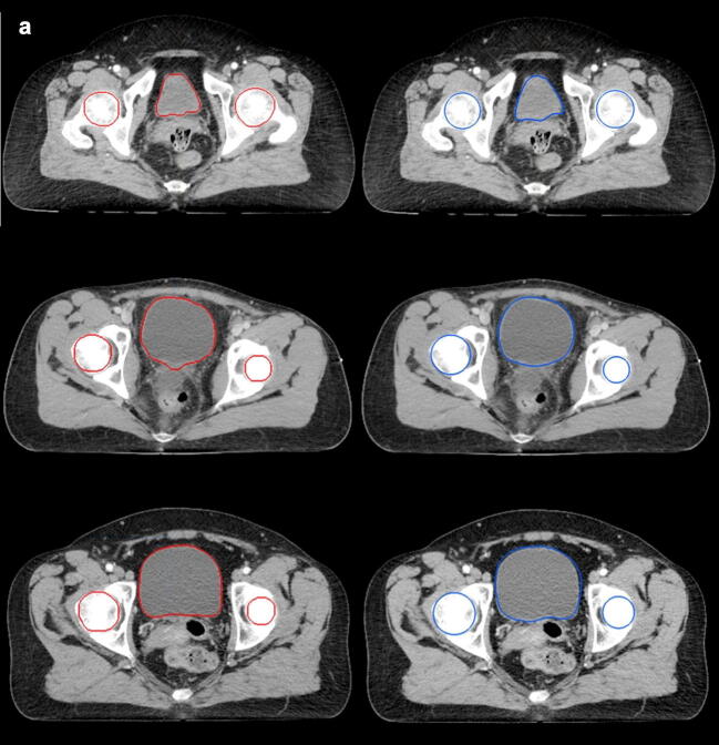
The CNN is a standard segmentation network modified to minimize memory usage. We used CTs and structure sets from 75 cervical cancers and 191 anorectal cancers receiving radiation therapy at Skåne University Hospital 2014-2018. Five structures were investigated: left/right femoral heads, bladder, bowel bag, and clinical target volume of lymph nodes (CTVNs). Dice score and mean surface distance (MSD) (mm) evaluated accuracy, and one oncologist qualitatively evaluated auto-segmentations.

RESULTS

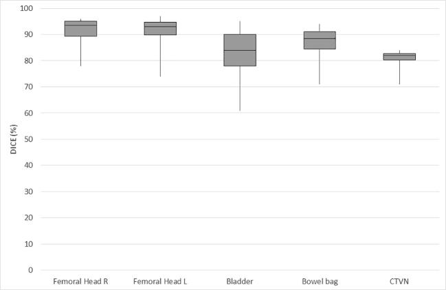
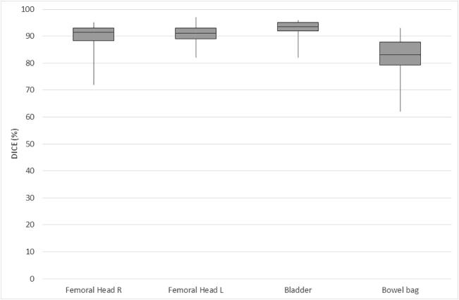
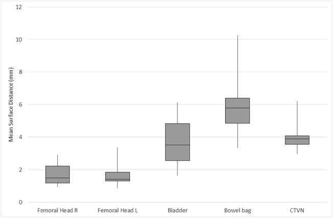
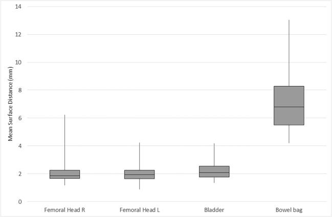
Median Dice/MSD scores for anorectal cancer: 0.91-0.92/1.93-1.86 femoral heads, 0.94/2.07 bladder, and 0.83/6.80 bowel bag. Median Dice scores for cervical cancer were 0.93-0.94/1.42-1.49 femoral heads, 0.84/3.51 bladder, 0.88/5.80 bowel bag, and 0.82/3.89 CTVNs. With qualitative evaluation, performance on femoral heads and bladder auto-segmentations was mostly excellent, but CTVN auto-segmentations were not acceptable to a larger extent.

DISCUSSION

It is possible to train a CNN with high overlap using structure sets as ground truth. Manually delineated pelvic volumes from structure sets do not always strictly follow volume boundaries and are sometimes inaccurately defined, which leads to similar inaccuracies in the CNN output. More data that is consistently annotated is needed to achieve higher CNN accuracy and to enable future clinical implementation.

摘要

背景

肿瘤学家在计算机断层扫描(CT)图像中勾画放射治疗体积非常耗时。基于图像处理的自动勾画方法虽然存在,但准确性各异且节省时间有限。使用卷积神经网络(CNN)进行体积勾画则更快且更准确。我们使用带有标注结构集的CT图像来训练和评估一个CNN。

材料与方法

该CNN是一个经过修改以最小化内存使用的标准分割网络。我们使用了2014年至2018年在斯科讷大学医院接受放射治疗的75例宫颈癌和191例肛管直肠癌的CT图像及结构集。研究了五个结构:左/右股骨头、膀胱、肠袋以及淋巴结临床靶体积(CTVN)。通过骰子系数和平均表面距离(MSD)(毫米)评估准确性,由一位肿瘤学家对自动分割结果进行定性评估。

结果

肛管直肠癌的骰子系数/平均表面距离中位数得分:股骨头为0.91 - 0.92/1.93 - 1.86,膀胱为0.94/2.07,肠袋为0.83/6.80。宫颈癌的骰子系数中位数得分:股骨头为0.93 - 0.94/1.42 - 1.49,膀胱为0.84/3.51,肠袋为0.88/5.80,CTVN为0.82/3.89。定性评估显示,股骨头和膀胱自动分割的性能大多出色,但CTVN自动分割在很大程度上不可接受。

讨论

使用结构集作为真值来训练具有高重叠率的CNN是可行的。从结构集中手动勾画的盆腔体积并非总是严格遵循体积边界,有时定义不准确,这导致CNN输出中出现类似的不准确情况。需要更多一致标注的数据来提高CNN的准确性并实现未来的临床应用。

https://cdn.ncbi.nlm.nih.gov/pmc/blobs/1424/7519211/4f5e80041aba/gr3a.jpg
https://cdn.ncbi.nlm.nih.gov/pmc/blobs/1424/7519211/ec4afb3f34a9/gr1a.jpg
https://cdn.ncbi.nlm.nih.gov/pmc/blobs/1424/7519211/ee68b76238e4/gr1b.jpg
https://cdn.ncbi.nlm.nih.gov/pmc/blobs/1424/7519211/7fc0988edc0d/gr2a.jpg
https://cdn.ncbi.nlm.nih.gov/pmc/blobs/1424/7519211/c69a935e2e30/gr2b.jpg
https://cdn.ncbi.nlm.nih.gov/pmc/blobs/1424/7519211/4f5e80041aba/gr3a.jpg

相似文献

1
Auto-segmentations by convolutional neural network in cervical and anorectal cancer with clinical structure sets as the ground truth.以临床结构集为基准真值,通过卷积神经网络对宫颈癌和肛管癌进行自动分割。
Clin Transl Radiat Oncol. 2020 Sep 14;25:37-45. doi: 10.1016/j.ctro.2020.09.004. eCollection 2020 Nov.
2
Cascaded deep learning-based auto-segmentation for head and neck cancer patients: Organs at risk on T2-weighted magnetic resonance imaging.基于级联深度学习的头颈部癌症患者自动分割:T2 加权磁共振成像上的危险器官。
Med Phys. 2021 Dec;48(12):7757-7772. doi: 10.1002/mp.15290. Epub 2021 Nov 1.
3
Development and validation of a deep learning algorithm for auto-delineation of clinical target volume and organs at risk in cervical cancer radiotherapy.开发和验证一种深度学习算法,用于自动勾画宫颈癌放射治疗的临床靶区和危及器官。
Radiother Oncol. 2020 Dec;153:172-179. doi: 10.1016/j.radonc.2020.09.060. Epub 2020 Oct 8.
4
Automatic segmentation of the clinical target volume and organs at risk in the planning CT for rectal cancer using deep dilated convolutional neural networks.使用深度扩张卷积神经网络在直肠癌计划 CT 中自动分割临床靶区和危及器官。
Med Phys. 2017 Dec;44(12):6377-6389. doi: 10.1002/mp.12602. Epub 2017 Oct 28.
5
A comparison of methods for fully automatic segmentation of tumors and involved nodes in PET/CT of head and neck cancers.头颈部癌症正电子发射断层扫描/计算机断层扫描中肿瘤和累及淋巴结全自动分割方法的比较。
Phys Med Biol. 2021 Mar 4;66(6):065012. doi: 10.1088/1361-6560/abe553.
6
Male pelvic multi-organ segmentation using token-based transformer Vnet.基于令牌的 Transformer Vnet 进行男性骨盆多器官分割。
Phys Med Biol. 2022 Oct 14;67(20). doi: 10.1088/1361-6560/ac95f7.
7
Deep learning-based auto-delineation of gross tumour volumes and involved nodes in PET/CT images of head and neck cancer patients.基于深度学习的头颈部癌症患者 PET/CT 图像中大体肿瘤体积和累及淋巴结的自动勾画。
Eur J Nucl Med Mol Imaging. 2021 Aug;48(9):2782-2792. doi: 10.1007/s00259-020-05125-x. Epub 2021 Feb 9.
8
Volumetric Pancreas Segmentation on Computed Tomography: Accuracy and Efficiency of a Convolutional Neural Network Versus Manual Segmentation in 3D Slicer in the Context of Interreader Variability of Expert Radiologists.体素胰腺 CT 分割:卷积神经网络与专家放射科医生 3D Slicer 中手动分割在读者间变异性方面的准确性和效率比较。
J Comput Assist Tomogr. 2022;46(6):841-847. doi: 10.1097/RCT.0000000000001374. Epub 2022 Sep 1.
9
Convolutional neural network-based automatic liver delineation on contrast-enhanced and non-contrast-enhanced CT images for radiotherapy planning.基于卷积神经网络的对比增强和非对比增强CT图像上的肝脏自动勾画用于放射治疗计划
Rep Pract Oncol Radiother. 2020 Nov-Dec;25(6):981-986. doi: 10.1016/j.rpor.2020.09.005. Epub 2020 Oct 2.
10
Pelvic U-Net: multi-label semantic segmentation of pelvic organs at risk for radiation therapy anal cancer patients using a deeply supervised shuffle attention convolutional neural network.盆腔 U-Net:使用深度监督洗牌注意力卷积神经网络对接受放射治疗的肛门癌患者的危险盆腔器官进行多标签语义分割。
Radiat Oncol. 2022 Jun 28;17(1):114. doi: 10.1186/s13014-022-02088-1.

引用本文的文献

1
Evaluation and failure analysis of four commercial deep learning-based autosegmentation software for abdominal organs at risk.四种基于深度学习的腹部危险器官自动分割商用软件的评估与故障分析
J Appl Clin Med Phys. 2025 Apr;26(4):e70010. doi: 10.1002/acm2.70010. Epub 2025 Feb 13.
2
A student trained convolutional neural network competing with a commercial AI software and experts in organ at risk segmentation.学生训练的卷积神经网络与商业 AI 软件和器官危险分割专家竞争。
Sci Rep. 2024 Oct 29;14(1):25929. doi: 10.1038/s41598-024-76288-y.
3
Deep learning for autosegmentation for radiotherapy treatment planning: State-of-the-art and novel perspectives.

本文引用的文献

1
RECOMIA-a cloud-based platform for artificial intelligence research in nuclear medicine and radiology.RECOMIA——一个用于核医学与放射学人工智能研究的基于云的平台。
EJNMMI Phys. 2020 Aug 4;7(1):51. doi: 10.1186/s40658-020-00316-9.
2
Auto-segmentation of organs at risk for head and neck radiotherapy planning: From atlas-based to deep learning methods.头颈部放射治疗计划中危及器官的自动分割:从基于图谱的方法到深度学习方法。
Med Phys. 2020 Sep;47(9):e929-e950. doi: 10.1002/mp.14320. Epub 2020 Jul 28.
3
Bone segmentation on whole-body CT using convolutional neural network with novel data augmentation techniques.
用于放射治疗计划自动分割的深度学习:现状与新视角。
Strahlenther Onkol. 2025 Mar;201(3):236-254. doi: 10.1007/s00066-024-02262-2. Epub 2024 Aug 6.
4
Cervical cancer segmentation based on medical images: a literature review.基于医学图像的宫颈癌分割:文献综述
Quant Imaging Med Surg. 2024 Jul 1;14(7):5176-5204. doi: 10.21037/qims-24-369. Epub 2024 Jun 11.
5
Artificial Intelligence-Based Organ Delineation for Radiation Treatment Planning of Prostate Cancer on Computed Tomography.基于人工智能的前列腺癌计算机断层扫描放射治疗计划中的器官勾画
Adv Radiat Oncol. 2023 Oct 14;9(3):101383. doi: 10.1016/j.adro.2023.101383. eCollection 2024 Mar.
6
Deep learning for automated segmentation in radiotherapy: a narrative review.深度学习在放射治疗中的自动分割:叙述性综述。
Br J Radiol. 2024 Jan 23;97(1153):13-20. doi: 10.1093/bjr/tqad018.
7
NRG Oncology Assessment of Artificial Intelligence Deep Learning-Based Auto-segmentation for Radiation Therapy: Current Developments, Clinical Considerations, and Future Directions.NRG 肿瘤学评估人工智能深度学习的自动分割在放射治疗中的应用:当前进展、临床考虑因素和未来方向。
Int J Radiat Oncol Biol Phys. 2024 May 1;119(1):261-280. doi: 10.1016/j.ijrobp.2023.10.033. Epub 2023 Nov 14.
8
Systematic Review of Tumor Segmentation Strategies for Bone Metastases.骨转移瘤分割策略的系统评价
Cancers (Basel). 2023 Mar 14;15(6):1750. doi: 10.3390/cancers15061750.
9
A dual deep neural network for auto-delineation in cervical cancer radiotherapy with clinical validation.一种具有临床验证的宫颈癌放疗自动勾画的双重深度神经网络。
Radiat Oncol. 2022 Nov 15;17(1):182. doi: 10.1186/s13014-022-02157-5.
10
Deep learning in CT image segmentation of cervical cancer: a systematic review and meta-analysis.深度学习在宫颈癌 CT 图像分割中的应用:系统评价和荟萃分析。
Radiat Oncol. 2022 Nov 7;17(1):175. doi: 10.1186/s13014-022-02148-6.
使用具有新型数据增强技术的卷积神经网络对全身CT进行骨分割。
Comput Biol Med. 2020 Jun;121:103767. doi: 10.1016/j.compbiomed.2020.103767. Epub 2020 Apr 20.
4
Segmentation of organs-at-risk in cervical cancer CT images with a convolutional neural network.利用卷积神经网络对宫颈癌 CT 图像中的危险器官进行分割。
Phys Med. 2020 Jan;69:184-191. doi: 10.1016/j.ejmp.2019.12.008. Epub 2020 Jan 6.
5
Benefits of deep learning for delineation of organs at risk in head and neck cancer.深度学习在头颈部癌症危险器官勾画中的应用优势。
Radiother Oncol. 2019 Sep;138:68-74. doi: 10.1016/j.radonc.2019.05.010. Epub 2019 May 27.
6
Applications and limitations of machine learning in radiation oncology.机器学习在放射肿瘤学中的应用与局限
Br J Radiol. 2019 Aug;92(1100):20190001. doi: 10.1259/bjr.20190001. Epub 2019 Jun 5.
7
Advances in Auto-Segmentation.自动分割技术的进展
Semin Radiat Oncol. 2019 Jul;29(3):185-197. doi: 10.1016/j.semradonc.2019.02.001.
8
Rapid advances in auto-segmentation of organs at risk and target volumes in head and neck cancer.头颈部癌症中危及器官和靶区自动勾画的快速进展。
Radiother Oncol. 2019 Jun;135:130-140. doi: 10.1016/j.radonc.2019.03.004. Epub 2019 Mar 22.
9
Use of Crowd Innovation to Develop an Artificial Intelligence-Based Solution for Radiation Therapy Targeting.利用众包创新开发基于人工智能的放射治疗靶向解决方案。
JAMA Oncol. 2019 May 1;5(5):654-661. doi: 10.1001/jamaoncol.2019.0159.
10
Deep learning for segmentation of 49 selected bones in CT scans: First step in automated PET/CT-based 3D quantification of skeletal metastases.深度学习在 CT 扫描中对 49 块选定骨骼的分割:基于 PET/CT 的骨骼转移全自动 3D 定量的第一步。
Eur J Radiol. 2019 Apr;113:89-95. doi: 10.1016/j.ejrad.2019.01.028. Epub 2019 Feb 1.