Department of Radiation Oncology and Winship Cancer Institute, Emory University, Atlanta, GA 30322, United States of America.
Department of Biomedical Informatics, Emory University, Atlanta, GA 30322, United States of America.
Phys Med Biol. 2022 Oct 14;67(20). doi: 10.1088/1361-6560/ac95f7.
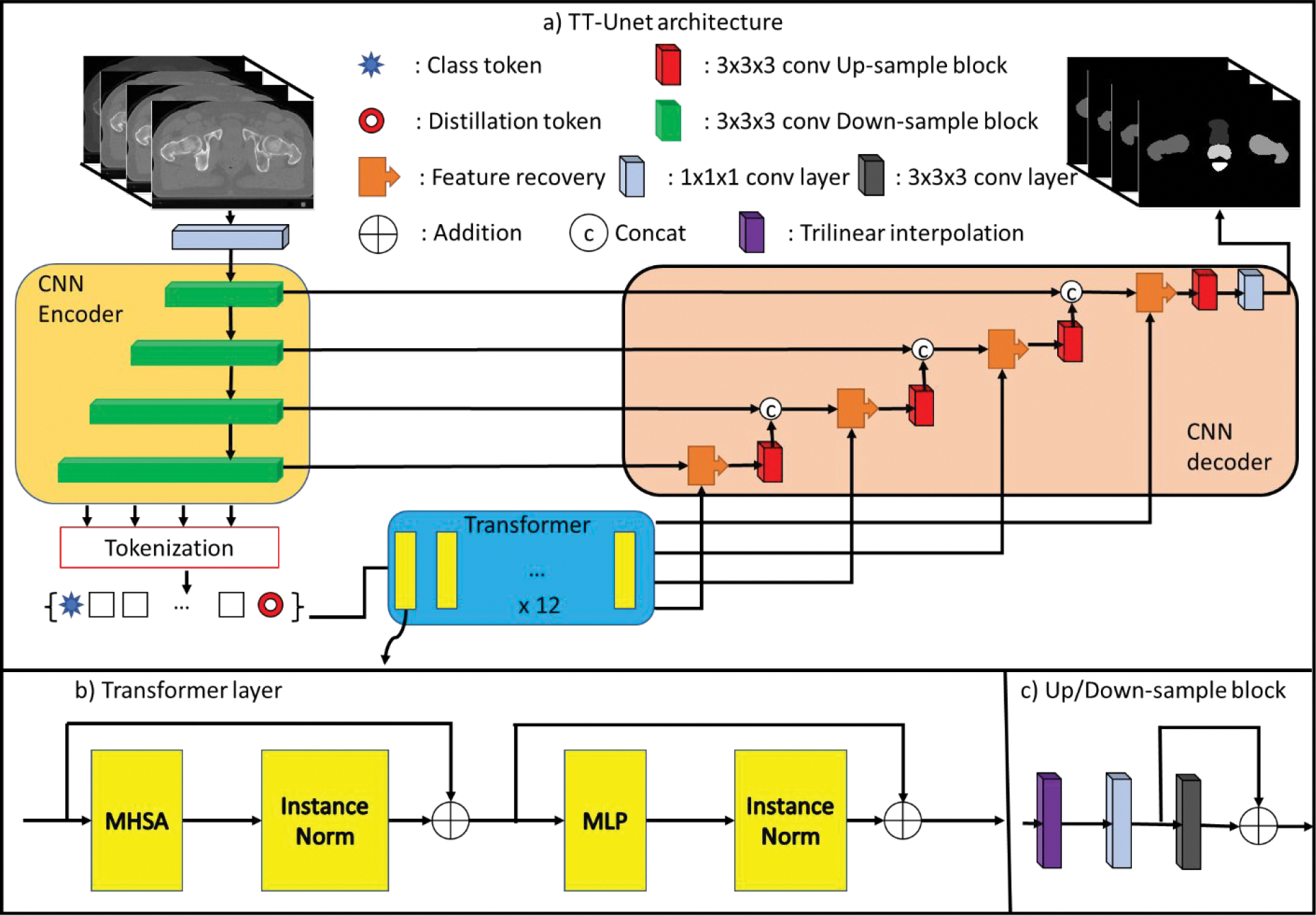
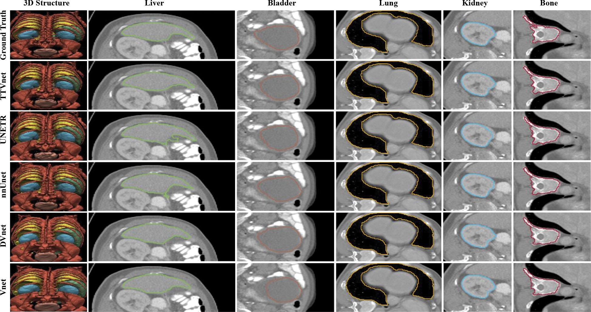
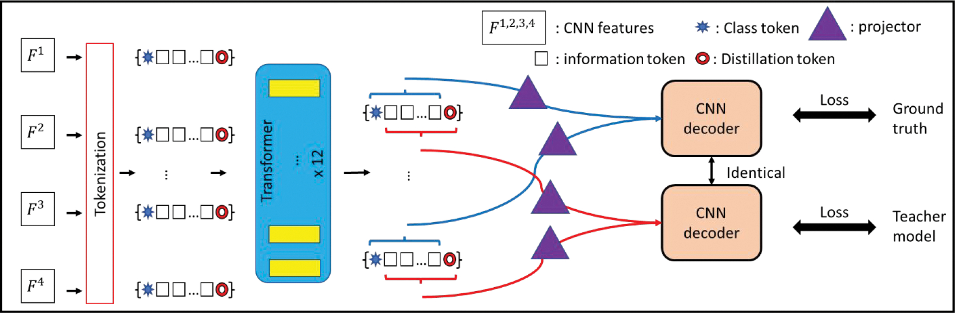
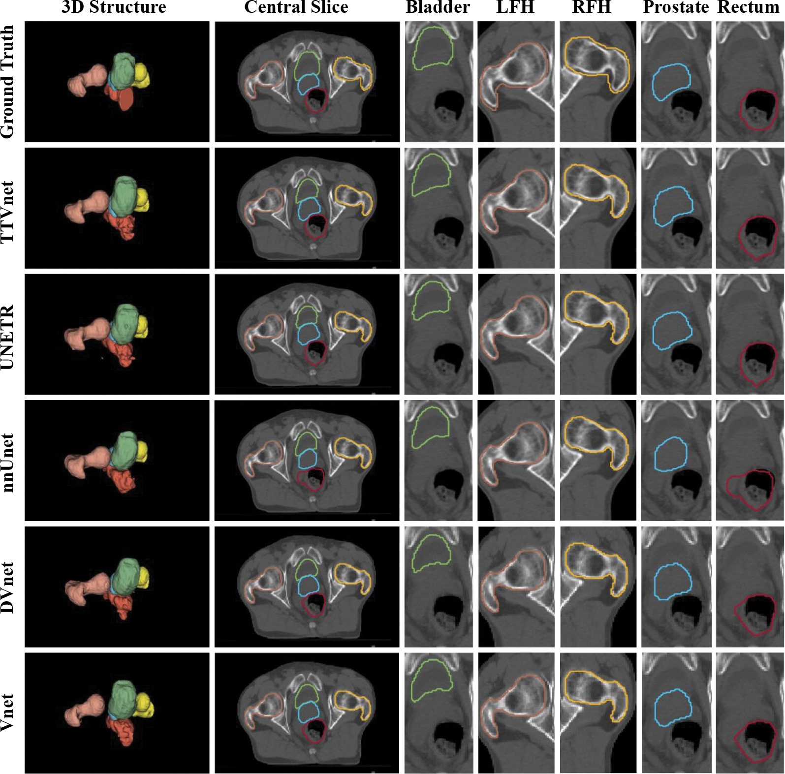
. This work aims to develop an automated segmentation method for the prostate and its surrounding organs-at-risk in pelvic computed tomography to facilitate prostate radiation treatment planning.. In this work, we propose a novel deep learning algorithm combining a U-shaped convolutional neural network (CNN) and vision transformer (VIT) for multi-organ (i.e. bladder, prostate, rectum, left and right femoral heads) segmentation in male pelvic CT images. The U-shaped model consists of three components: a CNN-based encoder for local feature extraction, a token-based VIT for capturing global dependencies from the CNN features, and a CNN-based decoder for predicting the segmentation outcome from the VIT's output. The novelty of our network is a token-based multi-head self-attention mechanism used in the transformer, which encourages long-range dependencies and forwards informative high-resolution feature maps from the encoder to the decoder. In addition, a knowledge distillation strategy is deployed to further enhance the learning capability of the proposed network.. We evaluated the network using: (1) a dataset collected from 94 patients with prostate cancer; (2) and a public dataset CT-ORG. A quantitative evaluation of the proposed network's performance was performed on each organ based on (1) volume similarity between the segmented contours and ground truth using Dice score, segmentation sensitivity, and precision, (2) surface similarity evaluated by Hausdorff distance (HD), mean surface distance (MSD) and residual mean square distance (RMS), (3) and percentage volume difference (PVD). The performance was then compared against other state-of-art methods. Average volume similarity measures obtained by the network overall organs were Dice score = 0.91, sensitivity = 0.90, precision = 0.92, average surface similarities were HD = 3.78 mm, MSD = 1.24 mm, RMS = 2.03 mm; average percentage volume difference was PVD = 9.9% on the first dataset. The network also obtained Dice score = 0.93, sensitivity = 0.93, precision = 0.93, average surface similarities were HD = 5.82 mm, MSD = 1.16 mm, RMS = 1.24 mm; average percentage volume difference was PVD = 6.6% on the CT-ORG dataset.. In summary, we propose a token-based transformer network with knowledge distillation for multi-organ segmentation using CT images. This method provides accurate and reliable segmentation results for each organ using CT imaging, facilitating the prostate radiation clinical workflow.
. 本研究旨在开发一种自动分割盆腔 CT 中前列腺及其周围危及器官的方法,以辅助前列腺放射治疗计划。. 在本研究中,我们提出了一种新的深度学习算法,该算法结合了 U 形卷积神经网络 (CNN) 和视觉转换器 (VIT),用于对男性盆腔 CT 图像中的多器官 (即膀胱、前列腺、直肠、左右股骨头) 进行分割。U 形模型由三个部分组成:基于 CNN 的编码器用于局部特征提取、基于令牌的 VIT 用于从 CNN 特征中捕获全局依赖关系,以及基于 CNN 的解码器用于从 VIT 的输出中预测分割结果。我们的网络的新颖之处在于在转换器中使用基于令牌的多头自注意力机制,该机制鼓励长程依赖关系,并从编码器向解码器传递有信息的高分辨率特征图。此外,还部署了知识蒸馏策略来进一步增强所提出的网络的学习能力。. 我们使用以下方法评估了网络:(1) 从 94 例前列腺癌患者中收集的数据集;(2) 和一个公共数据集 CT-ORG。基于 (1) 分割轮廓与地面实况之间的体积相似性,使用 Dice 评分、分割灵敏度和精度,(2) 基于 Hausdorff 距离 (HD)、平均表面距离 (MSD) 和残余均方距离 (RMS) 评估的表面相似性,以及 (3) 体积百分比差异 (PVD),对每个器官的网络性能进行了定量评估。然后将性能与其他最先进的方法进行了比较。网络对所有器官的平均体积相似性度量分别为 Dice 评分 = 0.91、灵敏度 = 0.90、精度 = 0.92、平均表面相似性分别为 HD = 3.78mm、MSD = 1.24mm、RMS = 2.03mm、平均体积百分比差异为 PVD = 9.9%。网络还在 CT-ORG 数据集上获得了 Dice 评分 = 0.93、灵敏度 = 0.93、精度 = 0.93、平均表面相似性分别为 HD = 5.82mm、MSD = 1.16mm、RMS = 1.24mm、平均体积百分比差异为 PVD = 6.6%。. 总之,我们提出了一种基于令牌的带有知识蒸馏的转换器网络,用于使用 CT 图像进行多器官分割。该方法使用 CT 成像对每个器官提供准确可靠的分割结果,从而简化了前列腺放射临床工作流程。