• 文献检索
  • 文档翻译
  • 深度研究
  • 学术资讯
  • Suppr Zotero 插件Zotero 插件
  • 邀请有礼
  • 套餐&价格
  • 历史记录
应用&插件
Suppr Zotero 插件Zotero 插件浏览器插件Mac 客户端Windows 客户端微信小程序
定价
高级版会员购买积分包购买API积分包
服务
文献检索文档翻译深度研究API 文档MCP 服务
关于我们
关于 Suppr公司介绍联系我们用户协议隐私条款
关注我们

Suppr 超能文献

核心技术专利:CN118964589B侵权必究
粤ICP备2023148730 号-1Suppr @ 2026

文献检索

告别复杂PubMed语法,用中文像聊天一样搜索,搜遍4000万医学文献。AI智能推荐,让科研检索更轻松。

立即免费搜索

文件翻译

保留排版,准确专业,支持PDF/Word/PPT等文件格式,支持 12+语言互译。

免费翻译文档

深度研究

AI帮你快速写综述,25分钟生成高质量综述,智能提取关键信息,辅助科研写作。

立即免费体验

真实微观结构模拟器 (RMS):显微镜图像三维细胞分割的扩散蒙特卡罗模拟。

Realistic Microstructure Simulator (RMS): Monte Carlo simulations of diffusion in three-dimensional cell segmentations of microscopy images.

机构信息

Center for Biomedical Imaging, Department of Radiology, New York University School of Medicine, New York, NY, USA; Center for Advanced Imaging Innovation and Research (CAI2R), New York University School of Medicine, New York, NY, USA.

Center for Biomedical Imaging, Department of Radiology, New York University School of Medicine, New York, NY, USA; Center for Advanced Imaging Innovation and Research (CAI2R), New York University School of Medicine, New York, NY, USA.

出版信息

J Neurosci Methods. 2021 Feb 15;350:109018. doi: 10.1016/j.jneumeth.2020.109018. Epub 2020 Dec 3.

DOI:10.1016/j.jneumeth.2020.109018
PMID:33279478
原文链接:https://pmc.ncbi.nlm.nih.gov/articles/PMC8026575/
Abstract

BACKGROUND

Monte Carlo simulations of diffusion are commonly used as a model validation tool as they are especially suitable for generating the diffusion MRI signal in complicated tissue microgeometries.

NEW METHOD

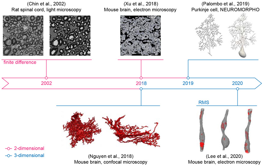
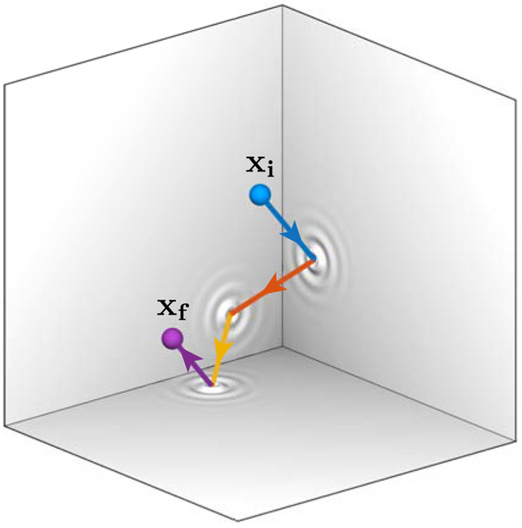
Here we describe the details of implementing Monte Carlo simulations in three-dimensional (3d) voxelized segmentations of cells in microscopy images. Using the concept of the corner reflector, we largely reduce the computational load of simulating diffusion within and exchange between multiple cells. Precision is further achieved by GPU-based parallel computations.

RESULTS

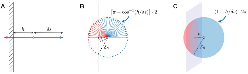
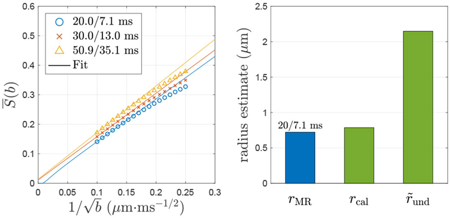
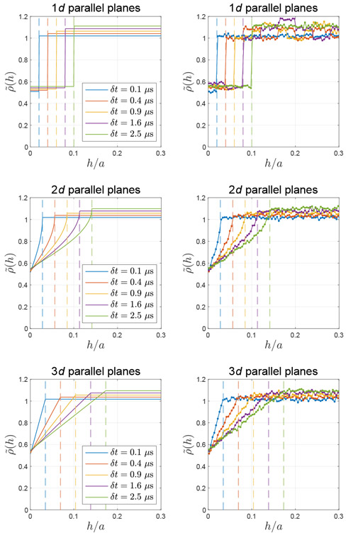
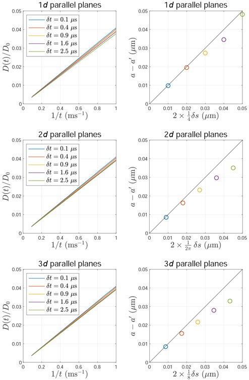
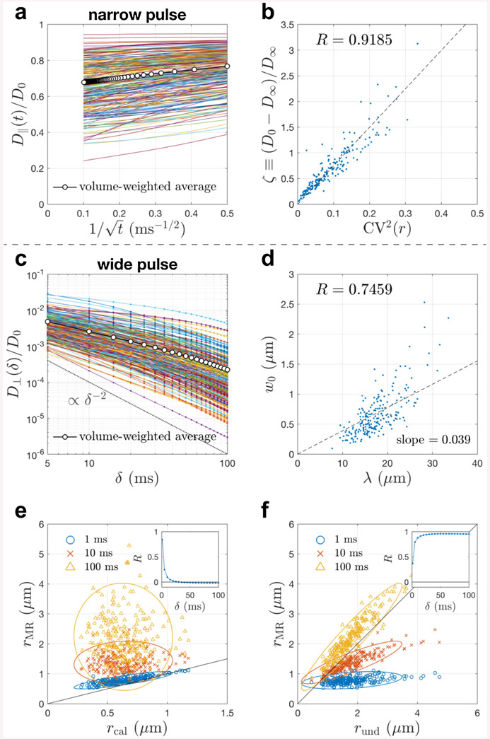
Our simulation of diffusion in white matter axons segmented from a mouse brain demonstrates its value in validating biophysical models. Furthermore, we provide the theoretical background for implementing a discretized diffusion process, and consider the finite-step effects of the particle-membrane reflection and permeation events, needed for efficient simulation of interactions with irregular boundaries, spatially variable diffusion coefficient, and exchange.

COMPARISON WITH EXISTING METHODS

To our knowledge, this is the first Monte Carlo pipeline for MR signal simulations in a substrate composed of numerous realistic cells, accounting for their permeable and irregularly-shaped membranes.

CONCLUSIONS

The proposed RMS pipeline makes it possible to achieve fast and accurate simulations of diffusion in realistic tissue microgeometry, as well as the interplay with other MR contrasts. Presently, RMS focuses on simulations of diffusion, exchange, and T and T NMR relaxation in static tissues, with a possibility to straightforwardly account for susceptibility-induced T effects and flow.

摘要

背景

扩散的蒙特卡罗模拟通常被用作模型验证工具,因为它们特别适合在复杂的组织微观结构中产生扩散 MRI 信号。

新方法

在这里,我们描述了在显微镜图像的三维(3d)体素化细胞分割中实现蒙特卡罗模拟的细节。我们利用角反射器的概念,大大降低了模拟多个细胞内和细胞间扩散的计算负荷。通过基于 GPU 的并行计算进一步提高了精度。

结果

我们对白质轴突的扩散模拟从老鼠大脑的分割中验证了生物物理模型的价值。此外,我们为离散扩散过程的实现提供了理论背景,并考虑了粒子-膜反射和渗透事件的有限步效应,这对于有效模拟不规则边界、空间变化扩散系数和交换的相互作用是必要的。

与现有方法的比较

据我们所知,这是第一个用于由大量真实细胞组成的基质中 MR 信号模拟的蒙特卡罗流水线,考虑了它们可渗透和不规则形状的膜。

结论

所提出的 RMS 流水线使得在真实组织微观结构中实现快速准确的扩散模拟以及与其他 MR 对比度的相互作用成为可能。目前,RMS 专注于静态组织中扩散、交换和 T 和 T NMR 弛豫的模拟,有可能直接考虑顺磁性诱导的 T 效应和流动。

https://cdn.ncbi.nlm.nih.gov/pmc/blobs/e5d3/8026575/3257501f1a6b/nihms-1651786-f0004.jpg
https://cdn.ncbi.nlm.nih.gov/pmc/blobs/e5d3/8026575/507c87034345/nihms-1651786-f0005.jpg
https://cdn.ncbi.nlm.nih.gov/pmc/blobs/e5d3/8026575/4c5e726b98c2/nihms-1651786-f0006.jpg
https://cdn.ncbi.nlm.nih.gov/pmc/blobs/e5d3/8026575/cc277bc39577/nihms-1651786-f0007.jpg
https://cdn.ncbi.nlm.nih.gov/pmc/blobs/e5d3/8026575/bb308892ddd0/nihms-1651786-f0009.jpg
https://cdn.ncbi.nlm.nih.gov/pmc/blobs/e5d3/8026575/e98c5f29f1bd/nihms-1651786-f0010.jpg
https://cdn.ncbi.nlm.nih.gov/pmc/blobs/e5d3/8026575/2b20263bc89b/nihms-1651786-f0008.jpg
https://cdn.ncbi.nlm.nih.gov/pmc/blobs/e5d3/8026575/1435c2f26739/nihms-1651786-f0011.jpg
https://cdn.ncbi.nlm.nih.gov/pmc/blobs/e5d3/8026575/a14126641158/nihms-1651786-f0001.jpg
https://cdn.ncbi.nlm.nih.gov/pmc/blobs/e5d3/8026575/6d2369033409/nihms-1651786-f0002.jpg
https://cdn.ncbi.nlm.nih.gov/pmc/blobs/e5d3/8026575/38ee4ef46a46/nihms-1651786-f0003.jpg
https://cdn.ncbi.nlm.nih.gov/pmc/blobs/e5d3/8026575/3257501f1a6b/nihms-1651786-f0004.jpg

相似文献

1
Realistic Microstructure Simulator (RMS): Monte Carlo simulations of diffusion in three-dimensional cell segmentations of microscopy images.真实微观结构模拟器 (RMS):显微镜图像三维细胞分割的扩散蒙特卡罗模拟。
J Neurosci Methods. 2021 Feb 15;350:109018. doi: 10.1016/j.jneumeth.2020.109018. Epub 2020 Dec 3.
2
Towards microstructure fingerprinting: Estimation of tissue properties from a dictionary of Monte Carlo diffusion MRI simulations.面向微观结构特征分析:从蒙特卡罗扩散 MRI 模拟字典中估计组织特性。
Neuroimage. 2019 Jan 1;184:964-980. doi: 10.1016/j.neuroimage.2018.09.076. Epub 2018 Sep 30.
3
Macromolecular crowding: chemistry and physics meet biology (Ascona, Switzerland, 10-14 June 2012).大分子拥挤现象:化学与物理邂逅生物学(瑞士阿斯科纳,2012年6月10日至14日)
Phys Biol. 2013 Aug;10(4):040301. doi: 10.1088/1478-3975/10/4/040301. Epub 2013 Aug 2.
4
MPEXS-DNA, a new GPU-based Monte Carlo simulator for track structures and radiation chemistry at subcellular scale.MPEXS-DNA,一种新的基于 GPU 的蒙特卡罗模拟程序,用于亚细胞尺度的径迹结构和辐射化学。
Med Phys. 2019 Mar;46(3):1483-1500. doi: 10.1002/mp.13370. Epub 2019 Jan 22.
5
Diffusion MRI simulation of realistic neurons with SpinDoctor and the Neuron Module.使用 SpinDoctor 和神经元模块对真实神经元进行扩散 MRI 模拟。
Neuroimage. 2020 Nov 15;222:117198. doi: 10.1016/j.neuroimage.2020.117198. Epub 2020 Jul 27.
6
Diffusion microscopist simulator: a general Monte Carlo simulation system for diffusion magnetic resonance imaging.扩散显微镜模拟器:一种用于扩散磁共振成像的通用蒙特卡罗模拟系统。
PLoS One. 2013 Oct 10;8(10):e76626. doi: 10.1371/journal.pone.0076626. eCollection 2013.
7
Monte Carlo simulation of water diffusion through cardiac tissue models.心脏组织模型中水扩散的蒙特卡罗模拟。
Med Eng Phys. 2023 Oct;120:104013. doi: 10.1016/j.medengphy.2023.104013. Epub 2023 Jun 24.
8
A generative model of realistic brain cells with application to numerical simulation of the diffusion-weighted MR signal.具有真实脑细胞生成模型的应用,用于扩散加权磁共振信号的数值模拟。
Neuroimage. 2019 Mar;188:391-402. doi: 10.1016/j.neuroimage.2018.12.025. Epub 2018 Dec 12.
9
Complex geometric models of diffusion and relaxation in healthy and damaged white matter.健康和受损白质中扩散和弛豫的复杂几何模型。
NMR Biomed. 2010 Feb;23(2):152-62. doi: 10.1002/nbm.1437.
10
Evaluating the accuracy and precision of a two-compartment Kärger model using Monte Carlo simulations.运用蒙特卡罗模拟评估双室 Kärger 模型的准确性和精密度。
J Magn Reson. 2010 Sep;206(1):59-67. doi: 10.1016/j.jmr.2010.06.002. Epub 2010 Jun 9.

引用本文的文献

1
SpinWalk: A Monte Carlo simulator for MR-signal formation in inhomogeneous tissue.自旋游走:一种用于非均匀组织中磁共振信号形成的蒙特卡罗模拟器。
Imaging Neurosci (Camb). 2025 Apr 15;3. doi: 10.1162/imag_a_00533. eCollection 2025.
2
Passive water exchange between multiple sites can explain why apparent exchange rate constants depend on ionic and osmotic conditions in gray matter.多个位点之间的被动水交换可以解释为什么表观交换速率常数取决于灰质中的离子和渗透条件。
bioRxiv. 2025 Jul 2:2025.05.27.655493. doi: 10.1101/2025.05.27.655493.
3
In vivo cortical microstructure mapping using high-gradient diffusion MRI accounting for intercompartmental water exchange effects.

本文引用的文献

1
The present and the future of microstructure MRI: From a paradigm shift to normal science.微观结构 MRI 的现状与未来:从范式转变到常规科学。
J Neurosci Methods. 2021 Mar 1;351:108947. doi: 10.1016/j.jneumeth.2020.108947. Epub 2020 Oct 21.
2
The impact of realistic axonal shape on axon diameter estimation using diffusion MRI.基于弥散磁共振成像的真实轴突形态对轴突直径评估的影响。
Neuroimage. 2020 Dec;223:117228. doi: 10.1016/j.neuroimage.2020.117228. Epub 2020 Aug 13.
3
A time-dependent diffusion MRI signature of axon caliber variations and beading.
利用考虑隔室间水交换效应的高梯度扩散磁共振成像进行体内皮质微结构映射。
Neuroimage. 2025 Jul 1;314:121258. doi: 10.1016/j.neuroimage.2025.121258. Epub 2025 May 9.
4
SpinFlowSim: A blood flow simulation framework for histology-informed diffusion MRI microvasculature mapping in cancer.SpinFlowSim:一种用于癌症中组织学信息扩散磁共振成像微血管映射的血流模拟框架。
Med Image Anal. 2025 May;102:103531. doi: 10.1016/j.media.2025.103531. Epub 2025 Mar 7.
5
Scattering approach to diffusion quantifies axonal damage in brain injury.扩散的散射方法可量化脑损伤中的轴突损伤。
ArXiv. 2025 Jan 30:arXiv:2501.18167v1.
6
Predicting Mesoscopic Larmor Frequency Shifts in White Matter With Diffusion MRI-A Monte Carlo Study in Axonal Phantoms.利用扩散磁共振成像预测白质中的介观拉莫尔频率偏移——轴突模型的蒙特卡罗研究
NMR Biomed. 2025 Mar;38(3):e70004. doi: 10.1002/nbm.70004.
7
Revealing membrane integrity and cell size from diffusion kurtosis time dependence.从扩散峰度时间依赖性揭示膜完整性和细胞大小。
Magn Reson Med. 2025 Mar;93(3):1329-1347. doi: 10.1002/mrm.30335. Epub 2024 Oct 29.
8
Exploring white matter dynamics and morphology through interactive numerical phantoms: the White Matter Generator.通过交互式数字模型探索白质动力学和形态学:白质生成器
Front Neuroinform. 2024 Jul 31;18:1354708. doi: 10.3389/fninf.2024.1354708. eCollection 2024.
9
In vivo mapping of cellular resolution neuropathology in brain ischemia with diffusion MRI.利用扩散磁共振成像对脑缺血中细胞分辨率神经病理学进行体内图谱绘制。
Sci Adv. 2024 Jul 19;10(29):eadk1817. doi: 10.1126/sciadv.adk1817. Epub 2024 Jul 17.
10
Tuned exchange imaging: Can the filter exchange imaging pulse sequence be adapted for applications with thin slices and restricted diffusion?调谐交换成像:滤波器交换成像脉冲序列是否可以适用于薄片和受限扩散的应用?
NMR Biomed. 2024 Nov;37(11):e5208. doi: 10.1002/nbm.5208. Epub 2024 Jul 4.
轴突管径变化和串珠样改变的时间依赖性扩散磁共振成像特征。
Commun Biol. 2020 Jul 7;3(1):354. doi: 10.1038/s42003-020-1050-x.
4
ConFiG: Contextual Fibre Growth to generate realistic axonal packing for diffusion MRI simulation.ConFiG:语境纤维生长生成用于扩散 MRI 模拟的真实轴突包装。
Neuroimage. 2020 Oct 15;220:117107. doi: 10.1016/j.neuroimage.2020.117107. Epub 2020 Jul 2.
5
In vivo observation and biophysical interpretation of time-dependent diffusion in human cortical gray matter.在体观察和生物物理解释人类皮质灰质中时变扩散。
Neuroimage. 2020 Nov 15;222:117054. doi: 10.1016/j.neuroimage.2020.117054. Epub 2020 Jun 22.
6
Nonivasive quantification of axon radii using diffusion MRI.利用弥散磁共振成像技术对轴突半径进行无创定量分析。
Elife. 2020 Feb 12;9:e49855. doi: 10.7554/eLife.49855.
7
Time-dependent diffusion in undulating thin fibers: Impact on axon diameter estimation.起伏细纤维中的时间相关扩散:对轴突直径估计的影响。
NMR Biomed. 2020 Mar;33(3):e4187. doi: 10.1002/nbm.4187. Epub 2019 Dec 23.
8
MEDUSA: A GPU-based tool to create realistic phantoms of the brain microstructure using tiny spheres.MEDUSA:一种基于 GPU 的工具,使用微小球体创建逼真的大脑微观结构的幻影。
Neuroimage. 2019 Jun;193:10-24. doi: 10.1016/j.neuroimage.2019.02.055. Epub 2019 Mar 5.
9
Along-axon diameter variation and axonal orientation dispersion revealed with 3D electron microscopy: implications for quantifying brain white matter microstructure with histology and diffusion MRI.利用 3D 电子显微镜揭示沿轴突直径变化和轴突取向离散:对组织学和扩散 MRI 定量分析脑白质微观结构的意义。
Brain Struct Funct. 2019 May;224(4):1469-1488. doi: 10.1007/s00429-019-01844-6. Epub 2019 Feb 21.
10
Intra-axonal diffusivity in brain white matter.脑白质内轴突弥散性。
Neuroimage. 2019 Apr 1;189:543-550. doi: 10.1016/j.neuroimage.2019.01.015. Epub 2019 Jan 16.