• 文献检索
  • 文档翻译
  • 深度研究
  • 学术资讯
  • Suppr Zotero 插件Zotero 插件
  • 邀请有礼
  • 套餐&价格
  • 历史记录
应用&插件
Suppr Zotero 插件Zotero 插件浏览器插件Mac 客户端Windows 客户端微信小程序
定价
高级版会员购买积分包购买API积分包
服务
文献检索文档翻译深度研究API 文档MCP 服务
关于我们
关于 Suppr公司介绍联系我们用户协议隐私条款
关注我们

Suppr 超能文献

核心技术专利:CN118964589B侵权必究
粤ICP备2023148730 号-1Suppr @ 2026

文献检索

告别复杂PubMed语法,用中文像聊天一样搜索,搜遍4000万医学文献。AI智能推荐,让科研检索更轻松。

立即免费搜索

文件翻译

保留排版,准确专业,支持PDF/Word/PPT等文件格式,支持 12+语言互译。

免费翻译文档

深度研究

AI帮你快速写综述,25分钟生成高质量综述,智能提取关键信息,辅助科研写作。

立即免费体验

相位成像与计算特异性(PICS)用于测量亚细胞区室中干物质变化。

Phase imaging with computational specificity (PICS) for measuring dry mass changes in sub-cellular compartments.

机构信息

Beckman Institute, University of Illinois at Urbana-Champaign, Urbana, IL, USA.

Department of Electrical and Computer Engineering, University of Illinois at Urbana-Champaign, Urbana, IL, USA.

出版信息

Nat Commun. 2020 Dec 7;11(1):6256. doi: 10.1038/s41467-020-20062-x.

DOI:10.1038/s41467-020-20062-x
PMID:33288761
原文链接:https://pmc.ncbi.nlm.nih.gov/articles/PMC7721808/
Abstract

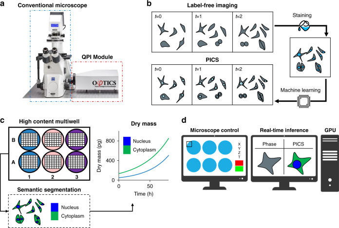
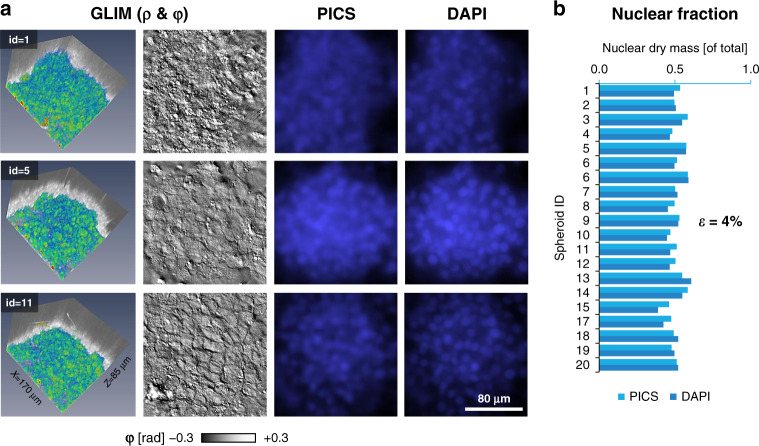
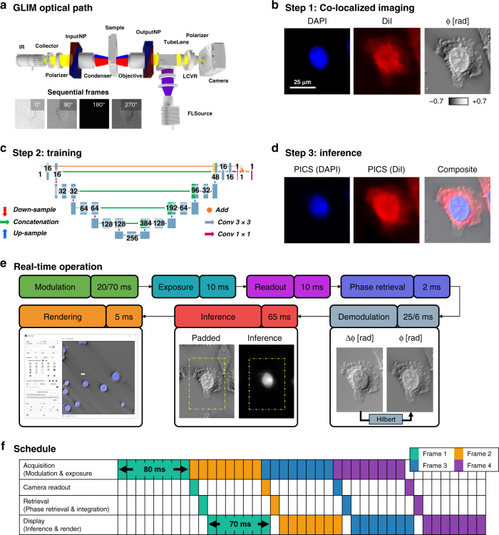
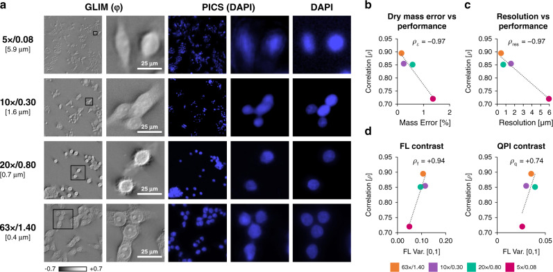
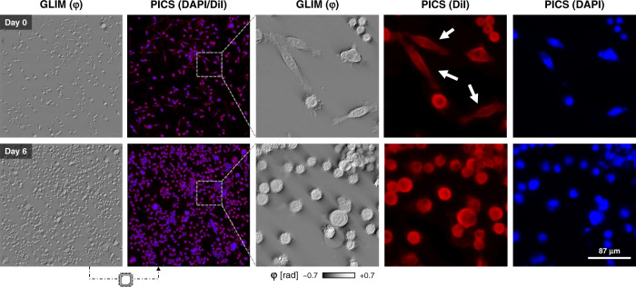
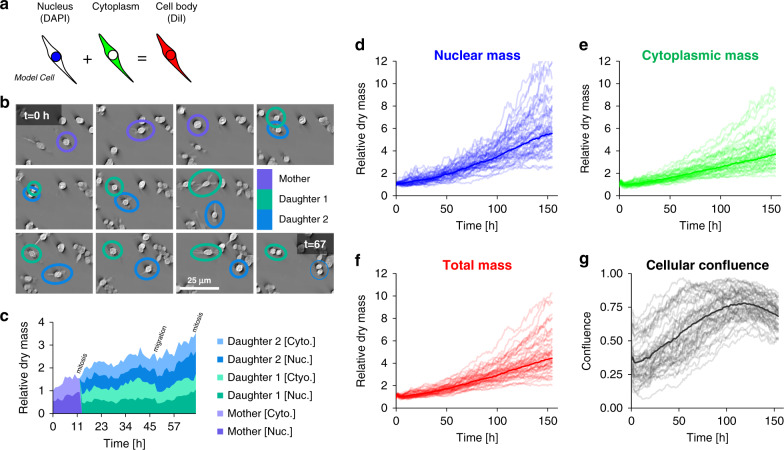
Due to its specificity, fluorescence microscopy has become a quintessential imaging tool in cell biology. However, photobleaching, phototoxicity, and related artifacts continue to limit fluorescence microscopy's utility. Recently, it has been shown that artificial intelligence (AI) can transform one form of contrast into another. We present phase imaging with computational specificity (PICS), a combination of quantitative phase imaging and AI, which provides information about unlabeled live cells with high specificity. Our imaging system allows for automatic training, while inference is built into the acquisition software and runs in real-time. Applying the computed fluorescence maps back to the quantitative phase imaging (QPI) data, we measured the growth of both nuclei and cytoplasm independently, over many days, without loss of viability. Using a QPI method that suppresses multiple scattering, we measured the dry mass content of individual cell nuclei within spheroids. In its current implementation, PICS offers a versatile quantitative technique for continuous simultaneous monitoring of individual cellular components in biological applications where long-term label-free imaging is desirable.

摘要

由于其特异性,荧光显微镜已成为细胞生物学中不可或缺的成像工具。然而,荧光漂白、光毒性和相关伪影仍然限制了荧光显微镜的应用。最近,已经证明人工智能 (AI) 可以将一种对比度转换为另一种对比度。我们提出了具有计算特异性的相衬成像 (PICS),这是定量相衬成像和人工智能的结合,它可以高特异性地提供关于未标记活细胞的信息。我们的成像系统允许自动训练,而推断则构建在采集软件中并实时运行。将计算出的荧光图应用回定量相衬成像 (QPI) 数据,我们可以在不丧失活力的情况下,独立测量细胞核和细胞质的生长情况,持续多天。使用一种抑制多次散射的 QPI 方法,我们测量了球体中单个人类细胞核的干物质含量。在当前的实现中,PICS 为生物应用中连续同时监测单个细胞成分提供了一种通用的定量技术,在这些应用中,长期无标记成像是理想的。

https://cdn.ncbi.nlm.nih.gov/pmc/blobs/e0a5/7721808/469b1ff625ff/41467_2020_20062_Fig6_HTML.jpg
https://cdn.ncbi.nlm.nih.gov/pmc/blobs/e0a5/7721808/da7fef6d445e/41467_2020_20062_Fig1_HTML.jpg
https://cdn.ncbi.nlm.nih.gov/pmc/blobs/e0a5/7721808/8ccbff0d1782/41467_2020_20062_Fig2_HTML.jpg
https://cdn.ncbi.nlm.nih.gov/pmc/blobs/e0a5/7721808/799d14e8e7d5/41467_2020_20062_Fig3_HTML.jpg
https://cdn.ncbi.nlm.nih.gov/pmc/blobs/e0a5/7721808/30d8b84e99f7/41467_2020_20062_Fig4_HTML.jpg
https://cdn.ncbi.nlm.nih.gov/pmc/blobs/e0a5/7721808/8a5850b4ebfd/41467_2020_20062_Fig5_HTML.jpg
https://cdn.ncbi.nlm.nih.gov/pmc/blobs/e0a5/7721808/469b1ff625ff/41467_2020_20062_Fig6_HTML.jpg

相似文献

1
Phase imaging with computational specificity (PICS) for measuring dry mass changes in sub-cellular compartments.相位成像与计算特异性(PICS)用于测量亚细胞区室中干物质变化。
Nat Commun. 2020 Dec 7;11(1):6256. doi: 10.1038/s41467-020-20062-x.
2
Single-shot quantitative phase imaging as an extension of differential interference contrast microscopy.单次定量相位成像作为微分干涉对比显微镜的扩展。
Genes Cells. 2021 Aug;26(8):596-610. doi: 10.1111/gtc.12876. Epub 2021 Jul 2.
3
Multiscale Assay of Unlabeled Neurite Dynamics Using Phase Imaging with Computational Specificity.使用具有计算特异性的相衬成像对未标记神经突动力学进行多尺度分析。
ACS Sens. 2021 May 28;6(5):1864-1874. doi: 10.1021/acssensors.1c00100. Epub 2021 Apr 21.
4
A quantitative method to track protein translocation between intracellular compartments in real-time in live cells using weighted local variance image analysis.一种使用加权局部方差图像分析在活细胞中实时追踪蛋白质在细胞内区室之间转运的定量方法。
PLoS One. 2013 Dec 20;8(12):e81988. doi: 10.1371/journal.pone.0081988. eCollection 2013.
5
Mathematical imaging methods for mitosis analysis in live-cell phase contrast microscopy.活细胞相差显微镜下有丝分裂分析的数学成像方法
Methods. 2017 Feb 15;115:91-99. doi: 10.1016/j.ymeth.2017.02.001. Epub 2017 Feb 9.
6
Time-lapse imaging of nuclear bodies.核小体的延时成像。
Methods Mol Biol. 2015;1262:55-67. doi: 10.1007/978-1-4939-2253-6_4.
7
Capturing cell morphology dynamics with high temporal resolution using single-shot quantitative phase gradient imaging.使用单次定量相梯度成像以高时间分辨率捕捉细胞形态动力学。
J Biomed Opt. 2024 Jun;29(Suppl 2):S22712. doi: 10.1117/1.JBO.29.S2.S22712. Epub 2024 Jul 16.
8
Label-free screening of brain tissue myelin content using phase imaging with computational specificity (PICS).使用具有计算特异性的相成像技术(PICS)对脑组织髓鞘含量进行无标记筛选。
APL Photonics. 2021 Jul 1;6(7):076103. doi: 10.1063/5.0050889. Epub 2021 Jul 12.
9
Label-Free Intracellular Multi-Specificity in Yeast Cells by Phase-Contrast Tomographic Flow Cytometry.通过相差断层扫描流式细胞术实现酵母细胞中无标记的细胞内多特异性
Small Methods. 2023 Nov;7(11):e2300447. doi: 10.1002/smtd.202300447. Epub 2023 Sep 5.
10
Live-dead assay on unlabeled cells using phase imaging with computational specificity.使用具有计算特异性的相差成像对未标记细胞进行死活检测。
Nat Commun. 2022 Feb 7;13(1):713. doi: 10.1038/s41467-022-28214-x.

引用本文的文献

1
Measuring Molecular Mass Densities at Subcellular Resolution Using Optical Diffraction Tomography.利用光学衍射层析成像技术在亚细胞分辨率下测量分子质量密度
Methods Mol Biol. 2025;2958:119-141. doi: 10.1007/978-1-0716-4714-1_8.
2
BlurryScope enables compact, cost-effective scanning microscopy for HER2 scoring using deep learning on blurry images.BlurryScope可通过对模糊图像进行深度学习,实现用于HER2评分的紧凑、经济高效的扫描显微镜技术。
NPJ Digit Med. 2025 Aug 6;8(1):506. doi: 10.1038/s41746-025-01882-x.
3
Chip-based label-free incoherent super-resolution optical microscopy.

本文引用的文献

1
Decellularized Matrix Produced by Mesenchymal Stem Cells Modulates Growth and Metabolic Activity of Hepatic Cell Cluster.间充质干细胞产生的脱细胞基质调节肝细胞簇的生长和代谢活性。
ACS Biomater Sci Eng. 2018 Feb 12;4(2):456-462. doi: 10.1021/acsbiomaterials.7b00494. Epub 2017 Nov 30.
2
Holographic virtual staining of individual biological cells.单个生物细胞的全像虚拟染色。
Proc Natl Acad Sci U S A. 2020 Apr 28;117(17):9223-9231. doi: 10.1073/pnas.1919569117. Epub 2020 Apr 13.
3
Epi-illumination gradient light interference microscopy for imaging opaque structures.
基于芯片的无标记非相干超分辨率光学显微镜。
Light Sci Appl. 2025 Aug 4;14(1):259. doi: 10.1038/s41377-025-01914-x.
4
Virtual staining of label-free tissue in imaging mass spectrometry.成像质谱中无标记组织的虚拟染色
Sci Adv. 2025 Aug;11(31):eadv0741. doi: 10.1126/sciadv.adv0741. Epub 2025 Aug 1.
5
Near-zero photon bioimaging by fusing deep learning and ultralow-light microscopy.通过融合深度学习与超弱光显微镜实现近零光子生物成像。
Proc Natl Acad Sci U S A. 2025 May 27;122(21):e2412261122. doi: 10.1073/pnas.2412261122. Epub 2025 May 19.
6
Enhancing Gamma Knife Cone-beam Computed Tomography Image Quality Using Pix2pix Generative Adversarial Networks: A Deep Learning Approach.使用Pix2pix生成对抗网络提高伽玛刀锥形束计算机断层扫描图像质量:一种深度学习方法
J Med Phys. 2025 Jan-Mar;50(1):30-37. doi: 10.4103/jmp.jmp_140_24. Epub 2025 Mar 24.
7
Deciphering structural complexity of brain, joint, and muscle tissues using Fourier Ptychographic Scattered Light Microscopy.利用傅里叶叠层散斑显微镜解析脑、关节和肌肉组织的结构复杂性。
bioRxiv. 2025 Apr 9:2024.11.28.625428. doi: 10.1101/2024.11.28.625428.
8
Biomaterials for Cell Manufacturing.细胞制造用生物材料。
ACS Macro Lett. 2024 Nov 19;13(11):1521-1530. doi: 10.1021/acsmacrolett.4c00634. Epub 2024 Oct 28.
9
GAN-based quantitative oblique back-illumination microscopy enables computationally efficient epi-mode refractive index tomography.基于生成对抗网络的定量斜后照明显微镜技术实现了计算效率高的落射式模式折射率断层扫描。
Biomed Opt Express. 2024 Jul 18;15(8):4764-4774. doi: 10.1364/BOE.528968. eCollection 2024 Aug 1.
10
Mantis: High-throughput 4D imaging and analysis of the molecular and physical architecture of cells.Mantis:细胞分子与物理结构的高通量4D成像及分析
PNAS Nexus. 2024 Aug 9;3(9):pgae323. doi: 10.1093/pnasnexus/pgae323. eCollection 2024 Sep.
基于梯度光干涉的 epi 照明显微镜用于观察不透明结构。
Nat Commun. 2019 Oct 16;10(1):4691. doi: 10.1038/s41467-019-12634-3.
4
Cell-to-cell influence on growth in large populations.细胞间对大量群体生长的影响。
Biomed Opt Express. 2019 Aug 20;10(9):4664-4675. doi: 10.1364/BOE.10.004664. eCollection 2019 Sep 1.
5
Virtual histological staining of unlabelled tissue-autofluorescence images via deep learning.基于深度学习的无标记组织自发荧光图像的虚拟组织学染色。
Nat Biomed Eng. 2019 Jun;3(6):466-477. doi: 10.1038/s41551-019-0362-y. Epub 2019 Mar 4.
6
Perspective: The promise of multi-cellular engineered living systems.观点:多细胞工程生命系统的前景。
APL Bioeng. 2018 Oct 11;2(4):040901. doi: 10.1063/1.5038337. eCollection 2018 Dec.
7
PhaseStain: the digital staining of label-free quantitative phase microscopy images using deep learning.相位染色:使用深度学习对无标记定量相显微镜图像进行数字染色。
Light Sci Appl. 2019 Feb 6;8:23. doi: 10.1038/s41377-019-0129-y. eCollection 2019.
8
Quantitative phase imaging reveals matrix stiffness-dependent growth and migration of cancer cells.定量相位成像揭示了基质硬度依赖性的癌细胞生长和迁移。
Sci Rep. 2019 Jan 22;9(1):248. doi: 10.1038/s41598-018-36551-5.
9
SLIM microscopy allows for visualization of DNA-containing liposomes designed for sperm-mediated gene transfer in cattle.SLIM 显微镜可用于观察设计用于牛精子介导基因转移的含 DNA 的脂质体。
Mol Biol Rep. 2019 Feb;46(1):695-703. doi: 10.1007/s11033-018-4525-9. Epub 2018 Dec 11.
10
Label-free prediction of three-dimensional fluorescence images from transmitted-light microscopy.无标记法预测透射光显微镜的三维荧光图像。
Nat Methods. 2018 Nov;15(11):917-920. doi: 10.1038/s41592-018-0111-2. Epub 2018 Sep 17.