• 文献检索
  • 文档翻译
  • 深度研究
  • 学术资讯
  • Suppr Zotero 插件Zotero 插件
  • 邀请有礼
  • 套餐&价格
  • 历史记录
应用&插件
Suppr Zotero 插件Zotero 插件浏览器插件Mac 客户端Windows 客户端微信小程序
定价
高级版会员购买积分包购买API积分包
服务
文献检索文档翻译深度研究API 文档MCP 服务
关于我们
关于 Suppr公司介绍联系我们用户协议隐私条款
关注我们

Suppr 超能文献

核心技术专利:CN118964589B侵权必究
粤ICP备2023148730 号-1Suppr @ 2026

文献检索

告别复杂PubMed语法,用中文像聊天一样搜索,搜遍4000万医学文献。AI智能推荐,让科研检索更轻松。

立即免费搜索

文件翻译

保留排版,准确专业,支持PDF/Word/PPT等文件格式,支持 12+语言互译。

免费翻译文档

深度研究

AI帮你快速写综述,25分钟生成高质量综述,智能提取关键信息,辅助科研写作。

立即免费体验

与 COVID-19 肺炎患者临床特征相关的胸部 CT 特征。

Chest CT features associated with the clinical characteristics of patients with COVID-19 pneumonia.

机构信息

Department of Respiratory Medicine, Xiangya Hospital, Central South University, Changsha, PR China.

Department of Respiratory Medicine, Wuhan First Hospital/Wuhan Hospital of Traditional Chinese and Western Medicine, Wuhan, PR China.

出版信息

Ann Med. 2021 Dec;53(1):169-180. doi: 10.1080/07853890.2020.1851044.

DOI:10.1080/07853890.2020.1851044
PMID:33426973
原文链接:https://pmc.ncbi.nlm.nih.gov/articles/PMC7877953/
Abstract

OBJECTIVES

Coronavirus disease 2019 (COVID-19) has rapidly swept across the world. This study aimed to explore the relationship between the chest CT findings and clinical characteristics of COVID-19 patients.

METHODS

Patients with COVID-19 confirmed by next-generation sequencing or RT-PCR who had undergone more than 4 serial chest CT procedures were retrospectively enrolled.

RESULTS

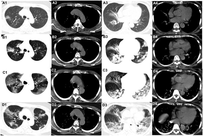
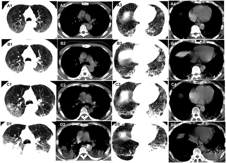
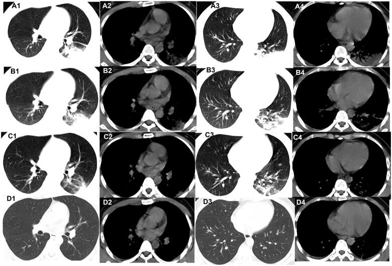
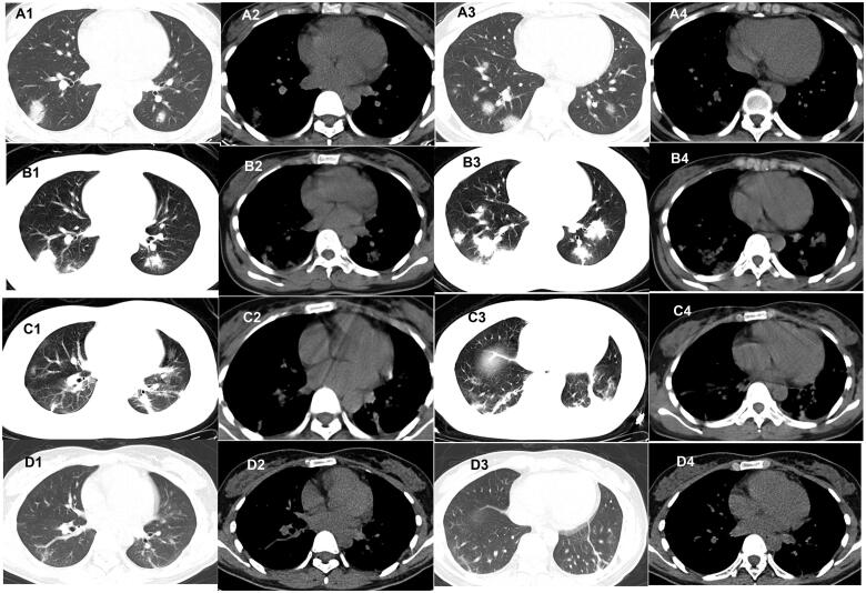
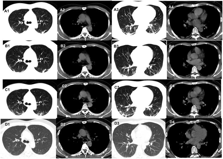
This study included 361 patients - 192 men and 169 women. On initial chest CT, more lesions were identified as multiple bilateral lungs lesions and localised in the peripheral lung. The predominant patterns of abnormality were ground-glass opacities (GGO) (28.5%), consolidation (13.0%), nodule (23.0%), fibrous stripes (5.3%) and mixed (30.2%). Severe cases were more common in patients with a mixed pattern (21.1%) and less common in patients with nodules (2.4%). During follow-up CT, the mediumtotal severity score (TSS) in patients with nodules and fibrous strips was significantly lower than that in patients with mixed patterns in all three stages ( < .01).

CONCLUSION

Chest CT plays an important role in diagnosing COVID-19. The CT features may vary by age. Different CT features are not only associated with clinical manifestation but also patient prognosis. Key messages The initial chest CT findings of COVID-19 could help us monitor and predict the outcome. Nodules were more common in non severe cases and had a favorable prognosis. The mixed pattern was more common in severe cases and usually had a relatively poor outcome.

摘要

目的

2019 年冠状病毒病(COVID-19)在全球迅速蔓延。本研究旨在探讨 COVID-19 患者的胸部 CT 表现与临床特征之间的关系。

方法

回顾性纳入经下一代测序或 RT-PCR 确诊为 COVID-19 且接受了 4 次以上连续胸部 CT 检查的患者。

结果

本研究共纳入 361 例患者,其中男 192 例,女 169 例。初次胸部 CT 时,更多病变被确定为多双侧肺部病变,且局限于外周肺部。异常的主要模式为磨玻璃密度影(GGO)(28.5%)、实变(13.0%)、结节(23.0%)、纤维条纹(5.3%)和混合(30.2%)。混合模式患者中严重病例更为常见(21.1%),而结节患者中则较为少见(2.4%)。在随访 CT 中,结节和纤维带患者的总严重程度评分(TSS)在所有三个阶段均明显低于混合模式患者(均<0.01)。

结论

胸部 CT 在 COVID-19 的诊断中具有重要作用。CT 特征可能因年龄而异。不同的 CT 特征不仅与临床表现有关,而且与患者预后有关。

关键信息

COVID-19 的初始胸部 CT 表现有助于我们监测和预测结果。结节在非重症病例中更为常见,且预后良好。混合模式在重症病例中更为常见,通常预后较差。

https://cdn.ncbi.nlm.nih.gov/pmc/blobs/3c01/7877953/4e17d20eba2b/IANN_A_1851044_F0005_B.jpg
https://cdn.ncbi.nlm.nih.gov/pmc/blobs/3c01/7877953/8d51d4bd95da/IANN_A_1851044_F0001_B.jpg
https://cdn.ncbi.nlm.nih.gov/pmc/blobs/3c01/7877953/8fb327aa80ff/IANN_A_1851044_F0002_B.jpg
https://cdn.ncbi.nlm.nih.gov/pmc/blobs/3c01/7877953/90f21f3b15e5/IANN_A_1851044_F0003_B.jpg
https://cdn.ncbi.nlm.nih.gov/pmc/blobs/3c01/7877953/ab735c928899/IANN_A_1851044_F0004_B.jpg
https://cdn.ncbi.nlm.nih.gov/pmc/blobs/3c01/7877953/4e17d20eba2b/IANN_A_1851044_F0005_B.jpg

相似文献

1
Chest CT features associated with the clinical characteristics of patients with COVID-19 pneumonia.与 COVID-19 肺炎患者临床特征相关的胸部 CT 特征。
Ann Med. 2021 Dec;53(1):169-180. doi: 10.1080/07853890.2020.1851044.
2
Temporal relationship between serial RT-PCR results and serial chest CT imaging, and serial CT changes in coronavirus 2019 (COVID-19) pneumonia: a descriptive study of 155 cases in China.新型冠状病毒(COVID-19)肺炎的实时逆转录聚合酶链反应(RT-PCR)结果与系列胸部 CT 影像学及系列 CT 改变的时间关系:中国 155 例的描述性研究。
Eur Radiol. 2021 Mar;31(3):1175-1184. doi: 10.1007/s00330-020-07268-9. Epub 2020 Sep 15.
3
The role of chest CT quantitative pulmonary inflammatory index in the evaluation of the course and treatment outcome of COVID-19 pneumonia.胸部 CT 定量肺炎症指数在评估 COVID-19 肺炎病程和治疗效果中的作用。
Sci Rep. 2021 Apr 8;11(1):7752. doi: 10.1038/s41598-021-87430-5.
4
Clinical manifestations and radiological features by chest computed tomographic findings of a novel coronavirus disease-19 pneumonia among 92 patients in Japan.日本 92 例新型冠状病毒病肺炎患者的胸部计算机断层扫描结果的临床表现和放射学特征。
J Microbiol Immunol Infect. 2021 Aug;54(4):748-751. doi: 10.1016/j.jmii.2020.07.011. Epub 2020 Aug 20.
5
Computed tomography features of COVID-19 in children: A systematic review and meta-analysis.儿童 COVID-19 的计算机断层扫描特征:系统评价和荟萃分析。
Medicine (Baltimore). 2021 Sep 24;100(38):e22571. doi: 10.1097/MD.0000000000022571.
6
COVID-19 pneumonia: CT findings of 122 patients and differentiation from influenza pneumonia.新型冠状病毒肺炎:122例患者的CT表现及与流感肺炎的鉴别诊断
Eur Radiol. 2020 Oct;30(10):5463-5469. doi: 10.1007/s00330-020-06928-0. Epub 2020 May 12.
7
Imaging and clinical features of patients with 2019 novel coronavirus SARS-CoV-2.新型冠状病毒 2019 年 SARS-CoV-2 患者的影像学和临床特征。
Eur J Nucl Med Mol Imaging. 2020 May;47(5):1275-1280. doi: 10.1007/s00259-020-04735-9. Epub 2020 Feb 28.
8
2019 novel coronavirus (COVID-19) pneumonia: CT manifestations and pattern of evolution in 110 patients in Jiangxi, China.2019 新型冠状病毒(COVID-19)肺炎:中国江西 110 例患者的 CT 表现和演变模式。
Eur Radiol. 2021 Feb;31(2):1059-1068. doi: 10.1007/s00330-020-07201-0. Epub 2020 Aug 27.
9
CT-scan findings of COVID-19 pneumonia based on the time elapsed from the beginning of symptoms to the CT imaging evaluation: a descriptive study in Iran.基于从症状出现到 CT 影像评估时间的 COVID-19 肺炎 CT 扫描结果:伊朗的一项描述性研究。
Rom J Intern Med. 2020 Dec 17;58(4):242-250. doi: 10.2478/rjim-2020-0019. Print 2020 Dec 1.
10
Relation Between Chest CT Findings and Clinical Conditions of Coronavirus Disease (COVID-19) Pneumonia: A Multicenter Study.新型冠状病毒肺炎的胸部 CT 表现与临床特征的关系:一项多中心研究。
AJR Am J Roentgenol. 2020 May;214(5):1072-1077. doi: 10.2214/AJR.20.22976. Epub 2020 Mar 3.

引用本文的文献

1
Pulmonary and renal long COVID at two-year revisit.两年复诊时的肺部和肾脏长期新冠症状
iScience. 2024 Jun 21;27(7):110344. doi: 10.1016/j.isci.2024.110344. eCollection 2024 Jul 19.
2
Flea-Borne Typhus as a COVID-19 Mimic: A Report of Four Cases.作为新冠病毒病模仿者的蚤传斑疹伤寒:4例报告
Case Rep Infect Dis. 2024 Feb 14;2024:9914306. doi: 10.1155/2024/9914306. eCollection 2024.
3
Detection method of viral pneumonia imaging features based on CT scan images in COVID-19 case study.基于CT扫描图像的新型冠状病毒肺炎病例研究中病毒性肺炎影像特征的检测方法

本文引用的文献

1
CT imaging changes of corona virus disease 2019(COVID-19): a multi-center study in Southwest China.COVID-19 的 CT 影像学改变:中国西南部多中心研究。
J Transl Med. 2020 Apr 6;18(1):154. doi: 10.1186/s12967-020-02324-w.
2
Frequency and Distribution of Chest Radiographic Findings in Patients Positive for COVID-19.COVID-19 阳性患者的胸部 X 线表现的频率和分布。
Radiology. 2020 Aug;296(2):E72-E78. doi: 10.1148/radiol.2020201160. Epub 2020 Mar 27.
3
CT image visual quantitative evaluation and clinical classification of coronavirus disease (COVID-19).
MethodsX. 2023 Dec 19;12:102507. doi: 10.1016/j.mex.2023.102507. eCollection 2024 Jun.
4
Segmentation of lung lobes and lesions in chest CT for the classification of COVID-19 severity.用于 COVID-19 严重程度分类的胸部 CT 中肺叶和病变的分割。
Sci Rep. 2023 Nov 28;13(1):20899. doi: 10.1038/s41598-023-47743-z.
5
Differences in Clinical and Imaging Features between Asymptomatic and Symptomatic COVID-19 Patients.无症状和有症状 COVID-19 患者的临床和影像学特征差异。
Int J Clin Pract. 2022 Dec 14;2022:4763953. doi: 10.1155/2022/4763953. eCollection 2022.
6
Registered Clinical Trials for Artificial Intelligence in Lung Disease: A Scoping Review on ClinicalTrials.gov.肺部疾病人工智能注册临床试验:ClinicalTrials.gov上的一项范围综述
Diagnostics (Basel). 2022 Dec 5;12(12):3046. doi: 10.3390/diagnostics12123046.
7
Impact of X-radiation in the management of COVID-19 disease.X射线辐射在新型冠状病毒肺炎疾病管理中的影响。
World J Radiol. 2022 Jul 28;14(7):219-228. doi: 10.4329/wjr.v14.i7.219.
8
A Case of Post-COVID-19 Fibrosis Mimicking Thoracic Manifestation of Ankylosing Spondylitis.1例模仿强直性脊柱炎胸部表现的新型冠状病毒肺炎后纤维化病例
Acta Med Litu. 2022;29(1):81-85. doi: 10.15388/Amed.2021.29.1.10. Epub 2022 Mar 29.
9
Multicenter Study on COVID-19 Lung Computed Tomography Segmentation with varying Glass Ground Opacities using Unseen Deep Learning Artificial Intelligence Paradigms: COVLIAS 1.0 Validation.多中心研究使用未见深度学习人工智能范例的 COVID-19 肺部计算机断层扫描分割,伴有不同玻璃状混浊:COVLIAS 1.0 验证。
J Med Syst. 2022 Aug 21;46(10):62. doi: 10.1007/s10916-022-01850-y.
10
The prognostic value of biomarker levels and chest imaging in patients with COVID-19 presenting to the emergency department.急诊科就诊的 COVID-19 患者生物标志物水平和胸部影像学的预后价值。
Am J Emerg Med. 2022 Sep;59:15-23. doi: 10.1016/j.ajem.2022.06.043. Epub 2022 Jun 26.
CT 影像视觉定量评估及新型冠状病毒肺炎(COVID-19)临床分类。
Eur Radiol. 2020 Aug;30(8):4407-4416. doi: 10.1007/s00330-020-06817-6. Epub 2020 Mar 25.
4
The progression of computed tomographic (CT) images in patients with coronavirus disease (COVID-19) pneumonia: Running title: The CT progression of COVID-19 pneumonia.新型冠状病毒肺炎患者的计算机断层扫描(CT)图像进展:标题:新型冠状病毒肺炎的 CT 进展。
J Infect. 2020 Jun;80(6):e30-e31. doi: 10.1016/j.jinf.2020.03.020. Epub 2020 Mar 20.
5
Chest CT manifestations of new coronavirus disease 2019 (COVID-19): a pictorial review.新型冠状病毒病 2019(COVID-19)的胸部 CT 表现:影像学综述。
Eur Radiol. 2020 Aug;30(8):4381-4389. doi: 10.1007/s00330-020-06801-0. Epub 2020 Mar 19.
6
Early Clinical and CT Manifestations of Coronavirus Disease 2019 (COVID-19) Pneumonia.新型冠状病毒肺炎(COVID-19)肺炎的早期临床和 CT 表现。
AJR Am J Roentgenol. 2020 Aug;215(2):338-343. doi: 10.2214/AJR.20.22961. Epub 2020 Mar 17.
7
Coronavirus Disease 2019 (COVID-19): A Systematic Review of Imaging Findings in 919 Patients.新型冠状病毒病 2019(COVID-19):919 例患者影像学表现的系统评价。
AJR Am J Roentgenol. 2020 Jul;215(1):87-93. doi: 10.2214/AJR.20.23034. Epub 2020 Mar 14.
8
The Clinical and Chest CT Features Associated With Severe and Critical COVID-19 Pneumonia.与严重和危重新冠肺炎相关的临床和胸部 CT 特征。
Invest Radiol. 2020 Jun;55(6):327-331. doi: 10.1097/RLI.0000000000000672.
9
Clinical Characteristics of Coronavirus Disease 2019 in China.《中国 2019 年冠状病毒病临床特征》
N Engl J Med. 2020 Apr 30;382(18):1708-1720. doi: 10.1056/NEJMoa2002032. Epub 2020 Feb 28.
10
Radiological findings from 81 patients with COVID-19 pneumonia in Wuhan, China: a descriptive study.中国武汉 81 例新冠肺炎患者的放射学特征:一项描述性研究。
Lancet Infect Dis. 2020 Apr;20(4):425-434. doi: 10.1016/S1473-3099(20)30086-4. Epub 2020 Feb 24.