• 文献检索
  • 文档翻译
  • 深度研究
  • 学术资讯
  • Suppr Zotero 插件Zotero 插件
  • 邀请有礼
  • 套餐&价格
  • 历史记录
应用&插件
Suppr Zotero 插件Zotero 插件浏览器插件Mac 客户端Windows 客户端微信小程序
定价
高级版会员购买积分包购买API积分包
服务
文献检索文档翻译深度研究API 文档MCP 服务
关于我们
关于 Suppr公司介绍联系我们用户协议隐私条款
关注我们

Suppr 超能文献

核心技术专利:CN118964589B侵权必究
粤ICP备2023148730 号-1Suppr @ 2026

文献检索

告别复杂PubMed语法,用中文像聊天一样搜索,搜遍4000万医学文献。AI智能推荐,让科研检索更轻松。

立即免费搜索

文件翻译

保留排版,准确专业,支持PDF/Word/PPT等文件格式,支持 12+语言互译。

免费翻译文档

深度研究

AI帮你快速写综述,25分钟生成高质量综述,智能提取关键信息,辅助科研写作。

立即免费体验

胸部X线片中的新型冠状病毒肺炎:从检测、严重程度评分到患者疾病监测

COVID-19 in CXR: From Detection and Severity Scoring to Patient Disease Monitoring.

作者信息

Frid-Adar Maayan, Amer Rula, Gozes Ophir, Nassar Jannette, Greenspan Hayit

出版信息

IEEE J Biomed Health Inform. 2021 Jun;25(6):1892-1903. doi: 10.1109/JBHI.2021.3069169. Epub 2021 Jun 3.

DOI:10.1109/JBHI.2021.3069169
PMID:33769939
原文链接:https://pmc.ncbi.nlm.nih.gov/articles/PMC8545163/
Abstract

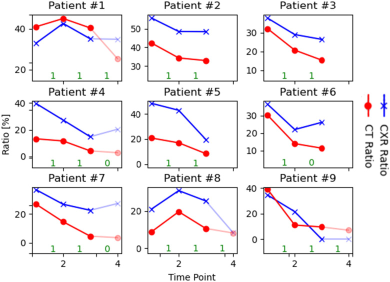
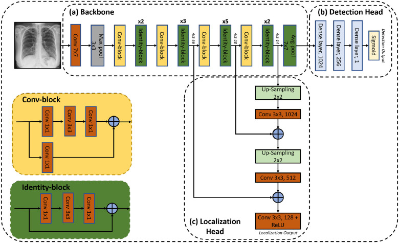
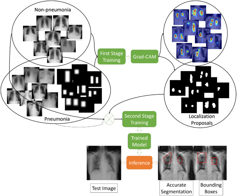
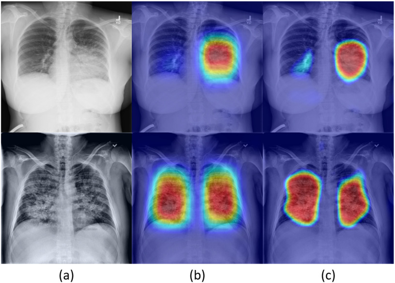
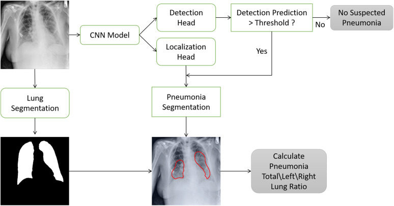
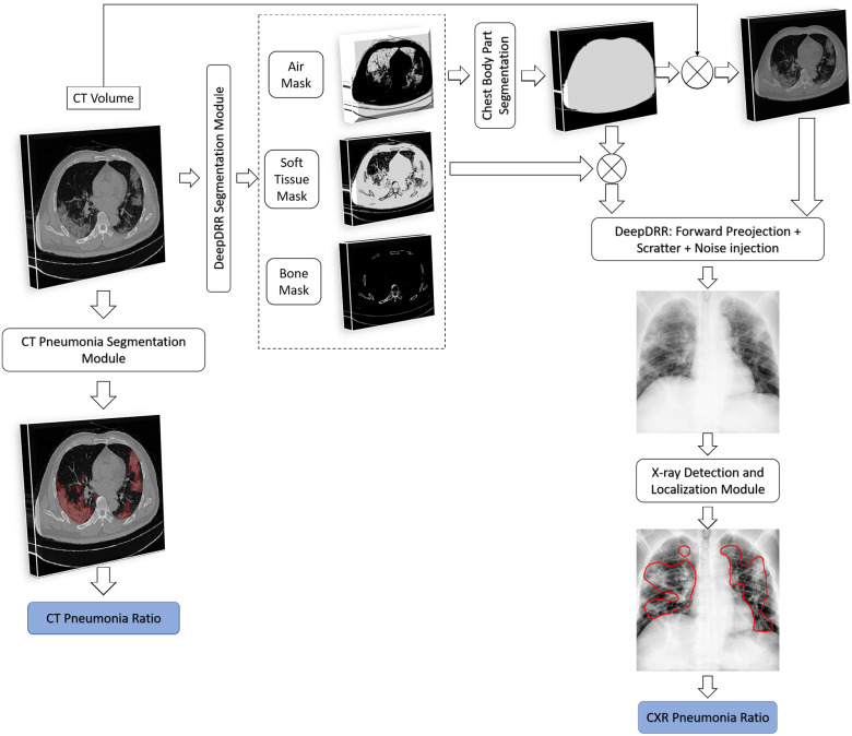
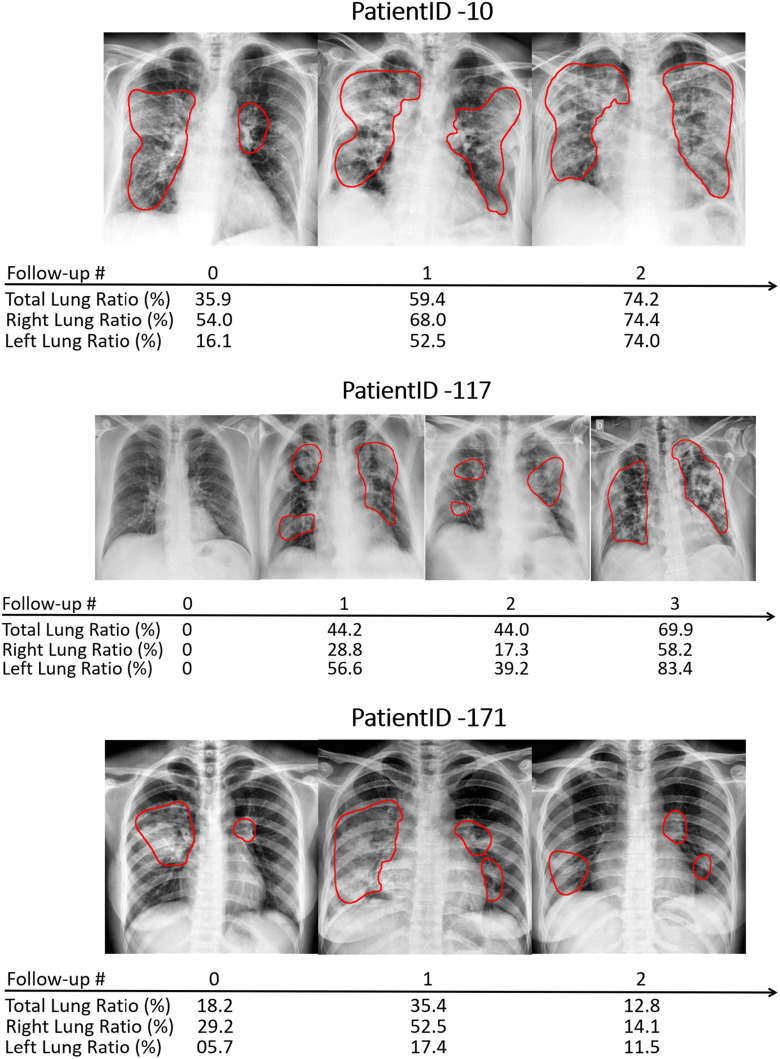
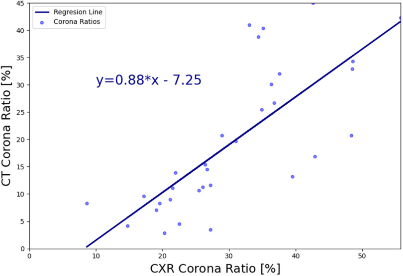
This work estimates the severity of pneumonia in COVID-19 patients and reports the findings of a longitudinal study of disease progression. It presents a deep learning model for simultaneous detection and localization of pneumonia in chest Xray (CXR) images, which is shown to generalize to COVID-19 pneumonia. The localization maps are utilized to calculate a "Pneumonia Ratio" which indicates disease severity. The assessment of disease severity serves to build a temporal disease extent profile for hospitalized patients. To validate the model's applicability to the patient monitoring task, we developed a validation strategy which involves a synthesis of Digital Reconstructed Radiographs (DRRs - synthetic Xray) from serial CT scans; we then compared the disease progression profiles that were generated from the DRRs to those that were generated from CT volumes.

摘要

这项工作评估了新冠肺炎患者肺炎的严重程度,并报告了一项关于疾病进展的纵向研究结果。它提出了一种深度学习模型,用于同时检测和定位胸部X光(CXR)图像中的肺炎,该模型已被证明可推广至新冠肺炎肺炎。利用定位图来计算“肺炎比率”,该比率可表明疾病的严重程度。疾病严重程度的评估有助于为住院患者建立疾病随时间变化的范围概况。为了验证该模型在患者监测任务中的适用性,我们制定了一种验证策略,该策略涉及从系列CT扫描中合成数字重建放射影像(DRRs——合成X光);然后,我们将从DRRs生成的疾病进展概况与从CT容积生成的疾病进展概况进行了比较。

https://cdn.ncbi.nlm.nih.gov/pmc/blobs/0449/8545163/f0abb433adfd/frida13-3069169.jpg
https://cdn.ncbi.nlm.nih.gov/pmc/blobs/0449/8545163/3c8ccf3c7848/frida1-3069169.jpg
https://cdn.ncbi.nlm.nih.gov/pmc/blobs/0449/8545163/f798d8f94a6a/frida2-3069169.jpg
https://cdn.ncbi.nlm.nih.gov/pmc/blobs/0449/8545163/bc153bb40fa7/frida3-3069169.jpg
https://cdn.ncbi.nlm.nih.gov/pmc/blobs/0449/8545163/c91da75aed7e/frida4-3069169.jpg
https://cdn.ncbi.nlm.nih.gov/pmc/blobs/0449/8545163/bbd6f7f92d3c/frida5-3069169.jpg
https://cdn.ncbi.nlm.nih.gov/pmc/blobs/0449/8545163/1e99fcb796a4/frida6-3069169.jpg
https://cdn.ncbi.nlm.nih.gov/pmc/blobs/0449/8545163/e722d95f3e94/frida7-3069169.jpg
https://cdn.ncbi.nlm.nih.gov/pmc/blobs/0449/8545163/e1412d97a5b0/frida8-3069169.jpg
https://cdn.ncbi.nlm.nih.gov/pmc/blobs/0449/8545163/82cd466d241c/frida9-3069169.jpg
https://cdn.ncbi.nlm.nih.gov/pmc/blobs/0449/8545163/9e36a12570d9/frida10-3069169.jpg
https://cdn.ncbi.nlm.nih.gov/pmc/blobs/0449/8545163/3530def3a50c/frida11-3069169.jpg
https://cdn.ncbi.nlm.nih.gov/pmc/blobs/0449/8545163/b95f396c2534/frida12-3069169.jpg
https://cdn.ncbi.nlm.nih.gov/pmc/blobs/0449/8545163/f0abb433adfd/frida13-3069169.jpg

相似文献

1
COVID-19 in CXR: From Detection and Severity Scoring to Patient Disease Monitoring.胸部X线片中的新型冠状病毒肺炎:从检测、严重程度评分到患者疾病监测
IEEE J Biomed Health Inform. 2021 Jun;25(6):1892-1903. doi: 10.1109/JBHI.2021.3069169. Epub 2021 Jun 3.
2
Progression and Resolution of COVID-19 Pneumonia on Chest Radiograph.COVID-19 肺炎在胸部 X 光片上的进展和转归。
J Coll Physicians Surg Pak. 2021 Mar;31(3):258-261. doi: 10.29271/jcpsp.2021.03.258.
3
A rapid screening classifier for diagnosing COVID-19.用于诊断 COVID-19 的快速筛选分类器。
Int J Biol Sci. 2021 Jan 9;17(2):539-548. doi: 10.7150/ijbs.53982. eCollection 2021.
4
COVID-19 outbreak in Italy: experimental chest X-ray scoring system for quantifying and monitoring disease progression.意大利的 COVID-19 疫情:用于定量和监测疾病进展的实验性胸部 X 射线评分系统。
Radiol Med. 2020 May;125(5):509-513. doi: 10.1007/s11547-020-01200-3. Epub 2020 May 1.
5
Implementation of a Deep Learning-Based Computer-Aided Detection System for the Interpretation of Chest Radiographs in Patients Suspected for COVID-19.基于深度学习的计算机辅助检测系统在疑似 COVID-19 患者胸部 X 线片中的应用。
Korean J Radiol. 2020 Oct;21(10):1150-1160. doi: 10.3348/kjr.2020.0536. Epub 2020 Jul 17.
6
COVID-19 severity detection using chest X-ray segmentation and deep learning.使用胸部 X 光分割和深度学习检测 COVID-19 严重程度。
Sci Rep. 2024 Aug 27;14(1):19846. doi: 10.1038/s41598-024-70801-z.
7
Clinical and laboratory data, radiological structured report findings and quantitative evaluation of lung involvement on baseline chest CT in COVID-19 patients to predict prognosis.对 COVID-19 患者基线胸部 CT 上的临床和实验室数据、放射学结构化报告结果以及肺部受累的定量评估,以预测其预后。
Radiol Med. 2021 Jan;126(1):29-39. doi: 10.1007/s11547-020-01293-w. Epub 2020 Oct 12.
8
Radiographic severity index in COVID-19 pneumonia: relationship to age and sex in 783 Italian patients.COVID-19 肺炎的放射学严重程度指数:783 例意大利患者的年龄和性别相关性。
Radiol Med. 2020 May;125(5):461-464. doi: 10.1007/s11547-020-01202-1. Epub 2020 May 1.
9
Chest x-ray severity score in COVID-19 patients on emergency department admission: a two-centre study.COVID-19患者急诊科入院时胸部X线严重程度评分:一项双中心研究。
Eur Radiol Exp. 2020 Dec 15;4(1):68. doi: 10.1186/s41747-020-00195-w.
10
Clinical and radiological findings of adult hospitalized patients with community-acquired pneumonia from SARS-CoV-2 and endemic human coronaviruses.成人社区获得性肺炎住院患者中 SARS-CoV-2 和地方性人冠状病毒的临床和影像学表现。
PLoS One. 2021 Jan 14;16(1):e0245547. doi: 10.1371/journal.pone.0245547. eCollection 2021.

引用本文的文献

1
The Role of AI in Hospitals and Clinics: Transforming Healthcare in the 21st Century.人工智能在医院和诊所中的作用:变革21世纪的医疗保健
Bioengineering (Basel). 2024 Mar 29;11(4):337. doi: 10.3390/bioengineering11040337.
2
Lung pneumonia severity scoring in chest X-ray images using transformers.基于变换模型的胸部 X 射线图像中肺炎严重程度评分
Med Biol Eng Comput. 2024 Aug;62(8):2389-2407. doi: 10.1007/s11517-024-03066-3. Epub 2024 Apr 9.
3
Detection of Severe Lung Infection on Chest Radiographs of COVID-19 Patients: Robustness of AI Models across Multi-Institutional Data.

本文引用的文献

1
Leveraging Data Science to Combat COVID-19: A Comprehensive Review.利用数据科学抗击新冠疫情:全面综述
IEEE Trans Artif Intell. 2020 Sep 2;1(1):85-103. doi: 10.1109/TAI.2020.3020521. eCollection 2020 Aug.
2
Multi-population generalizability of a deep learning-based chest radiograph severity score for COVID-19.基于深度学习的 COVID-19 胸片严重程度评分在多人群中的泛化能力。
Medicine (Baltimore). 2022 Jul 22;101(29):e29587. doi: 10.1097/MD.0000000000029587.
3
COVID-19 detection and disease progression visualization: Deep learning on chest X-rays for classification and coarse localization.
新型冠状病毒肺炎患者胸部X线片上严重肺部感染的检测:人工智能模型在多机构数据中的稳健性
Diagnostics (Basel). 2024 Feb 5;14(3):341. doi: 10.3390/diagnostics14030341.
4
Deep Learning Methods for Interpretation of Pulmonary CT and X-ray Images in Patients with COVID-19-Related Lung Involvement: A Systematic Review.用于解读2019冠状病毒病相关肺部受累患者肺部CT和X线图像的深度学习方法:一项系统综述
J Clin Med. 2023 May 13;12(10):3446. doi: 10.3390/jcm12103446.
5
Prognosis Prediction in COVID-19 Patients through Deep Feature Space Reasoning.通过深度特征空间推理对COVID-19患者进行预后预测
Diagnostics (Basel). 2023 Apr 11;13(8):1387. doi: 10.3390/diagnostics13081387.
6
An XAI approach for COVID-19 detection using transfer learning with X-ray images.一种使用X射线图像的迁移学习进行COVID-19检测的可解释人工智能方法。
Heliyon. 2023 Apr;9(4):e15137. doi: 10.1016/j.heliyon.2023.e15137. Epub 2023 Apr 7.
7
Automated prediction of COVID-19 severity upon admission by chest X-ray images and clinical metadata aiming at accuracy and explainability.基于胸部 X 光图像和临床元数据的 COVID-19 严重程度自动预测,旨在提高准确性和可解释性。
Sci Rep. 2023 Mar 14;13(1):4226. doi: 10.1038/s41598-023-30505-2.
8
Artificial intelligence and discrete-event simulation for capacity management of intensive care units during the Covid-19 pandemic: A case study.人工智能与离散事件模拟在新冠疫情期间重症监护病房容量管理中的应用:一项案例研究
J Bus Res. 2023 May;160:113806. doi: 10.1016/j.jbusres.2023.113806. Epub 2023 Mar 3.
9
In Search of an Efficient and Reliable Deep Learning Model for Identification of COVID-19 Infection from Chest X-ray Images.寻找一种高效可靠的深度学习模型,用于从胸部X光图像中识别新型冠状病毒肺炎感染情况。
Diagnostics (Basel). 2023 Feb 3;13(3):574. doi: 10.3390/diagnostics13030574.
10
Review on the Evaluation and Development of Artificial Intelligence for COVID-19 Containment.关于 COVID-19 防控人工智能评估与发展的综述。
Sensors (Basel). 2023 Jan 3;23(1):527. doi: 10.3390/s23010527.
新冠病毒疾病(COVID-19)检测与疾病进展可视化:基于胸部X光的深度学习用于分类和粗略定位。
Appl Intell (Dordr). 2021;51(2):1010-1021. doi: 10.1007/s10489-020-01867-1. Epub 2020 Sep 12.
4
A critic evaluation of methods for COVID-19 automatic detection from X-ray images.对从X射线图像中自动检测COVID-19的方法的批判性评估。
Inf Fusion. 2021 Dec;76:1-7. doi: 10.1016/j.inffus.2021.04.008. Epub 2021 Apr 30.
5
Automated Assessment and Tracking of COVID-19 Pulmonary Disease Severity on Chest Radiographs using Convolutional Siamese Neural Networks.使用卷积暹罗神经网络对胸部X光片上的COVID-19肺部疾病严重程度进行自动评估和跟踪。
Radiol Artif Intell. 2020 Jul 22;2(4):e200079. doi: 10.1148/ryai.2020200079. eCollection 2020 Jul.
6
COVID-Net: a tailored deep convolutional neural network design for detection of COVID-19 cases from chest X-ray images.COVID-Net:一种针对胸部 X 光图像中 COVID-19 病例检测的定制化深度卷积神经网络设计。
Sci Rep. 2020 Nov 11;10(1):19549. doi: 10.1038/s41598-020-76550-z.
7
Unveiling COVID-19 from CHEST X-Ray with Deep Learning: A Hurdles Race with Small Data.利用深度学习揭示 CHEST X-RAY 中的 COVID-19:小数据的障碍竞赛。
Int J Environ Res Public Health. 2020 Sep 22;17(18):6933. doi: 10.3390/ijerph17186933.
8
Predicting COVID-19 Pneumonia Severity on Chest X-ray With Deep Learning.利用深度学习通过胸部X光预测新冠肺炎肺炎严重程度
Cureus. 2020 Jul 28;12(7):e9448. doi: 10.7759/cureus.9448.
9
Iteratively Pruned Deep Learning Ensembles for COVID-19 Detection in Chest X-rays.用于胸部X光片中COVID-19检测的迭代剪枝深度学习集成模型
IEEE Access. 2020;8:115041-115050. doi: 10.1109/access.2020.3003810. Epub 2020 Jun 19.
10
Covid-19: automatic detection from X-ray images utilizing transfer learning with convolutional neural networks.新冠病毒(Covid-19):利用卷积神经网络的迁移学习从 X 光图像中自动检测。
Phys Eng Sci Med. 2020 Jun;43(2):635-640. doi: 10.1007/s13246-020-00865-4. Epub 2020 Apr 3.