• 文献检索
  • 文档翻译
  • 深度研究
  • 学术资讯
  • Suppr Zotero 插件Zotero 插件
  • 邀请有礼
  • 套餐&价格
  • 历史记录
应用&插件
Suppr Zotero 插件Zotero 插件浏览器插件Mac 客户端Windows 客户端微信小程序
定价
高级版会员购买积分包购买API积分包
服务
文献检索文档翻译深度研究API 文档MCP 服务
关于我们
关于 Suppr公司介绍联系我们用户协议隐私条款
关注我们

Suppr 超能文献

核心技术专利:CN118964589B侵权必究
粤ICP备2023148730 号-1Suppr @ 2026

文献检索

告别复杂PubMed语法,用中文像聊天一样搜索,搜遍4000万医学文献。AI智能推荐,让科研检索更轻松。

立即免费搜索

文件翻译

保留排版,准确专业,支持PDF/Word/PPT等文件格式,支持 12+语言互译。

免费翻译文档

深度研究

AI帮你快速写综述,25分钟生成高质量综述,智能提取关键信息,辅助科研写作。

立即免费体验

使用全卷积网络对增强和非增强计算机断层扫描肝脏图像进行自动轮廓提取

Automated Contouring of Contrast and Noncontrast Computed Tomography Liver Images With Fully Convolutional Networks.

作者信息

Anderson Brian M, Lin Ethan Y, Cardenas Carlos E, Gress Dustin A, Erwin William D, Odisio Bruno C, Koay Eugene J, Brock Kristy K

机构信息

Department of Imaging Physics, The University of Texas MD Anderson Cancer Center, Houston, Texas.

Department of Radiation Physics, The University of Texas MD Anderson Cancer Center, Houston, Texas.

出版信息

Adv Radiat Oncol. 2020 May 16;6(1):100464. doi: 10.1016/j.adro.2020.04.023. eCollection 2021 Jan-Feb.

DOI:10.1016/j.adro.2020.04.023
PMID:33490720
原文链接:https://pmc.ncbi.nlm.nih.gov/articles/PMC7807136/
Abstract

PURPOSE

The deformable nature of the liver can make focal treatment challenging and is not adequately addressed with simple rigid registration techniques. More advanced registration techniques can take deformations into account (eg, biomechanical modeling) but require segmentations of the whole liver for each scan, which is a time-intensive process. We hypothesize that fully convolutional networks can be used to rapidly and accurately autosegment the liver, removing the temporal bottleneck for biomechanical modeling.

METHODS AND MATERIALS

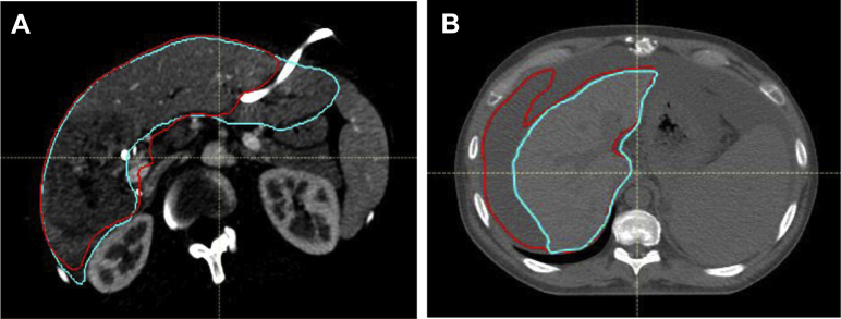
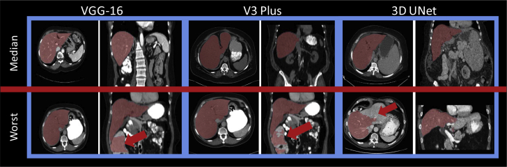
Manual liver segmentations on computed tomography scans from 183 patients treated at our institution and 30 scans from the Medical Image Computing & Computer Assisted Intervention challenges were collected for this study. Three architectures were investigated for rapid automated segmentation of the liver (VGG-16, DeepLabv3 +, and a 3-dimensional UNet). Fifty-six cases were set aside as a final test set for quantitative model evaluation. Accuracy of the autosegmentations was assessed using Dice similarity coefficient and mean surface distance. Qualitative evaluation was also performed by 3 radiation oncologists on 50 independent cases with previously clinically treated liver contours.

RESULTS

The mean (minimum-maximum) mean surface distance for the test groups with the final model, DeepLabv3 +, were as follows: μ: 0.99 mm (0.47-2.2), μ: 1.12 mm (0.41-2.87), and μ: 1.48 mm (0.82-3.96). The qualitative evaluation showed that 30 of 50 autosegmentations (60%) were preferred to manual contours (majority voting) in a blinded comparison, and 48 of 50 autosegmentations (96%) were deemed clinically acceptable by at least 1 reviewing physician.

CONCLUSIONS

The autosegmentations were preferred compared with manually defined contours in the majority of cases. The ability to rapidly segment the liver with high accuracy achieved in this investigation has the potential to enable the efficient integration of biomechanical model-based registration into a clinical workflow.

摘要

目的

肝脏的可变形特性会使局部治疗具有挑战性,而简单的刚性配准技术无法充分解决这一问题。更先进的配准技术可以考虑变形(例如生物力学建模),但每次扫描都需要对整个肝脏进行分割,这是一个耗时的过程。我们假设全卷积网络可用于快速、准确地自动分割肝脏,消除生物力学建模的时间瓶颈。

方法和材料

本研究收集了在我们机构接受治疗的183例患者的计算机断层扫描的手动肝脏分割数据,以及来自医学图像计算与计算机辅助干预挑战的30次扫描数据。研究了三种用于肝脏快速自动分割的架构(VGG-16、DeepLabv3 +和三维U-Net)。56例被留作最终测试集用于定量模型评估。使用骰子相似系数和平均表面距离评估自动分割的准确性。3名放射肿瘤学家还对50例具有先前临床治疗肝脏轮廓的独立病例进行了定性评估。

结果

最终模型DeepLabv3 +的测试组平均(最小-最大)平均表面距离如下:μ:0.99毫米(0.47-2.2),μ:1.12毫米(0.41-2.87),μ:1.48毫米(0.82-3.96)。定性评估显示,在盲法比较中,50例自动分割中有30例(60%)比手动轮廓更受青睐(多数投票),50例自动分割中有48例(96%)被至少1名审阅医生认为在临床上是可接受的。

结论

在大多数情况下,自动分割比手动定义的轮廓更受青睐。本研究中实现的快速、高精度分割肝脏的能力有可能使基于生物力学模型的配准有效地整合到临床工作流程中。

https://cdn.ncbi.nlm.nih.gov/pmc/blobs/cfab/7807136/86da73f6895c/gr3.jpg
https://cdn.ncbi.nlm.nih.gov/pmc/blobs/cfab/7807136/81869cd020b0/gr1.jpg
https://cdn.ncbi.nlm.nih.gov/pmc/blobs/cfab/7807136/8eb413fceb12/gr2.jpg
https://cdn.ncbi.nlm.nih.gov/pmc/blobs/cfab/7807136/86da73f6895c/gr3.jpg
https://cdn.ncbi.nlm.nih.gov/pmc/blobs/cfab/7807136/81869cd020b0/gr1.jpg
https://cdn.ncbi.nlm.nih.gov/pmc/blobs/cfab/7807136/8eb413fceb12/gr2.jpg
https://cdn.ncbi.nlm.nih.gov/pmc/blobs/cfab/7807136/86da73f6895c/gr3.jpg

相似文献

1
Automated Contouring of Contrast and Noncontrast Computed Tomography Liver Images With Fully Convolutional Networks.使用全卷积网络对增强和非增强计算机断层扫描肝脏图像进行自动轮廓提取
Adv Radiat Oncol. 2020 May 16;6(1):100464. doi: 10.1016/j.adro.2020.04.023. eCollection 2021 Jan-Feb.
2
Evaluation of Deformable Image Registration-Based Contour Propagation From Planning CT to Cone-Beam CT.基于可变形图像配准的轮廓从计划CT向锥形束CT传播的评估
Technol Cancer Res Treat. 2017 Dec;16(6):801-810. doi: 10.1177/1533034617697242. Epub 2017 Mar 10.
3
Machine-assisted interpolation algorithm for semi-automated segmentation of highly deformable organs.机器辅助插值算法,用于高度可变形器官的半自动分割。
Med Phys. 2022 Jan;49(1):41-51. doi: 10.1002/mp.15351. Epub 2021 Nov 27.
4
Automatic liver tumor localization using deep learning-based liver boundary motion estimation and biomechanical modeling (DL-Bio).基于深度学习的肝脏边界运动估计和生物力学建模(DL-Bio)的自动肝脏肿瘤定位。
Med Phys. 2021 Dec;48(12):7790-7805. doi: 10.1002/mp.15275. Epub 2021 Nov 19.
5
Evaluating the clinical acceptability of deep learning contours of prostate and organs-at-risk in an automated prostate treatment planning process.评估自动前列腺治疗计划流程中深度学习前列腺和危及器官轮廓的临床可接受性。
Med Phys. 2022 Apr;49(4):2570-2581. doi: 10.1002/mp.15525. Epub 2022 Feb 21.
6
Quantification of liver-Lung shunt fraction on 3D SPECT/CT images for selective internal radiation therapy of liver cancer using CNN-based segmentations and non-rigid registration.基于卷积神经网络(CNN)分割和非刚性配准的三维单光子发射计算机断层扫描/计算机断层扫描(SPECT/CT)图像定量肝脏-肺分流分数在肝癌选择性内放射治疗中的应用
Comput Methods Programs Biomed. 2023 May;233:107453. doi: 10.1016/j.cmpb.2023.107453. Epub 2023 Mar 7.
7
Quantitative evaluation of a cone-beam computed tomography-planning computed tomography deformable image registration method for adaptive radiation therapy.用于自适应放射治疗的锥束计算机断层扫描-计划计算机断层扫描可变形图像配准方法的定量评估
J Appl Clin Med Phys. 2007 Nov 5;8(4):96-113. doi: 10.1120/jacmp.v8i4.2432.
8
Evaluation of Atlas-based auto-segmentation of liver in MR images for Yttrium-90 selective internal radiation therapy.基于图谱的磁共振图像肝脏自动分割在钇-90 选择性内放射治疗中的评价。
J Appl Clin Med Phys. 2023 May;24(5):e13979. doi: 10.1002/acm2.13979. Epub 2023 Apr 17.
9
Comprehensive Evaluation of a Deep Learning Model for Automatic Organs-at-Risk Segmentation on Heterogeneous Computed Tomography Images for Abdominal Radiation Therapy.用于腹部放射治疗的异构计算机断层扫描图像上自动危及器官分割的深度学习模型的综合评估
Int J Radiat Oncol Biol Phys. 2023 Nov 15;117(4):994-1006. doi: 10.1016/j.ijrobp.2023.05.034. Epub 2023 May 26.
10
Fully automatic multi-organ segmentation for head and neck cancer radiotherapy using shape representation model constrained fully convolutional neural networks.使用基于形状表示模型约束的全卷积神经网络进行头颈部癌症放疗的全自动多器官分割。
Med Phys. 2018 Oct;45(10):4558-4567. doi: 10.1002/mp.13147. Epub 2018 Sep 19.

引用本文的文献

1
Evaluation of the liver load index as a predictor of liver exposure in right breast radiotherapy.评估肝脏负荷指数作为右乳放疗中肝脏暴露预测指标的价值。
Rep Pract Oncol Radiother. 2025 Aug 7;30(3):306-315. doi: 10.5603/rpor.105864. eCollection 2025.
2
Minimal Ablative Margin Quantification Using Hepatic Arterial Versus Portal Venous Phase CT for Colorectal Metastases Segmentation: A Dual-center, Retrospective Analysis.使用肝动脉期与门静脉期CT进行结直肠癌转移灶分割的最小消融切缘定量:一项双中心回顾性分析
J Comput Assist Tomogr. 2025 Jul 24. doi: 10.1097/RCT.0000000000001782.
3
Dimensionality Reduction and Nearest Neighbors for Improving Out-of-Distribution Detection in Medical Image Segmentation.

本文引用的文献

1
Fully Convolutional Neural Networks Improve Abdominal Organ Segmentation.全卷积神经网络改进腹部器官分割
Proc SPIE Int Soc Opt Eng. 2018 Mar;10574. doi: 10.1117/12.2293751.
2
An application of cascaded 3D fully convolutional networks for medical image segmentation.级联三维全卷积网络在医学图像分割中的应用。
Comput Med Imaging Graph. 2018 Jun;66:90-99. doi: 10.1016/j.compmedimag.2018.03.001. Epub 2018 Mar 16.
3
Deep learning of the sectional appearances of 3D CT images for anatomical structure segmentation based on an FCN voting method.
用于改善医学图像分割中分布外检测的降维和最近邻算法
J Mach Learn Biomed Imaging. 2024;2(UNSURE2023 Spec Iss):2006-2052. doi: 10.59275/j.melba.2024-g93a. Epub 2024 Oct 23.
4
Dimensionality Reduction for Improving Out-of-Distribution Detection in Medical Image Segmentation.用于改善医学图像分割中分布外检测的降维方法
Uncertain Safe Util Mach Learn Med Imaging (2023). 2023 Oct;14291:147-156. doi: 10.1007/978-3-031-44336-7_15. Epub 2023 Oct 7.
5
Quantitative Ablation Confirmation Methods in Percutaneous Thermal Ablation of Malignant Liver Tumors: Technical Insights, Clinical Evidence, and Future Outlook.恶性肝肿瘤经皮热消融术中的定量消融确认方法:技术见解、临床证据及未来展望
Radiol Imaging Cancer. 2025 May;7(3):e240293. doi: 10.1148/rycan.240293.
6
Fully automated deep learning based auto-contouring of liver segments and spleen on contrast-enhanced CT images.基于深度学习的全自动肝脏分段和脾脏自动勾画在增强 CT 图像上的应用。
Sci Rep. 2024 Feb 26;14(1):4678. doi: 10.1038/s41598-024-53997-y.
7
Fully-automated multi-organ segmentation tool applicable to both non-contrast and post-contrast abdominal CT: deep learning algorithm developed using dual-energy CT images.适用于非对比和对比增强腹部 CT 的全自动多器官分割工具:使用双能 CT 图像开发的深度学习算法。
Sci Rep. 2024 Feb 22;14(1):4378. doi: 10.1038/s41598-024-55137-y.
8
Ablative margin quantification using deformable versus rigid image registration in colorectal liver metastasis thermal ablation: a retrospective single-center study.使用可变形与刚性图像配准技术评估结直肠肝转移热消融术的消融边界:一项回顾性单中心研究。
Eur Radiol. 2024 Sep;34(9):5541-5550. doi: 10.1007/s00330-024-10632-8. Epub 2024 Feb 9.
9
Study Protocol STEREOLAB: Stereotactic Liver Ablation Assisted with Intra-Arterial CT Hepatic Arteriography and Ablation Confirmation Software Assessment.研究方案 STEREOLAB:立体定向肝脏消融术联合动脉内 CT 肝动脉造影和消融确认软件评估。
Cardiovasc Intervent Radiol. 2023 Dec;46(12):1748-1754. doi: 10.1007/s00270-023-03524-9. Epub 2023 Aug 10.
10
ENRICHing medical imaging training sets enables more efficient machine learning.丰富医学影像训练集可实现更高效的机器学习。
J Am Med Inform Assoc. 2023 May 19;30(6):1079-1090. doi: 10.1093/jamia/ocad055.
基于 FCN 投票方法的三维 CT 图像节段外观的深度学习用于解剖结构分割。
Med Phys. 2017 Oct;44(10):5221-5233. doi: 10.1002/mp.12480. Epub 2017 Aug 31.
4
Validation of biomechanical deformable image registration in the abdomen, thorax, and pelvis in a commercial radiotherapy treatment planning system.在商业放射治疗计划系统中验证腹部、胸部和骨盆的生物力学可变形图像配准。
Med Phys. 2017 Jul;44(7):3407-3417. doi: 10.1002/mp.12307. Epub 2017 Jun 1.
5
Automatic abdominal multi-organ segmentation using deep convolutional neural network and time-implicit level sets.基于深度卷积神经网络和时间隐式水平集的自动腹部多器官分割。
Int J Comput Assist Radiol Surg. 2017 Mar;12(3):399-411. doi: 10.1007/s11548-016-1501-5. Epub 2016 Nov 24.
6
Automatic 3D liver segmentation based on deep learning and globally optimized surface evolution.基于深度学习和全局优化表面演化的肝脏自动三维分割
Phys Med Biol. 2016 Dec 21;61(24):8676-8698. doi: 10.1088/1361-6560/61/24/8676. Epub 2016 Nov 23.
7
Evaluation of Six Registration Methods for the Human Abdomen on Clinically Acquired CT.临床获取的CT图像上六种人体腹部配准方法的评估
IEEE Trans Biomed Eng. 2016 Aug;63(8):1563-72. doi: 10.1109/TBME.2016.2574816. Epub 2016 Jun 1.
8
Fully Convolutional Networks for Semantic Segmentation.全卷积网络用于语义分割。
IEEE Trans Pattern Anal Mach Intell. 2017 Apr;39(4):640-651. doi: 10.1109/TPAMI.2016.2572683. Epub 2016 May 24.
9
Transfer learning improves supervised image segmentation across imaging protocols.迁移学习可提高不同成像协议下的监督式图像分割效果。
IEEE Trans Med Imaging. 2015 May;34(5):1018-30. doi: 10.1109/TMI.2014.2366792. Epub 2014 Nov 4.
10
Accuracy of finite element model-based multi-organ deformable image registration.基于有限元模型的多器官可变形图像配准的准确性。
Med Phys. 2005 Jun;32(6):1647-59. doi: 10.1118/1.1915012.