• 文献检索
  • 文档翻译
  • 深度研究
  • 学术资讯
  • Suppr Zotero 插件Zotero 插件
  • 邀请有礼
  • 套餐&价格
  • 历史记录
应用&插件
Suppr Zotero 插件Zotero 插件浏览器插件Mac 客户端Windows 客户端微信小程序
定价
高级版会员购买积分包购买API积分包
服务
文献检索文档翻译深度研究API 文档MCP 服务
关于我们
关于 Suppr公司介绍联系我们用户协议隐私条款
关注我们

Suppr 超能文献

核心技术专利:CN118964589B侵权必究
粤ICP备2023148730 号-1Suppr @ 2026

文献检索

告别复杂PubMed语法,用中文像聊天一样搜索,搜遍4000万医学文献。AI智能推荐,让科研检索更轻松。

立即免费搜索

文件翻译

保留排版,准确专业,支持PDF/Word/PPT等文件格式,支持 12+语言互译。

免费翻译文档

深度研究

AI帮你快速写综述,25分钟生成高质量综述,智能提取关键信息,辅助科研写作。

立即免费体验

分析和可视化挑战结果的方法和开源工具包。

Methods and open-source toolkit for analyzing and visualizing challenge results.

机构信息

Division of Biostatistics, German Cancer Research Center (DKFZ), Im Neuenheimer Feld 581, Heidelberg, 69120, Germany.

Division of Computer Assisted Medical Interventions (CAMI), German Cancer Research Center (DKFZ), Im Neuenheimer Feld 223, 69120, Heidelberg, Germany.

出版信息

Sci Rep. 2021 Jan 27;11(1):2369. doi: 10.1038/s41598-021-82017-6.

DOI:10.1038/s41598-021-82017-6
PMID:33504883
原文链接:https://pmc.ncbi.nlm.nih.gov/articles/PMC7841186/
Abstract

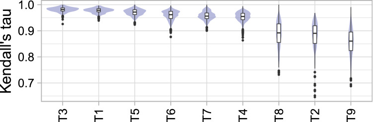
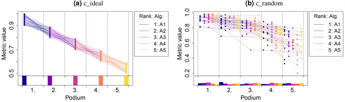
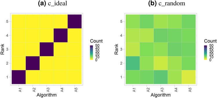
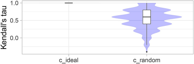
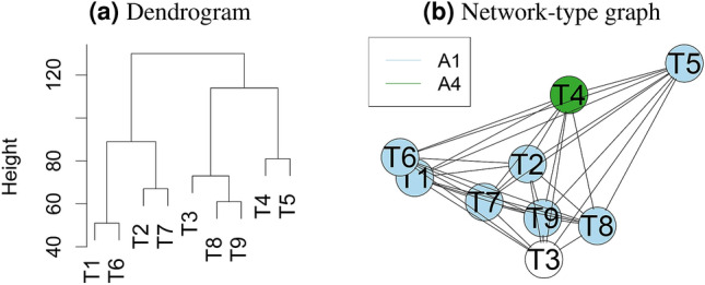
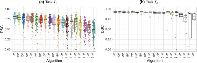
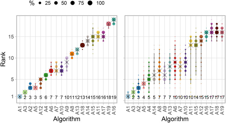
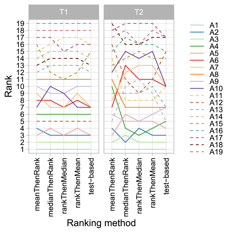
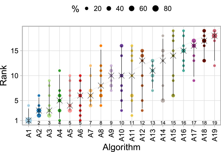
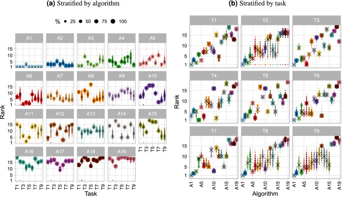
Grand challenges have become the de facto standard for benchmarking image analysis algorithms. While the number of these international competitions is steadily increasing, surprisingly little effort has been invested in ensuring high quality design, execution and reporting for these international competitions. Specifically, results analysis and visualization in the event of uncertainties have been given almost no attention in the literature. Given these shortcomings, the contribution of this paper is two-fold: (1) we present a set of methods to comprehensively analyze and visualize the results of single-task and multi-task challenges and apply them to a number of simulated and real-life challenges to demonstrate their specific strengths and weaknesses; (2) we release the open-source framework challengeR as part of this work to enable fast and wide adoption of the methodology proposed in this paper. Our approach offers an intuitive way to gain important insights into the relative and absolute performance of algorithms, which cannot be revealed by commonly applied visualization techniques. This is demonstrated by the experiments performed in the specific context of biomedical image analysis challenges. Our framework could thus become an important tool for analyzing and visualizing challenge results in the field of biomedical image analysis and beyond.

摘要

重大挑战已成为图像分析算法基准测试的事实上的标准。虽然这些国际竞赛的数量在稳步增加,但令人惊讶的是,几乎没有投入精力来确保这些国际竞赛的高质量设计、执行和报告。具体来说,在存在不确定性的情况下,结果分析和可视化在文献中几乎没有得到关注。鉴于这些缺点,本文的贡献有两个方面:(1)我们提出了一组方法来全面分析和可视化单任务和多任务挑战的结果,并将其应用于许多模拟和实际挑战,以展示它们的具体优势和劣势;(2)我们作为这项工作的一部分发布了开源框架 challengeR,以实现所提出方法的快速广泛采用。我们的方法提供了一种直观的方式,可以深入了解算法的相对和绝对性能,而这是通常应用的可视化技术无法揭示的。这通过在生物医学图像分析挑战的特定上下文中进行的实验得到了证明。因此,我们的框架可以成为分析和可视化生物医学图像分析领域及其他领域挑战结果的重要工具。

https://cdn.ncbi.nlm.nih.gov/pmc/blobs/c97e/7841186/cf796f743180/41598_2021_82017_Fig18_HTML.jpg
https://cdn.ncbi.nlm.nih.gov/pmc/blobs/c97e/7841186/2080c777e02a/41598_2021_82017_Fig1_HTML.jpg
https://cdn.ncbi.nlm.nih.gov/pmc/blobs/c97e/7841186/5c46211ddb23/41598_2021_82017_Fig2_HTML.jpg
https://cdn.ncbi.nlm.nih.gov/pmc/blobs/c97e/7841186/9e823fa5a782/41598_2021_82017_Fig3_HTML.jpg
https://cdn.ncbi.nlm.nih.gov/pmc/blobs/c97e/7841186/d17100b0eb26/41598_2021_82017_Fig4_HTML.jpg
https://cdn.ncbi.nlm.nih.gov/pmc/blobs/c97e/7841186/c2433b93945c/41598_2021_82017_Fig5_HTML.jpg
https://cdn.ncbi.nlm.nih.gov/pmc/blobs/c97e/7841186/a2eed7b6fe60/41598_2021_82017_Fig6_HTML.jpg
https://cdn.ncbi.nlm.nih.gov/pmc/blobs/c97e/7841186/db2057ff2e09/41598_2021_82017_Fig7_HTML.jpg
https://cdn.ncbi.nlm.nih.gov/pmc/blobs/c97e/7841186/1e564d431536/41598_2021_82017_Fig8_HTML.jpg
https://cdn.ncbi.nlm.nih.gov/pmc/blobs/c97e/7841186/3757c44a0730/41598_2021_82017_Fig9_HTML.jpg
https://cdn.ncbi.nlm.nih.gov/pmc/blobs/c97e/7841186/b77861f0a68b/41598_2021_82017_Fig10_HTML.jpg
https://cdn.ncbi.nlm.nih.gov/pmc/blobs/c97e/7841186/feb8cf7b0cc5/41598_2021_82017_Fig11_HTML.jpg
https://cdn.ncbi.nlm.nih.gov/pmc/blobs/c97e/7841186/019f6579aafe/41598_2021_82017_Fig12_HTML.jpg
https://cdn.ncbi.nlm.nih.gov/pmc/blobs/c97e/7841186/13db205d2a87/41598_2021_82017_Fig13_HTML.jpg
https://cdn.ncbi.nlm.nih.gov/pmc/blobs/c97e/7841186/5db0e17bcb7a/41598_2021_82017_Fig14_HTML.jpg
https://cdn.ncbi.nlm.nih.gov/pmc/blobs/c97e/7841186/ccfef3c32f78/41598_2021_82017_Fig15_HTML.jpg
https://cdn.ncbi.nlm.nih.gov/pmc/blobs/c97e/7841186/b86b5e2fc65c/41598_2021_82017_Fig16_HTML.jpg
https://cdn.ncbi.nlm.nih.gov/pmc/blobs/c97e/7841186/7ee4a0f5b82c/41598_2021_82017_Fig17_HTML.jpg
https://cdn.ncbi.nlm.nih.gov/pmc/blobs/c97e/7841186/cf796f743180/41598_2021_82017_Fig18_HTML.jpg

相似文献

1
Methods and open-source toolkit for analyzing and visualizing challenge results.分析和可视化挑战结果的方法和开源工具包。
Sci Rep. 2021 Jan 27;11(1):2369. doi: 10.1038/s41598-021-82017-6.
2
BIAS: Transparent reporting of biomedical image analysis challenges.偏差:生物医学图像分析挑战的透明报告
Med Image Anal. 2020 Dec;66:101796. doi: 10.1016/j.media.2020.101796. Epub 2020 Aug 21.
3
Folic acid supplementation and malaria susceptibility and severity among people taking antifolate antimalarial drugs in endemic areas.在流行地区,服用抗叶酸抗疟药物的人群中,叶酸补充剂与疟疾易感性和严重程度的关系。
Cochrane Database Syst Rev. 2022 Feb 1;2(2022):CD014217. doi: 10.1002/14651858.CD014217.
4
Ten years of image analysis and machine learning competitions in dementia.十年的痴呆症图像分析与机器学习竞赛
Neuroimage. 2022 Jun;253:119083. doi: 10.1016/j.neuroimage.2022.119083. Epub 2022 Mar 9.
5
Common Pitfalls and Recommendations for Grand Challenges in Medical Artificial Intelligence.医学人工智能大挑战中的常见陷阱和建议。
Eur Urol Focus. 2021 Jul;7(4):710-712. doi: 10.1016/j.euf.2021.05.008. Epub 2021 Jun 11.
6
Macromolecular crowding: chemistry and physics meet biology (Ascona, Switzerland, 10-14 June 2012).大分子拥挤现象:化学与物理邂逅生物学(瑞士阿斯科纳,2012年6月10日至14日)
Phys Biol. 2013 Aug;10(4):040301. doi: 10.1088/1478-3975/10/4/040301. Epub 2013 Aug 2.
7
Beyond rankings: Learning (more) from algorithm validation.超越排名:从算法验证中(更多地)学习。
Med Image Anal. 2023 May;86:102765. doi: 10.1016/j.media.2023.102765. Epub 2023 Mar 1.
8
FAST: framework for heterogeneous medical image computing and visualization.FAST:异构医学图像计算与可视化框架
Int J Comput Assist Radiol Surg. 2015 Nov;10(11):1811-22. doi: 10.1007/s11548-015-1158-5. Epub 2015 Feb 17.
9
The future of Cochrane Neonatal.考克兰新生儿协作网的未来。
Early Hum Dev. 2020 Nov;150:105191. doi: 10.1016/j.earlhumdev.2020.105191. Epub 2020 Sep 12.
10
Evaluation of current algorithms for segmentation of scar tissue from late gadolinium enhancement cardiovascular magnetic resonance of the left atrium: an open-access grand challenge.评估当前算法在左心房晚期钆增强心血管磁共振瘢痕组织分割中的应用:公开获取的大挑战。
J Cardiovasc Magn Reson. 2013 Dec 20;15(1):105. doi: 10.1186/1532-429X-15-105.

引用本文的文献

1
DeepISLES: a clinically validated ischemic stroke segmentation model from the ISLES'22 challenge.DeepISLES:一个来自ISLES'22挑战赛的经过临床验证的缺血性中风分割模型。
Nat Commun. 2025 Aug 9;16(1):7357. doi: 10.1038/s41467-025-62373-x.
2
GastroHUN an Endoscopy Dataset of Complete Systematic Screening Protocol for the Stomach.GastroHUN:一个用于胃部的完整系统筛查方案的内镜检查数据集。
Sci Data. 2025 Jan 17;12(1):102. doi: 10.1038/s41597-025-04401-5.
3
MICCAI-CDMRI 2023 QuantConn Challenge Findings on Achieving Robust Quantitative Connectivity through Harmonized Preprocessing of Diffusion MRI.

本文引用的文献

1
Comparative validation of multi-instance instrument segmentation in endoscopy: Results of the ROBUST-MIS 2019 challenge.多实例仪器分割在内窥镜中的比较验证:2019 年 ROBUST-MIS 挑战赛的结果。
Med Image Anal. 2021 May;70:101920. doi: 10.1016/j.media.2020.101920. Epub 2020 Nov 28.
2
nnU-Net: a self-configuring method for deep learning-based biomedical image segmentation.nnU-Net:一种基于深度学习的生物医学图像分割的自配置方法。
Nat Methods. 2021 Feb;18(2):203-211. doi: 10.1038/s41592-020-01008-z. Epub 2020 Dec 7.
3
Light source calibration for multispectral imaging in surgery.
MICCAI-CDMRI 2023定量连接挑战赛:通过扩散磁共振成像的统一预处理实现稳健定量连接的研究结果
ArXiv. 2024 Nov 14:arXiv:2411.09618v1.
4
Multi-Center Fetal Brain Tissue Annotation (FeTA) Challenge 2022 Results.2022年多中心胎儿脑组织标注(FeTA)挑战赛结果
IEEE Trans Med Imaging. 2025 Mar;44(3):1257-1272. doi: 10.1109/TMI.2024.3485554. Epub 2025 Mar 17.
5
WASPSYN: A Challenge for Domain Adaptive Synapse Detection in Microwasp Brain Connectomes.WASPSYN:微蜂大脑连接组中用于域自适应突触检测的挑战。
IEEE Trans Med Imaging. 2024 Nov;43(11):3719-3730. doi: 10.1109/TMI.2024.3400276. Epub 2024 Nov 4.
6
Welcome to the second issue of the (JMI) for the 2024 year!欢迎来到《日本医学影像杂志》(JMI)2024年的第二期!
J Med Imaging (Bellingham). 2024 Mar;11(2):020101. doi: 10.1117/1.JMI.11.2.020101. Epub 2024 Apr 29.
7
The multimodality cell segmentation challenge: toward universal solutions.多模态细胞分割挑战赛:迈向通用解决方案。
Nat Methods. 2024 Jun;21(6):1103-1113. doi: 10.1038/s41592-024-02233-6. Epub 2024 Mar 26.
8
DRAC 2022: A public benchmark for diabetic retinopathy analysis on ultra-wide optical coherence tomography angiography images.DRAC 2022:超广角光学相干断层扫描血管造影图像上糖尿病视网膜病变分析的公开基准。
Patterns (N Y). 2024 Feb 8;5(3):100929. doi: 10.1016/j.patter.2024.100929. eCollection 2024 Mar 8.
9
Metrics reloaded: recommendations for image analysis validation.重新加载指标:图像分析验证的建议。
Nat Methods. 2024 Feb;21(2):195-212. doi: 10.1038/s41592-023-02151-z. Epub 2024 Feb 12.
10
Understanding metric-related pitfalls in image analysis validation.理解图像分析验证中与度量相关的陷阱。
Nat Methods. 2024 Feb;21(2):182-194. doi: 10.1038/s41592-023-02150-0. Epub 2024 Feb 12.
手术中多光谱成像的光源校准。
Int J Comput Assist Radiol Surg. 2020 Jul;15(7):1117-1125. doi: 10.1007/s11548-020-02195-y. Epub 2020 Jun 13.
4
Why rankings of biomedical image analysis competitions should be interpreted with care.为什么要谨慎解读生物医学图像分析竞赛的排名。
Nat Commun. 2018 Dec 6;9(1):5217. doi: 10.1038/s41467-018-07619-7.
5
The challenge of mapping the human connectome based on diffusion tractography.基于弥散张量成像的人类连接组图谱绘制挑战。
Nat Commun. 2017 Nov 7;8(1):1349. doi: 10.1038/s41467-017-01285-x.
6
An objective comparison of cell-tracking algorithms.细胞追踪算法的客观比较。
Nat Methods. 2017 Dec;14(12):1141-1152. doi: 10.1038/nmeth.4473. Epub 2017 Oct 30.
7
Gland segmentation in colon histology images: The glas challenge contest.结肠组织学图像中的腺体分割:glas 挑战赛
Med Image Anal. 2017 Jan;35:489-502. doi: 10.1016/j.media.2016.08.008. Epub 2016 Sep 3.
8
Cloud-Based Evaluation of Anatomical Structure Segmentation and Landmark Detection Algorithms: VISCERAL Anatomy Benchmarks.基于云的解剖结构分割和地标检测算法评估:内脏解剖基准。
IEEE Trans Med Imaging. 2016 Nov;35(11):2459-2475. doi: 10.1109/TMI.2016.2578680. Epub 2016 Jun 9.
9
Quantitative evaluation of software packages for single-molecule localization microscopy.用于单分子定位显微镜的软件包的定量评估。
Nat Methods. 2015 Aug;12(8):717-24. doi: 10.1038/nmeth.3442. Epub 2015 Jun 15.
10
The Multimodal Brain Tumor Image Segmentation Benchmark (BRATS).多模态脑肿瘤图像分割基准(BRATS)。
IEEE Trans Med Imaging. 2015 Oct;34(10):1993-2024. doi: 10.1109/TMI.2014.2377694. Epub 2014 Dec 4.