Li Chang, Jin Rongbing, Liu Kaijun, Li Yang, Zuo Zhiwei, Tong Haipeng, Zhang Jingna, Zhang Junfeng, Guo Yu, Lai Yuqi, Sun Jinju, Wang Jian, Xiong Kunlin, Chen Xiao
Department of Radiology, Daping Hospital, Army Medical University, Chongqing, China.
Department of Radiology, Southwest Hospital, Army Medical University, Chongqing, China.
Front Neurosci. 2021 Jan 18;14:602501. doi: 10.3389/fnins.2020.602501. eCollection 2020.
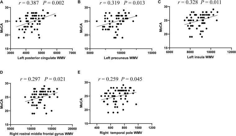
Type 2 diabetes mellitus (T2DM) patients are highly susceptible to developing dementia, especially for those with mild cognitive impairment (MCI), but its underlying cause is still unclear. In this study, we performed a battery of neuropsychological tests and high-resolution sagittal T1-weighted structural imaging to explore how T2DM affects white matter volume (WMV) and cognition in 30 T2DM-MCI patients, 30 T2DM with normal cognition (T2DM-NC) patients, and 30 age-, sex-, and education-matched healthy control (HC) individuals. The WMV of the whole brain was obtained with automated segmentation methods. Correlations between the WMV of each brain region and neuropsychological tests were analyzed in the T2DM patients. The T2DM-NC patients and HC individuals did not reveal any significant differences in WMV. Compared with the T2DM-NC group, the T2DM-MCI group showed statistically significant reduction in the WMV of seven brain regions, mainly located in the frontotemporal lobe and limbic system, five of which significantly correlated with Montreal Cognitive Assessment (MoCA) scores. Subsequently, we evaluated the discriminative ability of these five regions for MCI in T2DM patients. The WMV of four regions, including left posterior cingulate, precuneus, insula, and right rostral middle frontal gyrus had high diagnostic value for MCI detection in T2DM patients (AUC > 0.7). Among these four regions, left precuneus WMV presented the best diagnostic value (AUC: 0.736; sensitivity: 70.00%; specificity: 73.33%; Youden index: 0.4333), but with no significant difference relative to the minimum AUC. In conclusion, T2DM could give rise to the white matter atrophy of several brain regions. Each WMV of left posterior cingulate, precuneus, insula, and right rostral middle frontal gyrus could be an independent imaging biomarker to detect cognitive impairment at the early stage in T2DM patients and play an important role in its pathophysiological mechanism.
2型糖尿病(T2DM)患者极易患痴呆症,尤其是那些患有轻度认知障碍(MCI)的患者,但其潜在病因仍不清楚。在本研究中,我们对30例T2DM-MCI患者、30例认知正常的T2DM(T2DM-NC)患者和30例年龄、性别和教育程度相匹配的健康对照(HC)个体进行了一系列神经心理学测试和高分辨率矢状面T1加权结构成像,以探讨T2DM如何影响白质体积(WMV)和认知。采用自动分割方法获取全脑WMV。分析了T2DM患者各脑区WMV与神经心理学测试之间的相关性。T2DM-NC患者和HC个体在WMV方面未显示出任何显著差异。与T2DM-NC组相比,T2DM-MCI组在七个脑区的WMV出现统计学显著降低,主要位于额颞叶和边缘系统,其中五个脑区与蒙特利尔认知评估(MoCA)评分显著相关。随后,我们评估了这五个区域对T2DM患者MCI的鉴别能力。包括左侧后扣带回、楔前叶、岛叶和右侧额中回喙部在内的四个区域的WMV对T2DM患者MCI的检测具有较高的诊断价值(AUC>0.7)。在这四个区域中,左侧楔前叶WMV的诊断价值最佳(AUC:0.736;敏感性:70.00%;特异性:73.33%;约登指数:0.4333),但与最小AUC相比无显著差异。总之,T2DM可导致多个脑区的白质萎缩。左侧后扣带回、楔前叶、岛叶和右侧额中回喙部的每个WMV都可能是检测T2DM患者早期认知障碍的独立影像学生物标志物,并在其病理生理机制中发挥重要作用。