• 文献检索
  • 文档翻译
  • 深度研究
  • 学术资讯
  • Suppr Zotero 插件Zotero 插件
  • 邀请有礼
  • 套餐&价格
  • 历史记录
应用&插件
Suppr Zotero 插件Zotero 插件浏览器插件Mac 客户端Windows 客户端微信小程序
定价
高级版会员购买积分包购买API积分包
服务
文献检索文档翻译深度研究API 文档MCP 服务
关于我们
关于 Suppr公司介绍联系我们用户协议隐私条款
关注我们

Suppr 超能文献

核心技术专利:CN118964589B侵权必究
粤ICP备2023148730 号-1Suppr @ 2026

文献检索

告别复杂PubMed语法,用中文像聊天一样搜索,搜遍4000万医学文献。AI智能推荐,让科研检索更轻松。

立即免费搜索

文件翻译

保留排版,准确专业,支持PDF/Word/PPT等文件格式,支持 12+语言互译。

免费翻译文档

深度研究

AI帮你快速写综述,25分钟生成高质量综述,智能提取关键信息,辅助科研写作。

立即免费体验

中度和重度 COVID-19 肺炎对短期右心室功能的影响:前瞻性观察性单流行中心分析。

The effect of moderate and severe COVID-19 pneumonia on short-term right ventricular functions: a prospective observational single pandemic center analysis.

机构信息

Department of Cardiology, Umraniye Training and Research Hospital, University of Health Sciences, Elmalıkent, Adem Yavuz Cd., Ümraniye, 34764, Istanbul, Turkey.

Department of Cardiology, Gaziantep Abdülkadir Yüksel State Hospital, Gaziantep, Turkey.

出版信息

Int J Cardiovasc Imaging. 2021 Jun;37(6):1883-1890. doi: 10.1007/s10554-021-02171-w. Epub 2021 Feb 8.

DOI:10.1007/s10554-021-02171-w
PMID:33555535
原文链接:https://pmc.ncbi.nlm.nih.gov/articles/PMC7868518/
Abstract

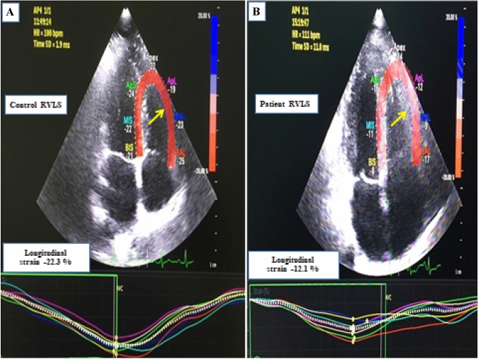
It has been reported that myocardial damage and heart failure are more common in COVID-19 patients with severe symptoms. The aim of our study was to measure the right ventricular functions of COVID-19 patients 30 days after their discharge, and compare them to the right ventricular functions of healthy volunteers. Fifty one patients with COVID-19 and 32 healthy volunteers who underwent echocardiographic examinations were enrolled in our study. 29 patients were treated for severe and 22 patients were treated for moderate COVID-19 pneumonia. The study was conducted prospectively, in a single center, between 15 May 2020 and 15 July 2020. We analyzed the right ventricular functions of the patients using conventional techniques and two-dimensional speckle-tracking. Right ventricular end-diastolic and end-systolic area were statistically higher than control group. The right ventricular fractional area change (RVFAC) was significantly lesser in the patient group compared to the control group. Tricuspid annular plane systolic motion (TAPSE) was within normal limits in both groups, it was lower in the patient group compared to the control group. Pulmonary artery pressure was found to be significantly higher in the patient group. Right ventricular global longitudinal strain (RV-GLS) was lesser than the control group (- 15.7 [(- 12.6)-(- 18.7)] vs. - 18.1 [(- 14.8)-(- 21)]; p 0.011). Right ventricular free wall strain (RV-FWS) was lesser in the patient group compared to the control group (- 16 [(- 12.7)-(- 19)] vs - 21.6 [(- 17)-(- 25.3)]; p < 0.001). We found subclinical right ventricular dysfunction in the echocardiographies of COVID-19 patients although there were no risk factors.

摘要

据报道,重症 COVID-19 患者的心肌损伤和心力衰竭更为常见。本研究旨在测量 COVID-19 患者出院后 30 天的右心室功能,并将其与健康志愿者的右心室功能进行比较。我们纳入了 51 例 COVID-19 患者和 32 例健康志愿者进行超声心动图检查。29 例患者接受重症 COVID-19 肺炎治疗,22 例患者接受中度 COVID-19 肺炎治疗。这项研究是在 2020 年 5 月 15 日至 7 月 15 日期间在一家单中心前瞻性进行的。我们使用常规技术和二维斑点追踪技术分析患者的右心室功能。右心室舒张末期和收缩末期面积明显高于对照组。与对照组相比,患者组的右心室射血分数(RVFAC)明显降低。两组三尖瓣环平面收缩期运动(TAPSE)均在正常范围内,但患者组低于对照组。患者组肺动脉压明显升高。右心室整体纵向应变(RV-GLS)低于对照组(-15.7[-12.6 到-18.7] 比 -18.1[-14.8 到-21];p=0.011)。与对照组相比,患者组右心室游离壁应变(RV-FWS)较低(-16[-12.7 到-19]比 -16[-17 到-25.3];p<0.001)。尽管没有危险因素,但我们在 COVID-19 患者的超声心动图中发现了亚临床右心室功能障碍。

https://cdn.ncbi.nlm.nih.gov/pmc/blobs/a5c0/7868518/edb1cafa4577/10554_2021_2171_Fig1_HTML.jpg

相似文献

1
The effect of moderate and severe COVID-19 pneumonia on short-term right ventricular functions: a prospective observational single pandemic center analysis.中度和重度 COVID-19 肺炎对短期右心室功能的影响:前瞻性观察性单流行中心分析。
Int J Cardiovasc Imaging. 2021 Jun;37(6):1883-1890. doi: 10.1007/s10554-021-02171-w. Epub 2021 Feb 8.
2
Prognostic Value of Right Ventricular Longitudinal Strain in Patients With COVID-19.新冠肺炎患者右心室纵向应变的预后价值。
JACC Cardiovasc Imaging. 2020 Nov;13(11):2287-2299. doi: 10.1016/j.jcmg.2020.04.014. Epub 2020 Apr 28.
3
Long term effects of mild severity COVID‑19 on right ventricular functions.轻度严重 COVID-19 对右心室功能的长期影响。
Int J Cardiovasc Imaging. 2021 Dec;37(12):3451-3457. doi: 10.1007/s10554-021-02340-x. Epub 2021 Jul 12.
4
Impairment of right ventricular longitudinal strain associated with severity of pneumonia in patients recovered from COVID-19.新冠肺炎康复患者右心室纵向应变与肺炎严重程度的关系。
Int J Cardiovasc Imaging. 2021 Aug;37(8):2387-2397. doi: 10.1007/s10554-021-02214-2. Epub 2021 Apr 11.
5
The value of speckle-tracking echocardiography in identifying right heart dysfunction in patients with chronic thromboembolic pulmonary hypertension.斑点追踪超声心动图在识别慢性血栓栓塞性肺动脉高压患者右心功能不全中的价值。
Int J Cardiovasc Imaging. 2018 Dec;34(12):1895-1904. doi: 10.1007/s10554-018-1423-0. Epub 2018 Jul 30.
6
Right ventricular speckle tracking assessment for differentiation of pressure- versus volume-overloaded right ventricle.右心室斑点追踪评估用于鉴别压力负荷过重与容量负荷过重的右心室。
Clin Physiol Funct Imaging. 2018 Sep;38(5):763-771. doi: 10.1111/cpf.12477. Epub 2017 Oct 26.
7
Could strain echocardiography help to assess systolic function in critically ill COVID-19 patients?应变超声心动图能否帮助评估危重症 COVID-19 患者的收缩功能?
J Clin Monit Comput. 2021 Oct;35(5):1229-1234. doi: 10.1007/s10877-021-00677-1. Epub 2021 Feb 27.
8
Obstructive sleep apnoea and right ventricular function: A combined assessment by speckle tracking and three-dimensional echocardiography.阻塞性睡眠呼吸暂停与右心室功能:斑点追踪与三维超声心动图联合评估
Int J Cardiol. 2017 Sep 15;243:544-549. doi: 10.1016/j.ijcard.2017.05.002. Epub 2017 May 4.
9
Echocardiographic predictors of intraoperative right ventricular dysfunction: a 2D and speckle tracking echocardiography study.术中右心室功能障碍的超声心动图预测指标:一项二维和斑点追踪超声心动图研究
Cardiovasc Ultrasound. 2019 Jun 7;17(1):11. doi: 10.1186/s12947-019-0161-3.
10
Three-dimensional echocardiography and 2D-3D speckle-tracking imaging in chronic pulmonary hypertension: diagnostic accuracy in detecting hemodynamic signs of right ventricular (RV) failure.三维超声心动图和二维-三维斑点追踪成像在慢性肺动脉高压中的应用:检测右心室(RV)衰竭血流动力学征象的诊断准确性
J Am Heart Assoc. 2015 Mar 19;4(3):e001584. doi: 10.1161/JAHA.114.001584.

引用本文的文献

1
A deeper look at long-term effects of COVID-19 on myocardial function in survivors with no prior heart diseases: a GRADE approach systematic review and meta-analysis.深入研究新冠病毒病对无既往心脏病幸存者心肌功能的长期影响:采用GRADE方法进行的系统评价和荟萃分析
Front Cardiovasc Med. 2024 Nov 19;11:1458389. doi: 10.3389/fcvm.2024.1458389. eCollection 2024.
2
COVID-19 is associated with cardiac structural and functional remodelling in healthy middle-aged and older individuals.新冠病毒肺炎与健康的中老年人心脏结构和功能重塑有关。
Clin Physiol Funct Imaging. 2025 Jan;45(1):e12909. doi: 10.1111/cpf.12909. Epub 2024 Oct 8.
3

本文引用的文献

1
SARS-CoV-2 Infects Human Pluripotent Stem Cell-Derived Cardiomyocytes, Impairing Electrical and Mechanical Function.SARS-CoV-2 感染人多能干细胞衍生的心肌细胞,损害电和机械功能。
Stem Cell Reports. 2021 Mar 9;16(3):478-492. doi: 10.1016/j.stemcr.2021.02.008. Epub 2021 Feb 13.
2
Patients Recovered From COVID-19 Show Ongoing Subclinical Myocarditis as Revealed by Cardiac Magnetic Resonance Imaging.心脏磁共振成像显示,从新冠病毒肺炎中康复的患者存在持续的亚临床心肌炎。
JACC Cardiovasc Imaging. 2020 Nov;13(11):2476-2478. doi: 10.1016/j.jcmg.2020.08.012. Epub 2020 Aug 28.
3
Evaluation of biventricular function in patients with COVID-19 using speckle tracking echocardiography.
Rhythm Disturbances in Post-Acute COVID-19 Syndrome in Young Men without Pre-Existing Known Cardiovascular Disease-A Case Series.
无已知心血管疾病史的年轻男性急性 COVID-19 综合征后的节律紊乱——病例系列
Biomedicines. 2023 Apr 11;11(4):1146. doi: 10.3390/biomedicines11041146.
4
Cardiac Manifestations of Post-Acute COVID-19 Infection.急性 COVID-19 感染后心脏表现。
Curr Cardiol Rep. 2022 Dec;24(12):1775-1783. doi: 10.1007/s11886-022-01793-3. Epub 2022 Nov 2.
5
ARDS in the time of corona: context and perspective.新冠时期的 ARDS:背景与展望。
Am J Physiol Lung Cell Mol Physiol. 2022 Oct 1;323(4):L431-L437. doi: 10.1152/ajplung.00432.2021. Epub 2022 Aug 23.
6
Cardiac impairments in postacute COVID-19 with sustained symptoms: A review of the literature and proof of concept.急性新冠感染后持续性症状患者的心脏损伤:文献回顾与概念验证。
Physiol Rep. 2022 Aug;10(16):e15430. doi: 10.14814/phy2.15430.
7
Role of Cardiac Imaging Modalities in the Evaluation of COVID-19-Related Cardiomyopathy.心脏成像模态在新型冠状病毒肺炎相关心肌病评估中的作用
Diagnostics (Basel). 2022 Apr 4;12(4):896. doi: 10.3390/diagnostics12040896.
8
Combined Use of Electrocardiography and Ultrasound to Detect Cardiac and Pulmonary Involvement after Recovery from COVID-19 Pneumonia: A Case Series.心电图与超声联合应用检测新冠病毒肺炎康复后心脏和肺部受累情况:病例系列
J Cardiovasc Dev Dis. 2021 Oct 17;8(10):133. doi: 10.3390/jcdd8100133.
9
Echocardiographic assessment of the right ventricle in COVID-19: a systematic review.超声心动图评估 COVID-19 患者右心室:系统评价。
Int J Cardiovasc Imaging. 2021 Dec;37(12):3499-3512. doi: 10.1007/s10554-021-02353-6. Epub 2021 Jul 22.
10
Long term effects of mild severity COVID‑19 on right ventricular functions.轻度严重 COVID-19 对右心室功能的长期影响。
Int J Cardiovasc Imaging. 2021 Dec;37(12):3451-3457. doi: 10.1007/s10554-021-02340-x. Epub 2021 Jul 12.
应用斑点追踪超声心动图评价 COVID-19 患者的双心室功能。
Int J Cardiovasc Imaging. 2021 Jan;37(1):135-144. doi: 10.1007/s10554-020-01968-5. Epub 2020 Aug 15.
4
Biventricular strain by speckle tracking echocardiography in COVID-19: findings and possible prognostic implications.斑点追踪超声心动图对 COVID-19 患者双心室应变的评估:结果与可能的预后意义。
Future Cardiol. 2021 Jul;17(4):663-667. doi: 10.2217/fca-2020-0100. Epub 2020 Aug 4.
5
Outcomes of Cardiovascular Magnetic Resonance Imaging in Patients Recently Recovered From Coronavirus Disease 2019 (COVID-19).COVID-19 患者近期康复后的心血管磁共振成像结果。
JAMA Cardiol. 2020 Nov 1;5(11):1265-1273. doi: 10.1001/jamacardio.2020.3557.
6
COVID-19-associated coagulopathy.新型冠状病毒肺炎相关凝血病
Diagnosis (Berl). 2020 Nov 18;7(4):357-363. doi: 10.1515/dx-2020-0078.
7
In the eye of the storm: the right ventricle in COVID-19.风暴之眼:新型冠状病毒肺炎中的右心室
Pulm Circ. 2020 Jul 2;10(3):2045894020936660. doi: 10.1177/2045894020936660. eCollection 2020 Jul-Sep.
8
Prognostic Value of Right Ventricular Longitudinal Strain in Patients With COVID-19.新冠肺炎患者右心室纵向应变的预后价值。
JACC Cardiovasc Imaging. 2020 Nov;13(11):2287-2299. doi: 10.1016/j.jcmg.2020.04.014. Epub 2020 Apr 28.
9
Global evaluation of echocardiography in patients with COVID-19.全球范围内对 COVID-19 患者的超声心动图评估。
Eur Heart J Cardiovasc Imaging. 2020 Sep 1;21(9):949-958. doi: 10.1093/ehjci/jeaa178.
10
Novel insights on the pulmonary vascular consequences of COVID-19.新型冠状病毒肺炎对肺血管影响的新认识。
Am J Physiol Lung Cell Mol Physiol. 2020 Aug 1;319(2):L277-L288. doi: 10.1152/ajplung.00195.2020. Epub 2020 Jun 17.