• 文献检索
  • 文档翻译
  • 深度研究
  • 学术资讯
  • Suppr Zotero 插件Zotero 插件
  • 邀请有礼
  • 套餐&价格
  • 历史记录
应用&插件
Suppr Zotero 插件Zotero 插件浏览器插件Mac 客户端Windows 客户端微信小程序
定价
高级版会员购买积分包购买API积分包
服务
文献检索文档翻译深度研究API 文档MCP 服务
关于我们
关于 Suppr公司介绍联系我们用户协议隐私条款
关注我们

Suppr 超能文献

核心技术专利:CN118964589B侵权必究
粤ICP备2023148730 号-1Suppr @ 2026

文献检索

告别复杂PubMed语法,用中文像聊天一样搜索,搜遍4000万医学文献。AI智能推荐,让科研检索更轻松。

立即免费搜索

文件翻译

保留排版,准确专业,支持PDF/Word/PPT等文件格式,支持 12+语言互译。

免费翻译文档

深度研究

AI帮你快速写综述,25分钟生成高质量综述,智能提取关键信息,辅助科研写作。

立即免费体验

基于 594 项指标的肺癌 CT 扫描定量成像决策支持(QIDS)工具一致性评估和放射组学分析。

Quantitative imaging decision support (QIDS) tool consistency evaluation and radiomic analysis by means of 594 metrics in lung carcinoma on chest CT scan.

机构信息

Radiology Division, "Istituto Nazionale Tumori IRCCS Fondazione Pascale-IRCCS di Napoli", Naples, Italy.

Department of Radiological Sciences, Diagnostic Imaging Unit, "Azienda Ospedaliera Universitaria Senese," Siena, Italy.

出版信息

Cancer Control. 2021 Jan-Dec;28:1073274820985786. doi: 10.1177/1073274820985786.

DOI:10.1177/1073274820985786
PMID:33567876
原文链接:https://pmc.ncbi.nlm.nih.gov/articles/PMC8482708/
Abstract

OBJECTIVE

To evaluate the consistency of the quantitative imaging decision support (QIDS) tool and radiomic analysis using 594 metrics in lung carcinoma on chest CT scan.

MATERIALS AND METHODS

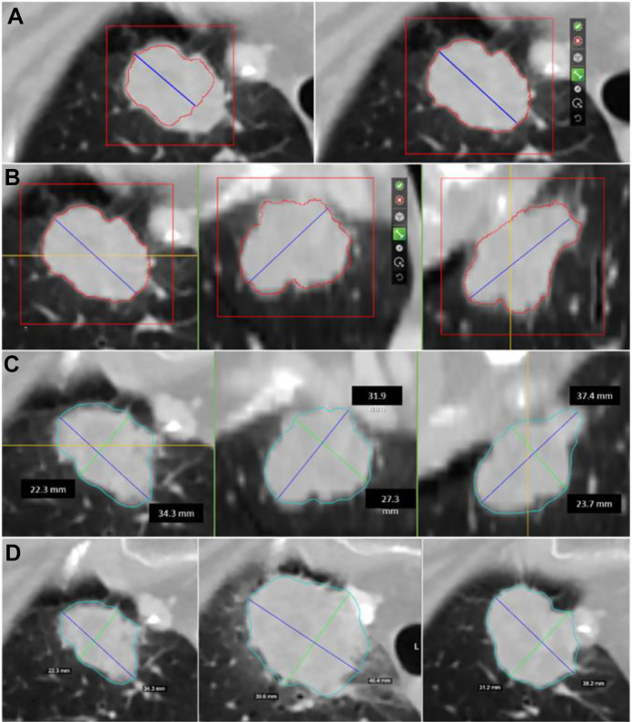
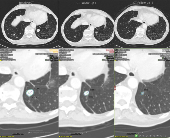
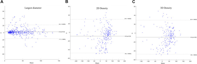
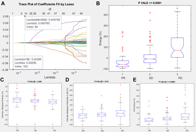
We included, retrospectively, 150 patients with histologically confirmed lung cancer who underwent chemotherapy and baseline and follow-ups CT scans. Using the QIDS platform, 3 radiologists segmented each lesion and automatically collected the longest diameter and the density mean value. Inter-observer variability, Bland Altman analysis and Spearman's correlation coefficient were performed. QIDS tool consistency was assessed in terms of agreement rate in the treatment response classification. Kruskal Wallis test and the least absolute shrinkage and selection operator (LASSO) method with 10-fold cross validation were used to identify radiomic metrics correlated with lesion size change.

RESULTS

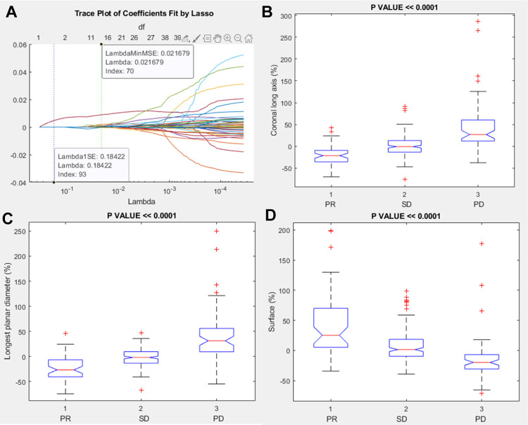
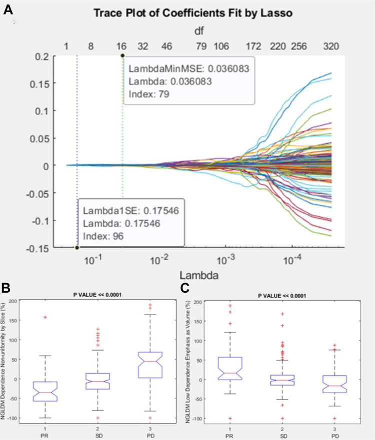
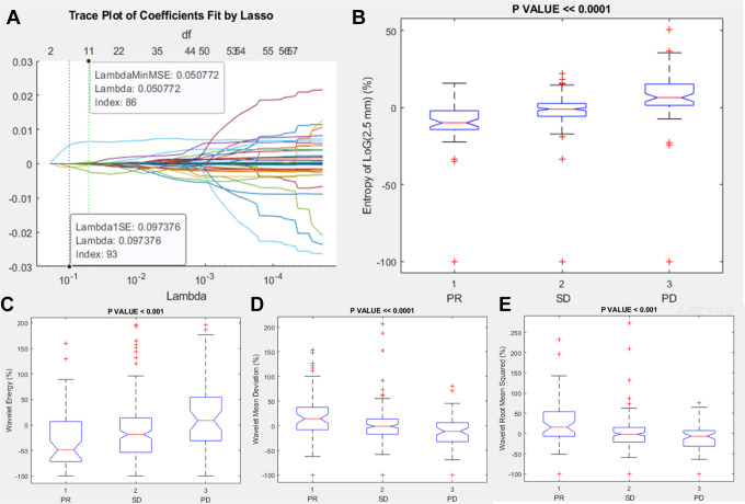
Good and significant correlation was obtained between the measurements of largest diameter and of density among the QIDS tool and the radiologists measurements. Inter-observer variability values were over 0.85. HealthMyne QIDS tool quantitative volumetric delineation was consistent and matched with each radiologist measurement considering the RECIST classification (80-84%) while a lower concordance among QIDS and the radiologists CHOI classification was observed (58-63%). Among 594 extracted metrics, significant and robust predictors of RECIST response were energy, histogram entropy and uniformity, Kurtosis, coronal long axis, longest planar diameter, surface, Neighborhood Grey-Level Different Matrix (NGLDM) dependence nonuniformity and low dependence emphasis as Volume, entropy of Log(2.5 mm), wavelet energy, deviation and root man squared.

CONCLUSION

In conclusion, we demonstrated that HealthMyne quantitative volumetric delineation was consistent and that several radiomic metrics extracted by QIDS were significant and robust predictors of RECIST response.

摘要

目的

评估定量成像决策支持(QIDS)工具与使用 594 项指标对胸部 CT 扫描肺癌的放射组学分析的一致性。

材料与方法

我们回顾性纳入了 150 名经组织学证实患有肺癌且接受化疗以及基线和随访 CT 扫描的患者。使用 QIDS 平台,3 名放射科医生对每个病变进行分割并自动采集最长直径和密度平均值。进行了观察者间变异性、Bland Altman 分析和 Spearman 相关系数分析。QIDS 工具一致性通过治疗反应分类中的一致性率进行评估。Kruskal Wallis 检验和 10 折交叉验证的最小绝对收缩和选择算子(LASSO)方法用于识别与病变大小变化相关的放射组学指标。

结果

QIDS 工具和放射科医生测量的最大直径和密度测量之间获得了良好且显著的相关性。观察者间变异性值超过 0.85。考虑到 RECIST 分类(80-84%),HealthMyne QIDS 工具定量体积描绘是一致的并且与每个放射科医生的测量值相匹配,而 QIDS 和放射科医生 CHOI 分类之间的一致性较低(58-63%)。在提取的 594 项指标中,与 RECIST 反应相关的显著且稳健的预测因子为能量、直方图熵和均匀性、峰度、冠状长轴、最长平面直径、表面、邻域灰度差异矩阵(NGLDM)依赖非均匀性和低依赖强调作为体积、对数(2.5mm)的熵、小波能量、偏差和根均方。

结论

总之,我们证明了 HealthMyne 定量体积描绘是一致的,并且 QIDS 提取的几项放射组学指标是 RECIST 反应的显著且稳健的预测因子。

https://cdn.ncbi.nlm.nih.gov/pmc/blobs/11bc/8482708/cd31554e797f/10.1177_1073274820985786-fig7.jpg
https://cdn.ncbi.nlm.nih.gov/pmc/blobs/11bc/8482708/7e0466ccfe24/10.1177_1073274820985786-fig1.jpg
https://cdn.ncbi.nlm.nih.gov/pmc/blobs/11bc/8482708/df52e4feb646/10.1177_1073274820985786-fig2.jpg
https://cdn.ncbi.nlm.nih.gov/pmc/blobs/11bc/8482708/c9fabf5647ba/10.1177_1073274820985786-fig3.jpg
https://cdn.ncbi.nlm.nih.gov/pmc/blobs/11bc/8482708/24c36f64f2f0/10.1177_1073274820985786-fig4.jpg
https://cdn.ncbi.nlm.nih.gov/pmc/blobs/11bc/8482708/3b69d3f110b0/10.1177_1073274820985786-fig5.jpg
https://cdn.ncbi.nlm.nih.gov/pmc/blobs/11bc/8482708/1076626f170d/10.1177_1073274820985786-fig6.jpg
https://cdn.ncbi.nlm.nih.gov/pmc/blobs/11bc/8482708/cd31554e797f/10.1177_1073274820985786-fig7.jpg

相似文献

1
Quantitative imaging decision support (QIDS) tool consistency evaluation and radiomic analysis by means of 594 metrics in lung carcinoma on chest CT scan.基于 594 项指标的肺癌 CT 扫描定量成像决策支持(QIDS)工具一致性评估和放射组学分析。
Cancer Control. 2021 Jan-Dec;28:1073274820985786. doi: 10.1177/1073274820985786.
2
Inter-Reader Variability of Volumetric Subsolid Pulmonary Nodule Radiomic Features.容积性亚实性肺结节放射组学特征的读者间可变性。
Acad Radiol. 2022 Feb;29 Suppl 2:S98-S107. doi: 10.1016/j.acra.2021.01.026. Epub 2021 Feb 18.
3
A comparative study to evaluate CT-based semantic and radiomic features in preoperative diagnosis of invasive pulmonary adenocarcinomas manifesting as subsolid nodules.一项基于 CT 的语义和放射组学特征在术前诊断表现为亚实性结节的浸润性肺腺癌的对比研究。
Sci Rep. 2021 Jan 18;11(1):66. doi: 10.1038/s41598-020-79690-4.
4
Doubling time calculations for lung cancer by three-dimensional computer-aided volumetry: effects of inter-observer differences and nodule characteristics.通过三维计算机辅助容积测定法计算肺癌倍增时间:观察者间差异和结节特征的影响
J Med Imaging Radiat Oncol. 2014 Feb;58(1):82-8. doi: 10.1111/1754-9485.12128. Epub 2013 Oct 21.
5
Pretherapy 18F-fluorodeoxyglucose positron emission tomography/computed tomography robust radiomic features predict overall survival in non-small cell lung cancer.治疗前 18F-氟代脱氧葡萄糖正电子发射断层扫描/计算机断层扫描的稳健放射组学特征可预测非小细胞肺癌的总生存期。
Nucl Med Commun. 2022 May 1;43(5):540-548. doi: 10.1097/MNM.0000000000001541.
6
Deep Learning-based Image Conversion of CT Reconstruction Kernels Improves Radiomics Reproducibility for Pulmonary Nodules or Masses.基于深度学习的 CT 重建核图像转换可提高肺结节或肿块的放射组学可重复性。
Radiology. 2019 Aug;292(2):365-373. doi: 10.1148/radiol.2019181960. Epub 2019 Jun 18.
7
Comparison of CT volumetric measurement with RECIST response in patients with lung cancer.肺癌患者CT容积测量与RECIST反应的比较。
Eur J Radiol. 2016 Mar;85(3):524-33. doi: 10.1016/j.ejrad.2015.12.019. Epub 2016 Jan 2.
8
Volume-based response evaluation with consensual lesion selection: a pilot study by using cloud solutions and comparison to RECIST 1.1.基于体积的反应评估与共识性病灶选择:一项使用云解决方案并与RECIST 1.1进行比较的试点研究。
Acad Radiol. 2015 Feb;22(2):217-25. doi: 10.1016/j.acra.2014.09.008. Epub 2014 Dec 2.
9
Three-dimensional lung tumor segmentation from x-ray computed tomography using sparse field active models.基于稀疏域主动模型的 X 射线计算机断层扫描三维肺肿瘤分割。
Med Phys. 2012 Feb;39(2):851-65. doi: 10.1118/1.3676687.
10
Solid, part-solid, or non-solid?: classification of pulmonary nodules in low-dose chest computed tomography by a computer-aided diagnosis system.实性、部分实性或非实性?:计算机辅助诊断系统在低剂量胸部 CT 中对肺结节的分类。
Invest Radiol. 2015 Mar;50(3):168-73. doi: 10.1097/RLI.0000000000000121.

引用本文的文献

1
Visual Perception and Pre-Attentive Attributes in Oncological Data Visualisation.肿瘤学数据可视化中的视觉感知与前注意属性
Bioengineering (Basel). 2025 Jul 18;12(7):782. doi: 10.3390/bioengineering12070782.
2
Low expression of SGCA promotes lung squamous cell carcinoma malignant progression.SGCA低表达促进肺鳞状细胞癌的恶性进展。
Sci Rep. 2025 Jul 2;15(1):22578. doi: 10.1038/s41598-025-05312-6.
3
Scientific Status Quo of Small Renal Lesions: Diagnostic Assessment and Radiomics.小肾病变的科学现状:诊断评估与放射组学

本文引用的文献

1
Measuring Efficiency of Semi-automated Brain Tumor Segmentation by Simulating User Interaction.通过模拟用户交互来测量半自动脑肿瘤分割的效率
Front Comput Neurosci. 2020 Apr 16;14:32. doi: 10.3389/fncom.2020.00032. eCollection 2020.
2
Textural radiomic features and time-intensity curve data analysis by dynamic contrast-enhanced MRI for early prediction of breast cancer therapy response: preliminary data.基于动态对比增强 MRI 的纹理放射组学特征和时间-强度曲线数据分析对乳腺癌治疗反应的早期预测:初步数据。
Eur Radiol Exp. 2020 Feb 5;4(1):8. doi: 10.1186/s41747-019-0141-2.
3
A Roadmap for Foundational Research on Artificial Intelligence in Medical Imaging: From the 2018 NIH/RSNA/ACR/The Academy Workshop.
J Clin Med. 2024 Jan 18;13(2):547. doi: 10.3390/jcm13020547.
4
Overview of approaches to estimate real-world disease progression in lung cancer.肺癌真实世界疾病进展评估方法概述。
JNCI Cancer Spectr. 2023 Oct 31;7(6). doi: 10.1093/jncics/pkad074.
5
Radiomics and machine learning analysis by computed tomography and magnetic resonance imaging in colorectal liver metastases prognostic assessment.基于计算机断层扫描和磁共振成像的影像组学与机器学习分析在结直肠癌肝转移预后评估中的应用
Radiol Med. 2023 Nov;128(11):1310-1332. doi: 10.1007/s11547-023-01710-w. Epub 2023 Sep 11.
6
Artificial intelligence and radiation effects on brain tissue in glioblastoma patient: preliminary data using a quantitative tool.人工智能和辐射对胶质母细胞瘤患者脑组织的影响:使用定量工具的初步数据。
Radiol Med. 2023 Jul;128(7):813-827. doi: 10.1007/s11547-023-01655-0. Epub 2023 Jun 8.
7
Qualitative and semi-quantitative ultrasound assessment in delta and Omicron Covid-19 patients: data from high volume reference center.德尔塔和奥密克戎新冠患者的定性和半定量超声评估:来自大容量参考中心的数据。
Infect Agent Cancer. 2023 May 27;18(1):34. doi: 10.1186/s13027-023-00515-w.
8
Colorectal liver metastases patients prognostic assessment: prospects and limits of radiomics and radiogenomics.结直肠癌肝转移患者的预后评估:影像组学和影像基因组学的前景与局限
Infect Agent Cancer. 2023 Mar 16;18(1):18. doi: 10.1186/s13027-023-00495-x.
9
Radiomics in Lung Metastases: A Systematic Review.肺转移瘤的影像组学:一项系统综述。
J Pers Med. 2023 Jan 27;13(2):225. doi: 10.3390/jpm13020225.
10
Gender Medicine in Clinical Radiology Practice.临床放射学实践中的性别医学
J Pers Med. 2023 Jan 27;13(2):223. doi: 10.3390/jpm13020223.
人工智能在医学影像领域基础研究路线图:来自 2018 年 NIH/RSNA/ACR/美国学院联合研讨会
Radiology. 2019 Jun;291(3):781-791. doi: 10.1148/radiol.2019190613. Epub 2019 Apr 16.
4
Looking for Lepidic Component inside Invasive Adenocarcinomas Appearing as CT Solid Solitary Pulmonary Nodules (SPNs): CT Morpho-Densitometric Features and 18-FDG PET Findings.寻找表现为 CT 实性孤立性肺结节(SPN)的侵袭性腺癌内的 Lepidic 成分:CT 形态密度特征和 18-FDG PET 表现。
Biomed Res Int. 2019 Jan 13;2019:7683648. doi: 10.1155/2019/7683648. eCollection 2019.
5
Predicting survival time of lung cancer patients using radiomic analysis.利用影像组学分析预测肺癌患者的生存时间。
Oncotarget. 2017 Nov 1;8(61):104393-104407. doi: 10.18632/oncotarget.22251. eCollection 2017 Nov 28.
6
Delta-radiomics features for the prediction of patient outcomes in non-small cell lung cancer.Delta 放射组学特征预测非小细胞肺癌患者的预后。
Sci Rep. 2017 Apr 3;7(1):588. doi: 10.1038/s41598-017-00665-z.
7
Radiomic-Based Pathological Response Prediction from Primary Tumors and Lymph Nodes in NSCLC.基于影像组学的非小细胞肺癌原发肿瘤和淋巴结病理反应预测
J Thorac Oncol. 2017 Mar;12(3):467-476. doi: 10.1016/j.jtho.2016.11.2226. Epub 2016 Nov 27.
8
Pathologic stratification of operable lung adenocarcinoma using radiomics features extracted from dual energy CT images.利用从双能CT图像中提取的影像组学特征对可手术肺腺癌进行病理分层。
Oncotarget. 2017 Jan 3;8(1):523-535. doi: 10.18632/oncotarget.13476.
9
Radiomics Signature: A Potential Biomarker for the Prediction of Disease-Free Survival in Early-Stage (I or II) Non-Small Cell Lung Cancer.放射组学特征:预测早期(I 期或 II 期)非小细胞肺癌无病生存的潜在生物标志物。
Radiology. 2016 Dec;281(3):947-957. doi: 10.1148/radiol.2016152234. Epub 2016 Jun 27.
10
Radiomic phenotype features predict pathological response in non-small cell lung cancer.影像组学表型特征可预测非小细胞肺癌的病理反应。
Radiother Oncol. 2016 Jun;119(3):480-6. doi: 10.1016/j.radonc.2016.04.004. Epub 2016 Apr 13.