Ayalew Yodit Abebe, Fante Kinde Anlay, Mohammed Mohammed Aliy
Department of Biomedical Engineering, Hawassa Institute of Technology, Hawassa University, Hawassa, Ethiopia.
Faculty of Electrical and Computer Engineering, Jimma Institute of Technology, Jimma University, Jimma, Ethiopia.
BMC Biomed Eng. 2021 Mar 1;3(1):4. doi: 10.1186/s42490-021-00050-y.
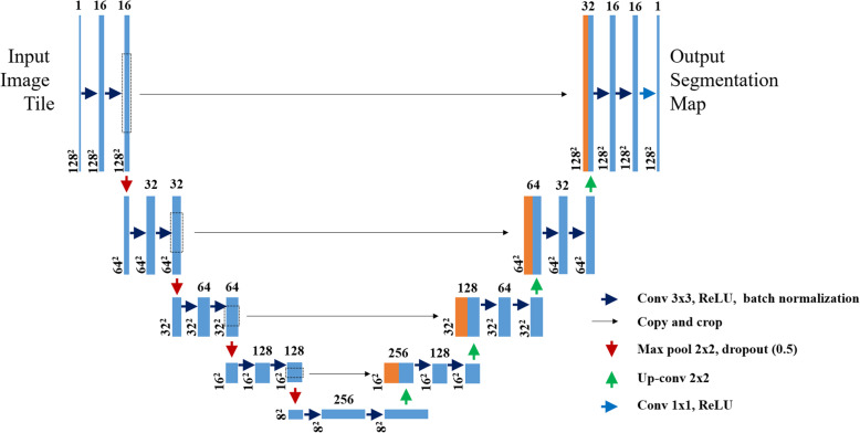
Liver cancer is the sixth most common cancer worldwide. It is mostly diagnosed with a computed tomography scan. Nowadays deep learning methods have been used for the segmentation of the liver and its tumor from the computed tomography (CT) scan images. This research mainly focused on segmenting liver and tumor from the abdominal CT scan images using a deep learning method and minimizing the effort and time used for a liver cancer diagnosis. The algorithm is based on the original UNet architecture. But, here in this paper, the numbers of filters on each convolutional block were reduced and new batch normalization and a dropout layer were added after each convolutional block of the contracting path.
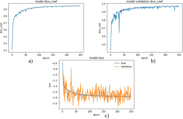
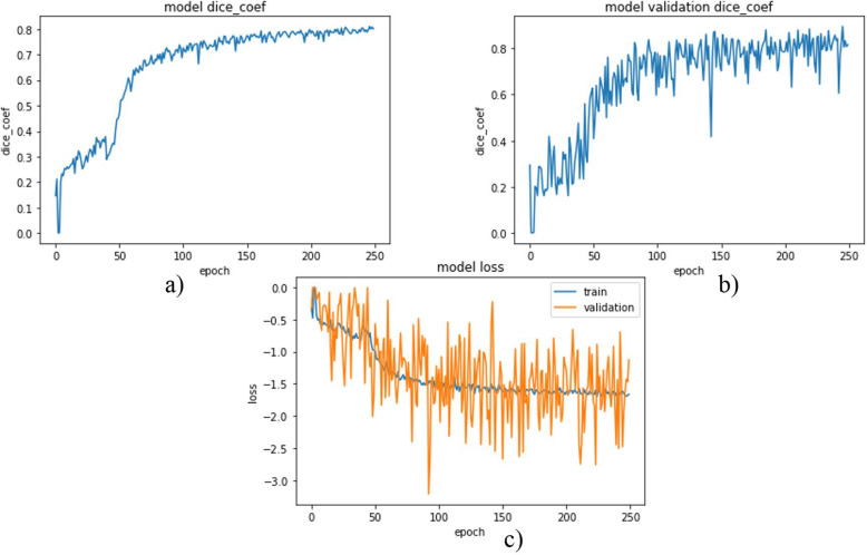
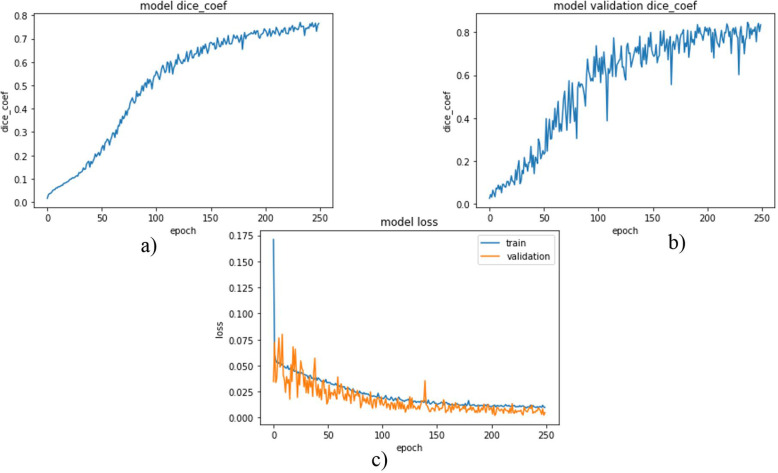
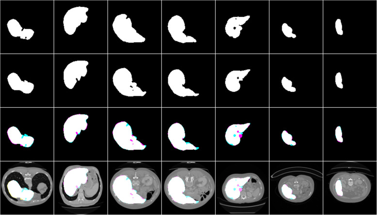
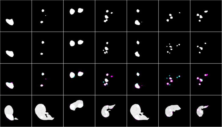
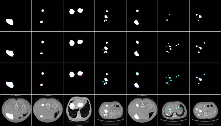
Using this algorithm a dice score of 0.96, 0.74, and 0.63 were obtained for liver segmentation, segmentation of tumors from the liver, and the segmentation of tumor from abdominal CT scan images respectively. The segmentation results of liver and tumor from the liver showed an improvement of 0.01 and 0.11 respectively from other works.
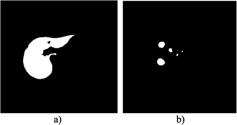
This work proposed a liver and a tumor segmentation method using a UNet architecture as a baseline. Modification regarding the number of filters and network layers were done on the original UNet model to reduce the network complexity and improve segmentation performance. A new class balancing method is also introduced to minimize the class imbalance problem. Through these, the algorithm attained better segmentation results and showed good improvement. However, it faced difficulty in segmenting small and irregular tumors.
肝癌是全球第六大常见癌症。其诊断主要依靠计算机断层扫描。如今,深度学习方法已被用于从计算机断层扫描(CT)图像中分割肝脏及其肿瘤。本研究主要聚焦于使用深度学习方法从腹部CT扫描图像中分割肝脏和肿瘤,并减少肝癌诊断所需的工作量和时间。该算法基于原始的U-Net架构。但是,在本文中,每个卷积块上的滤波器数量减少了,并且在收缩路径的每个卷积块之后添加了新的批量归一化和随机失活层。
使用该算法,肝脏分割、从肝脏中分割肿瘤以及从腹部CT扫描图像中分割肿瘤的骰子系数分别为0.96、0.74和0.63。从肝脏中分割肝脏和肿瘤的结果分别比其他研究提高了0.01和0.11。
本研究提出了一种以U-Net架构为基础的肝脏和肿瘤分割方法。对原始U-Net模型在滤波器数量和网络层方面进行了修改,以降低网络复杂度并提高分割性能。还引入了一种新的类别平衡方法来最小化类别不平衡问题。通过这些措施,该算法获得了更好的分割结果并显示出良好的改进。然而,在分割小的和不规则的肿瘤时面临困难。