Research and Practical Clinical Center for Diagnostics and Telemedicine Technologies of the Moscow Healthcare Department, Moscow, Russia.
Research and Practical Clinical Center for Diagnostics and Telemedicine Technologies of the Moscow Healthcare Department, Moscow, Russia.
Magn Reson Imaging. 2021 Jun;79:13-19. doi: 10.1016/j.mri.2021.03.005. Epub 2021 Mar 13.
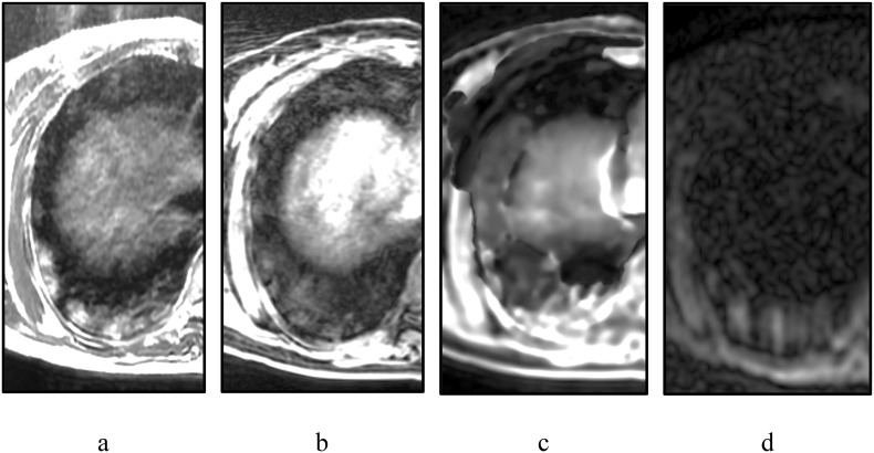
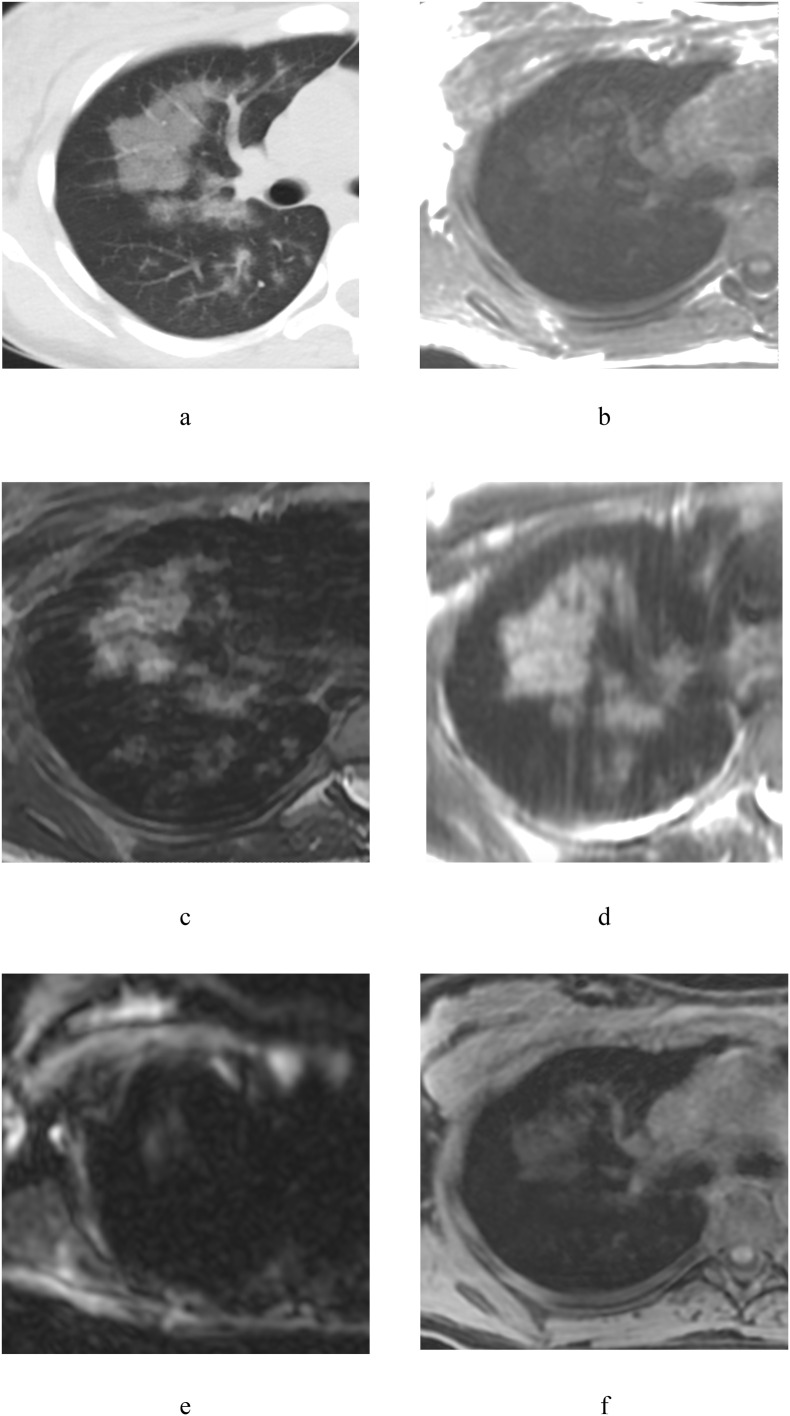
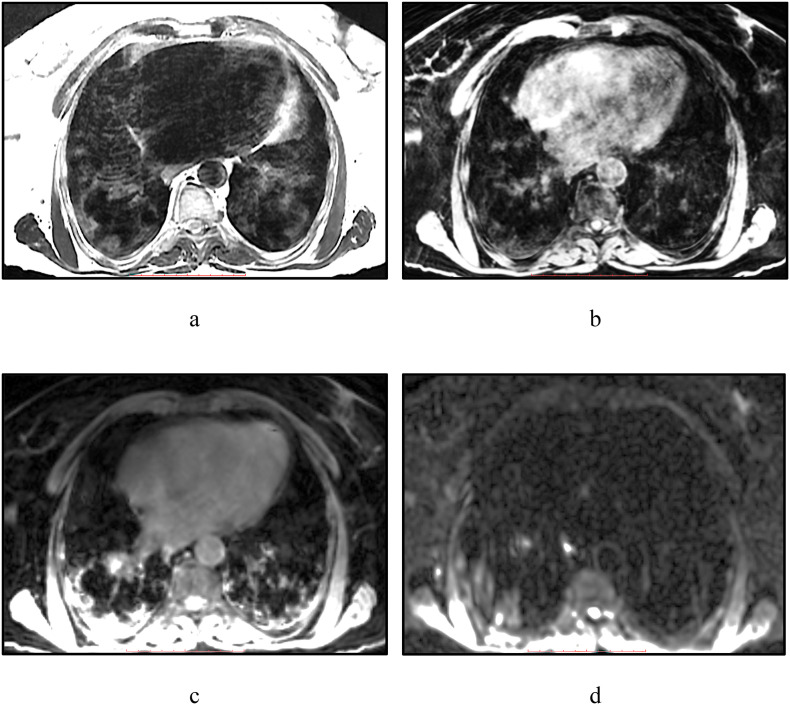
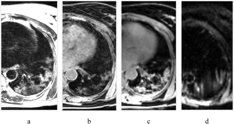
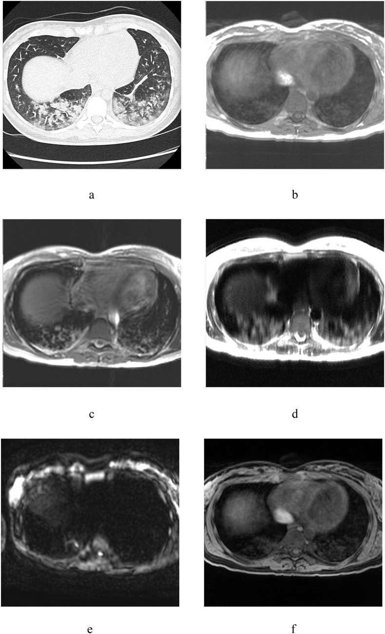
During the pandemic of novel coronavirus infection (COVID-19), computed tomography (CT) showed its effectiveness in diagnosis of coronavirus infection. However, ionizing radiation during CT studies causes concern for patients who require dynamic observation, as well as for examination of children and young people. For this retrospective study, we included 15 suspected for COVID-19 patients who were hospitalized in April 2020, Russia. There were 4 adults with positive polymerase chain reaction (PCR) test for COVID-19. All patients underwent magnetic resonance imaging (MRI) examinations using MR-LUND PROTOCOL: Single-shot Fast Spin Echo (SSFSE), LAVA 3D and IDEAL 3D, Echo-planar imaging (EPI) diffusion-weighted imaging (DWI) and Fast Spin Echo (FSE) T2 weighted imaging (T2WI). On T2WI changes were identified in 9 (60,0%) patients, on DWI - in 5 (33,3%) patients. In 5 (33,3%) patients lesions of the parenchyma were visualized on T2WI and DWI simultaneously. At the same time, 4 (26.7%) patients had changes in lung tissue only on T2WI. (P(McNemar) = 0,125; OR = 0,00 (95%); kappa = 0,500). In those patients who had CT scan, the changes were comparable to MRI. The results showed that in case of CT is not available, it is advisable to conduct a chest MRI for patients with suspected or confirmed COVID-19. Considering that T2WI is a fluid-sensitive sequence, if imaging for the lung infiltration is required, we can recommend the abbreviated MRI protocol consisting of T2 and T1 WI. These data may be applicable for interpreting other studies, such as thoracic spine MRI, detecting signs of viral pneumonia of asymptomatic patients. MRI can detect features of viral pneumonia.
在新型冠状病毒感染(COVID-19)大流行期间,计算机断层扫描(CT)在冠状病毒感染的诊断中显示出其有效性。然而,CT 检查过程中的电离辐射引起了需要动态观察的患者以及儿童和年轻人检查的关注。在这项回顾性研究中,我们纳入了 2020 年 4 月在俄罗斯住院的 15 名疑似 COVID-19 患者。其中 4 名成人的 COVID-19 聚合酶链反应(PCR)检测呈阳性。所有患者均接受了磁共振成像(MRI)检查,使用 MR-LUND PROTOCOL:单次激发快速自旋回波(SSFSE)、LAVA 3D 和 IDEAL 3D、回波平面成像(EPI)扩散加权成像(DWI)和快速自旋回波(FSE)T2 加权成像(T2WI)。在 9 名(60.0%)患者中发现 T2WI 改变,在 5 名(33.3%)患者中发现 DWI 改变。在 5 名(33.3%)患者中,T2WI 和 DWI 同时显示实质病变。同时,4 名(26.7%)患者仅在 T2WI 上观察到肺组织改变。(P(McNemar)= 0.125;OR= 0.00(95%);kappa= 0.500)。在那些进行了 CT 扫描的患者中,改变与 MRI 相似。结果表明,在无法进行 CT 检查的情况下,建议对疑似或确诊 COVID-19 的患者进行胸部 MRI 检查。鉴于 T2WI 是一种液体敏感序列,如果需要对肺部浸润进行成像,我们可以推荐包含 T2 和 T1 WI 的简化 MRI 方案。这些数据可能适用于解释其他研究,例如检测无症状患者的病毒性肺炎的胸椎 MRI。MRI 可以检测病毒性肺炎的特征。