Department of Biological Sciences, Graduate School of Science, The University of Tokyo, Tokyo, Japan.
Graduate School of Life Science, University of Hyogo, Hyogo, Japan.
Elife. 2021 Mar 23;10:e62389. doi: 10.7554/eLife.62389.
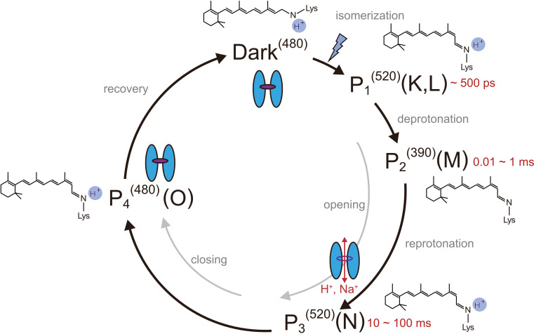
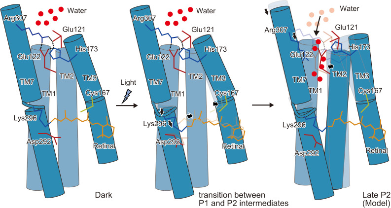
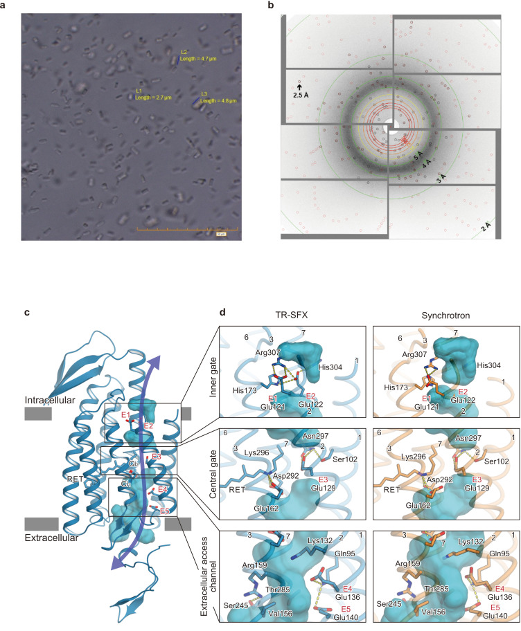
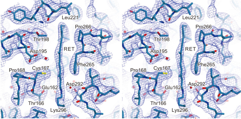
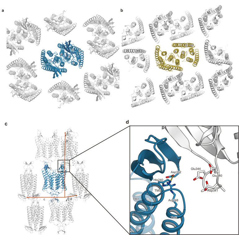
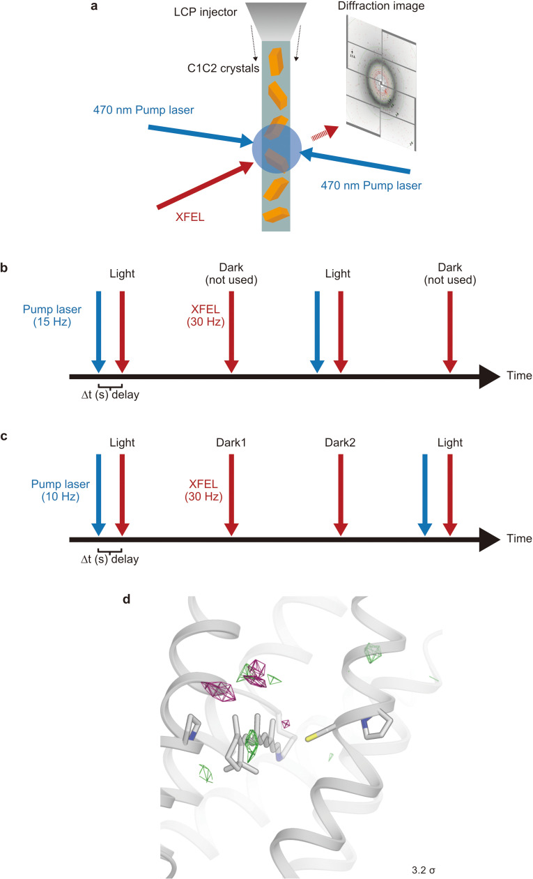
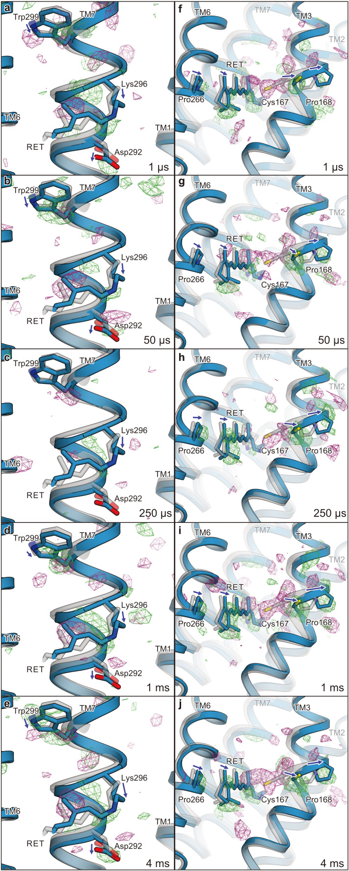
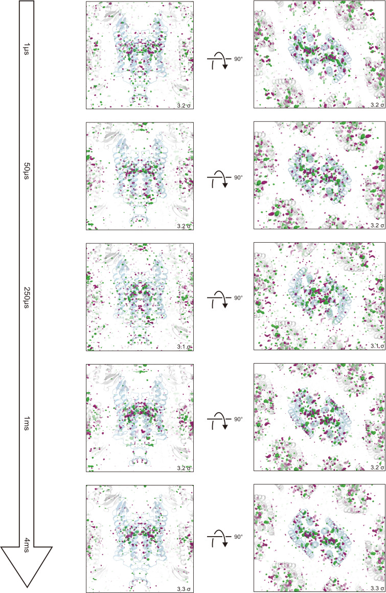
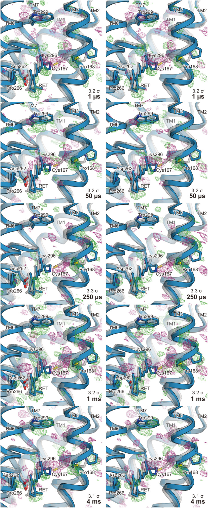
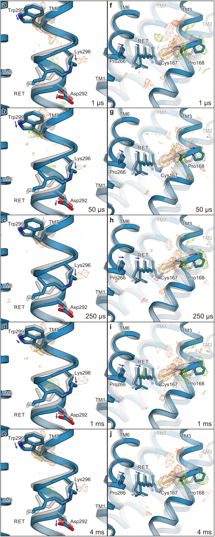
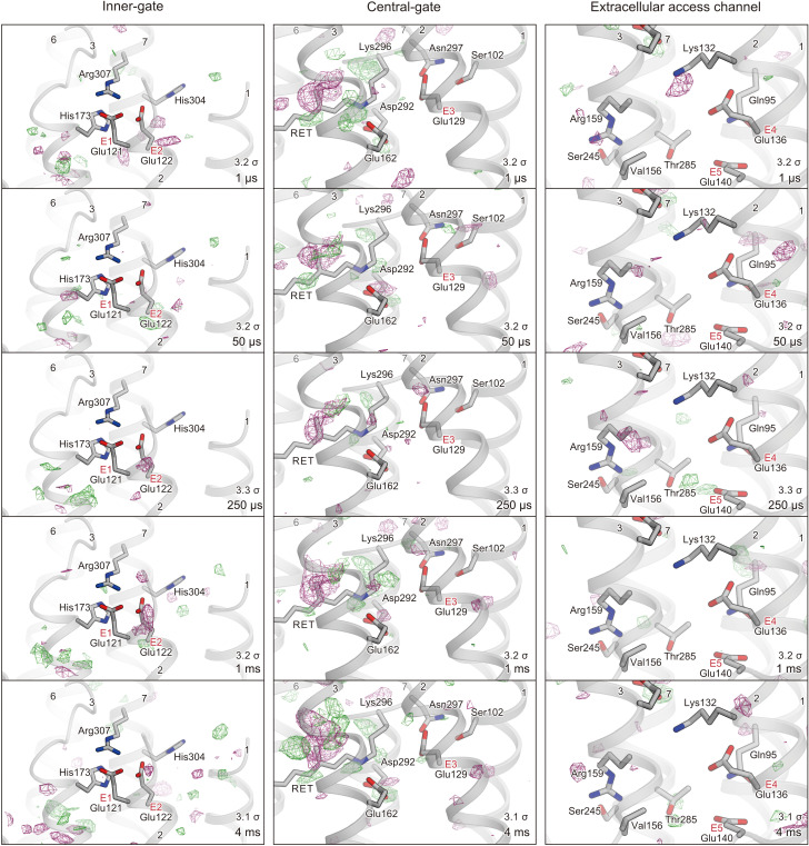
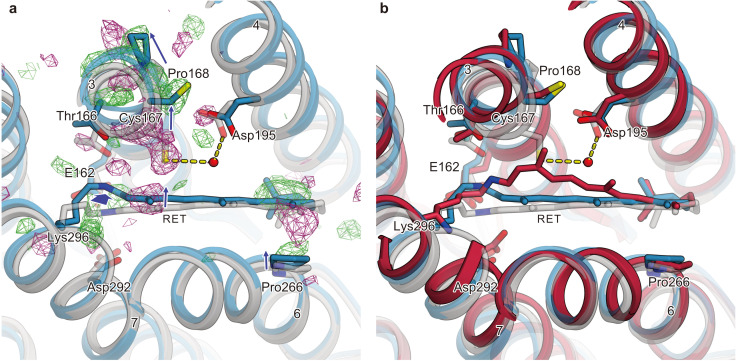
Channelrhodopsins (ChRs) are microbial light-gated ion channels utilized in optogenetics to control neural activity with light . Light absorption causes retinal chromophore isomerization and subsequent protein conformational changes visualized as optically distinguished intermediates, coupled with channel opening and closing. However, the detailed molecular events underlying channel gating remain unknown. We performed time-resolved serial femtosecond crystallographic analyses of ChR by using an X-ray free electron laser, which revealed conformational changes following photoactivation. The isomerized retinal adopts a twisted conformation and shifts toward the putative internal proton donor residues, consequently inducing an outward shift of TM3, as well as a local deformation in TM7. These early conformational changes in the pore-forming helices should be the triggers that lead to opening of the ion conducting pore.
通道视紫红质(ChRs)是微生物光门控离子通道,用于光遗传学以光控制神经活动。光吸收导致视黄醛发色团异构化,随后的蛋白质构象变化可视化为光区分的中间体,与通道的开启和关闭相关联。然而,通道门控的详细分子事件仍不清楚。我们使用自由电子激光进行了通道视紫红质的时间分辨连续飞秒晶体学分析,揭示了光激活后的构象变化。异构化的视黄醛采用扭曲构象,并向假定的内部质子供体残基移动,从而导致 TM3 的向外移动,以及 TM7 中的局部变形。这些孔形成螺旋中的早期构象变化应该是导致离子传导孔打开的触发因素。