Matamala Ella, Castillo Cristian, Vivar Juan P, Rojas Patricio A, Brauchi Sebastian E
Physiology Institute, Universidad Austral de Chile, Valdivia, Chile.
Millennium Nucleus of Ion Channel-Associated Diseases (MiNICAD), Valdivia, Chile.
Commun Biol. 2021 Mar 23;4(1):389. doi: 10.1038/s42003-021-01916-6.
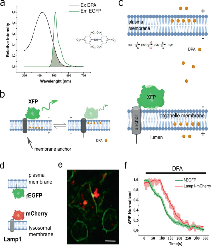
Eukaryotic cells are complex systems compartmentalized in membrane-bound organelles. Visualization of organellar electrical activity in living cells requires both a suitable reporter and non-invasive imaging at high spatiotemporal resolution. Here we present hVoS, an optical method to monitor changes in the membrane potential of subcellular membranes. This method takes advantage of a FRET pair consisting of a membrane-bound voltage-insensitive fluorescent donor and a non-fluorescent voltage-dependent acceptor that rapidly moves across the membrane in response to changes in polarity. Compared to the currently available techniques, hVoS has advantages including simple and precise subcellular targeting, the ability to record from individual organelles, and the potential for optical multiplexing of organellar activity.
真核细胞是在膜结合细胞器中分隔的复杂系统。对活细胞中细胞器电活动的可视化需要合适的报告分子和高时空分辨率的非侵入性成像。在此,我们展示了hVoS,一种监测亚细胞膜电位变化的光学方法。该方法利用了一个荧光共振能量转移(FRET)对,它由一个与膜结合的电压不敏感荧光供体和一个非荧光电压依赖性受体组成,该受体响应极性变化而迅速跨膜移动。与目前可用的技术相比,hVoS具有优势,包括简单而精确的亚细胞靶向、从单个细胞器进行记录的能力以及细胞器活动光学复用的潜力。