• 文献检索
  • 文档翻译
  • 深度研究
  • 学术资讯
  • Suppr Zotero 插件Zotero 插件
  • 邀请有礼
  • 套餐&价格
  • 历史记录
应用&插件
Suppr Zotero 插件Zotero 插件浏览器插件Mac 客户端Windows 客户端微信小程序
定价
高级版会员购买积分包购买API积分包
服务
文献检索文档翻译深度研究API 文档MCP 服务
关于我们
关于 Suppr公司介绍联系我们用户协议隐私条款
关注我们

Suppr 超能文献

核心技术专利:CN118964589B侵权必究
粤ICP备2023148730 号-1Suppr @ 2026

文献检索

告别复杂PubMed语法,用中文像聊天一样搜索,搜遍4000万医学文献。AI智能推荐,让科研检索更轻松。

立即免费搜索

文件翻译

保留排版,准确专业,支持PDF/Word/PPT等文件格式,支持 12+语言互译。

免费翻译文档

深度研究

AI帮你快速写综述,25分钟生成高质量综述,智能提取关键信息,辅助科研写作。

立即免费体验

使用3D活动轮廓和机器学习的自动脾脏损伤检测

Automated Spleen Injury Detection Using 3D Active Contours and Machine Learning.

作者信息

Wang Julie, Wood Alexander, Gao Chao, Najarian Kayvan, Gryak Jonathan

机构信息

Department of Electrical Engineering and Computer Science, University of Michigan, Ann Arbor, MI 48109, USA.

Department of Computational Medicine and Bioinformatics, University of Michigan, Ann Arbor, MI 48109, USA.

出版信息

Entropy (Basel). 2021 Mar 24;23(4):382. doi: 10.3390/e23040382.

DOI:10.3390/e23040382
PMID:33804831
原文链接:https://pmc.ncbi.nlm.nih.gov/articles/PMC8063804/
Abstract

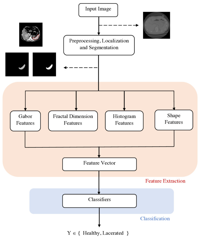
The spleen is one of the most frequently injured organs in blunt abdominal trauma. Computed tomography (CT) is the imaging modality of choice to assess patients with blunt spleen trauma, which may include lacerations, subcapsular or parenchymal hematomas, active hemorrhage, and vascular injuries. While computer-assisted diagnosis systems exist for other conditions assessed using CT scans, the current method to detect spleen injuries involves the manual review of scans by radiologists, which is a time-consuming and repetitive process. In this study, we propose an automated spleen injury detection method using machine learning. CT scans from patients experiencing traumatic injuries were collected from Michigan Medicine and the Crash Injury Research Engineering Network (CIREN) dataset. Ninety-nine scans of healthy and lacerated spleens were split into disjoint training and test sets, with random forest (RF), naive Bayes, SVM, -nearest neighbors (-NN) ensemble, and subspace discriminant ensemble models trained via 5-fold cross validation. Of these models, random forest performed the best, achieving an Area Under the receiver operating characteristic Curve (AUC) of 0.91 and an F1 score of 0.80 on the test set. These results suggest that an automated, quantitative assessment of traumatic spleen injury has the potential to enable faster triage and improve patient outcomes.

摘要

脾脏是钝性腹部创伤中最常受损的器官之一。计算机断层扫描(CT)是评估钝性脾外伤患者的首选成像方式,钝性脾外伤可能包括撕裂伤、包膜下或实质内血肿、活动性出血以及血管损伤。虽然存在用于通过CT扫描评估的其他病症的计算机辅助诊断系统,但目前检测脾损伤的方法是由放射科医生手动查看扫描图像,这是一个耗时且重复的过程。在本研究中,我们提出了一种使用机器学习的自动脾损伤检测方法。从密歇根医学中心和碰撞损伤研究工程网络(CIREN)数据集中收集了遭受创伤性损伤患者的CT扫描图像。将99张健康脾脏和撕裂脾脏的扫描图像分为不相交的训练集和测试集,通过五折交叉验证训练随机森林(RF)、朴素贝叶斯、支持向量机、k近邻(k-NN)集成模型和子空间判别集成模型。在这些模型中,随机森林表现最佳,在测试集上的受试者操作特征曲线下面积(AUC)达到0.91,F1分数达到0.80。这些结果表明,对创伤性脾损伤进行自动、定量评估有可能实现更快的分诊并改善患者预后。

https://cdn.ncbi.nlm.nih.gov/pmc/blobs/6ef1/8063804/d63fb379f1b3/entropy-23-00382-g003.jpg
https://cdn.ncbi.nlm.nih.gov/pmc/blobs/6ef1/8063804/b97bc45a190a/entropy-23-00382-g001.jpg
https://cdn.ncbi.nlm.nih.gov/pmc/blobs/6ef1/8063804/76fcbedc4e81/entropy-23-00382-g002.jpg
https://cdn.ncbi.nlm.nih.gov/pmc/blobs/6ef1/8063804/d63fb379f1b3/entropy-23-00382-g003.jpg
https://cdn.ncbi.nlm.nih.gov/pmc/blobs/6ef1/8063804/b97bc45a190a/entropy-23-00382-g001.jpg
https://cdn.ncbi.nlm.nih.gov/pmc/blobs/6ef1/8063804/76fcbedc4e81/entropy-23-00382-g002.jpg
https://cdn.ncbi.nlm.nih.gov/pmc/blobs/6ef1/8063804/d63fb379f1b3/entropy-23-00382-g003.jpg

相似文献

1
Automated Spleen Injury Detection Using 3D Active Contours and Machine Learning.使用3D活动轮廓和机器学习的自动脾脏损伤检测
Entropy (Basel). 2021 Mar 24;23(4):382. doi: 10.3390/e23040382.
2
Computed tomography of blunt spleen injury: a pictorial review.钝性脾损伤的计算机断层扫描:图文综述
Malays J Med Sci. 2011 Jan;18(1):60-7.
3
Automated segmentation of the injured spleen.损伤脾脏的自动分割
Int J Comput Assist Radiol Surg. 2016 Mar;11(3):351-68. doi: 10.1007/s11548-015-1288-9. Epub 2015 Sep 4.
4
Machine learning-based CT texture analysis to predict HPV status in oropharyngeal squamous cell carcinoma: comparison of 2D and 3D segmentation.基于机器学习的 CT 纹理分析预测口咽鳞状细胞癌 HPV 状态:2D 与 3D 分割的比较。
Eur Radiol. 2020 Dec;30(12):6858-6866. doi: 10.1007/s00330-020-07011-4. Epub 2020 Jun 26.
5
Spleen Segmentation and Assessment in CT Images for Traumatic Abdominal Injuries.CT 图像用于创伤性腹部损伤的脾脏分割和评估。
J Med Syst. 2015 Sep;39(9):87. doi: 10.1007/s10916-015-0271-x. Epub 2015 Jul 25.
6
Comparison and validation of injury risk classifiers for advanced automated crash notification systems.先进自动碰撞通知系统损伤风险分类器的比较与验证
Traffic Inj Prev. 2014;15 Suppl 1:S126-33. doi: 10.1080/15389588.2014.927577.
7
Fully Automated Spleen Localization And Segmentation Using Machine Learning And 3D Active Contours.使用机器学习和3D活动轮廓的全自动脾脏定位与分割
Annu Int Conf IEEE Eng Med Biol Soc. 2018 Jul;2018:53-56. doi: 10.1109/EMBC.2018.8512182.
8
Automated Kidney Segmentation for Traumatic Injured Patients through Ensemble Learning and Active Contour Modeling.通过集成学习和主动轮廓模型对创伤性损伤患者进行自动肾脏分割
Annu Int Conf IEEE Eng Med Biol Soc. 2018 Jul;2018:3418-3421. doi: 10.1109/EMBC.2018.8512967.
9
Outcome prediction of intracranial aneurysm treatment by flow diverters using machine learning.使用机器学习对颅内动脉瘤血管内治疗的结果预测。
Neurosurg Focus. 2018 Nov 1;45(5):E7. doi: 10.3171/2018.8.FOCUS18332.
10
Automatic Valve Plane Localization in Myocardial Perfusion SPECT/CT by Machine Learning: Anatomic and Clinical Validation.基于机器学习的心肌灌注SPECT/CT自动瓣膜平面定位:解剖学与临床验证
J Nucl Med. 2017 Jun;58(6):961-967. doi: 10.2967/jnumed.116.179911. Epub 2016 Nov 3.

引用本文的文献

1
The RSNA Abdominal Traumatic Injury CT (RATIC) Dataset.RSNA腹部创伤性损伤CT(RATIC)数据集。
Radiol Artif Intell. 2024 Nov;6(6):e240101. doi: 10.1148/ryai.240101.
2
Artificial Intelligence-What to Expect From Machine Learning and Deep Learning in Hernia Surgery.人工智能——疝手术中机器学习和深度学习的展望
J Abdom Wall Surg. 2024 Sep 6;3:13059. doi: 10.3389/jaws.2024.13059. eCollection 2024.
3
Deep Learning for Automated Detection and Localization of Traumatic Abdominal Solid Organ Injuries on CT Scans.深度学习在 CT 扫描中自动检测和定位创伤性腹部实体器官损伤中的应用。

本文引用的文献

1
Lung cancer histology classification from CT images based on radiomics and deep learning models.基于放射组学和深度学习模型的 CT 图像肺癌组织学分类。
Med Biol Eng Comput. 2021 Jan;59(1):215-226. doi: 10.1007/s11517-020-02302-w. Epub 2021 Jan 7.
2
Accurate and Efficient Intracranial Hemorrhage Detection and Subtype Classification in 3D CT Scans with Convolutional and Long Short-Term Memory Neural Networks.基于卷积和长短时记忆神经网络的 3D CT 扫描中准确且高效的颅内出血检测和亚型分类。
Sensors (Basel). 2020 Oct 1;20(19):5611. doi: 10.3390/s20195611.
3
CT of blunt splenic injuries: what the trauma team wants to know from the radiologist.
J Imaging Inform Med. 2024 Jun;37(3):1113-1123. doi: 10.1007/s10278-024-01038-5. Epub 2024 Feb 16.
4
Toward automated interpretable AAST grading for blunt splenic injury.实现钝性脾损伤 AAST 分级的自动化可解释性。
Emerg Radiol. 2023 Feb;30(1):41-50. doi: 10.1007/s10140-022-02099-1. Epub 2022 Nov 12.
5
Importance of the neutrophil‑to‑lymphocyte ratio as a prognostic factor in patients with spleen trauma: A single center experience.中性粒细胞与淋巴细胞比值作为脾外伤患者预后因素的重要性:单中心经验
Biomed Rep. 2022 Aug 17;17(4):80. doi: 10.3892/br.2022.1563. eCollection 2022 Oct.
6
Entropy in Image Analysis III.图像分析中的熵III。
Entropy (Basel). 2021 Dec 8;23(12):1648. doi: 10.3390/e23121648.
CT 平扫诊断钝性脾损伤:创伤团队希望放射科医生提供的信息。
Clin Radiol. 2019 Dec;74(12):903-911. doi: 10.1016/j.crad.2019.07.017. Epub 2019 Aug 28.
4
Image-based Classification of Tumor Type and Growth Rate using Machine Learning: a preclinical study.基于图像的肿瘤类型和生长速度的机器学习分类:一项临床前研究。
Sci Rep. 2019 Aug 29;9(1):12529. doi: 10.1038/s41598-019-48738-5.
5
Radiological images and machine learning: Trends, perspectives, and prospects.放射影像学与机器学习:趋势、视角与展望。
Comput Biol Med. 2019 May;108:354-370. doi: 10.1016/j.compbiomed.2019.02.017. Epub 2019 Feb 27.
6
A Novel Method for Classifying Liver and Brain Tumors Using Convolutional Neural Networks, Discrete Wavelet Transform and Long Short-Term Memory Networks.基于卷积神经网络、离散小波变换和长短时记忆网络的肝脏和脑肿瘤分类新方法。
Sensors (Basel). 2019 Apr 28;19(9):1992. doi: 10.3390/s19091992.
7
Fully Automated Spleen Localization And Segmentation Using Machine Learning And 3D Active Contours.使用机器学习和3D活动轮廓的全自动脾脏定位与分割
Annu Int Conf IEEE Eng Med Biol Soc. 2018 Jul;2018:53-56. doi: 10.1109/EMBC.2018.8512182.
8
Ensemble of subspace discriminant classifiers for schistosomal liver fibrosis staging in mice microscopic images.用于小鼠显微图像中血吸虫性肝纤维化分期的子空间判别分类器集成
Health Inf Sci Syst. 2018 Nov 1;6(1):21. doi: 10.1007/s13755-018-0059-8. eCollection 2018 Dec.
9
Role of Big Data and Machine Learning in Diagnostic Decision Support in Radiology.大数据和机器学习在放射诊断决策支持中的作用。
J Am Coll Radiol. 2018 Mar;15(3 Pt B):569-576. doi: 10.1016/j.jacr.2018.01.028.
10
Deep Learning in Medical Imaging: General Overview.医学成像中的深度学习:概述
Korean J Radiol. 2017 Jul-Aug;18(4):570-584. doi: 10.3348/kjr.2017.18.4.570. Epub 2017 May 19.