Department of Geoinformatics, Faculty of Electronics, Telecommunication and Informatics, Gdańsk University of Technology, 80-233 Gdańsk, Poland.
Sensors (Basel). 2021 Mar 12;21(6):2027. doi: 10.3390/s21062027.
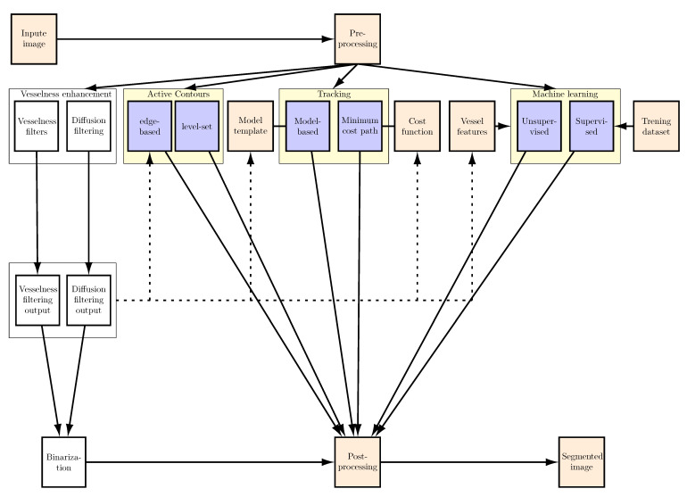
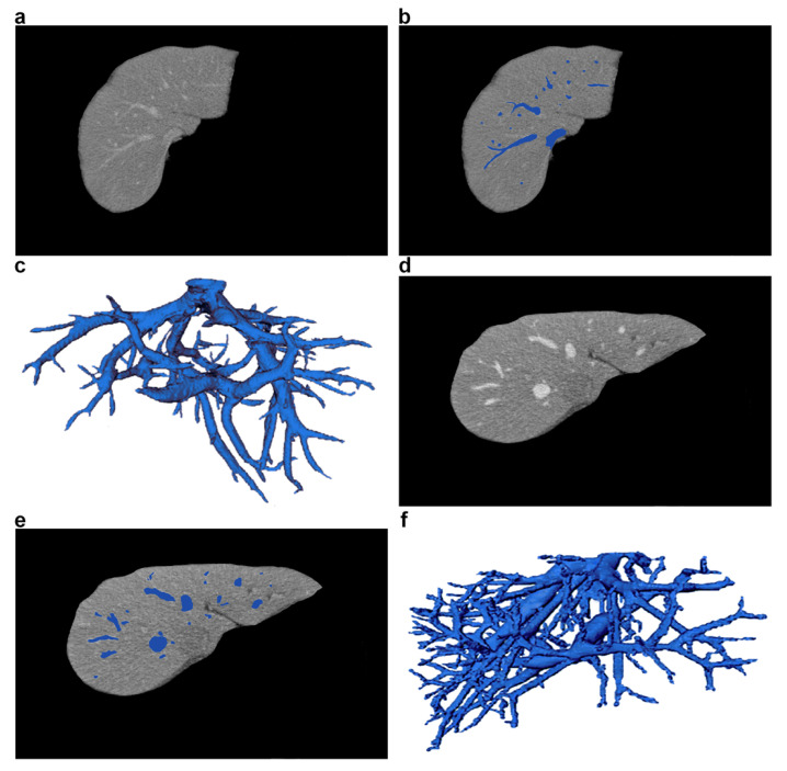
The segmentation of liver blood vessels is of major importance as it is essential for formulating diagnoses, planning and delivering treatments, as well as evaluating the results of clinical procedures. Different imaging techniques are available for application in clinical practice, so the segmentation methods should take into account the characteristics of the imaging technique. Based on the literature, this review paper presents the most advanced and effective methods of liver vessel segmentation, as well as their performance according to the metrics used. This paper includes results available for four imaging methods, namely: computed tomography (CT), computed tomography angiography (CTA), magnetic resonance (MR), and ultrasonography (USG). The publicly available datasets used in research are also presented. This paper may help researchers gain better insight into the available materials and methods, making it easier to develop new, more effective solutions, as well as to improve existing approaches. This article analyzes in detail various segmentation methods, which can be divided into three groups: active contours, tracking-based, and machine learning techniques. For each group of methods, their theoretical and practical characteristics are discussed, and the pros and cons are highlighted. The most advanced and promising approaches are also suggested. However, we conclude that liver vasculature segmentation is still an open problem, because of the various deficiencies and constraints researchers need to address and try to eliminate from the solutions used.
肝血管分割具有重要意义,因为它是制定诊断、规划和提供治疗以及评估临床程序结果的关键。不同的成像技术可用于临床实践,因此分割方法应考虑成像技术的特点。基于文献,本文介绍了最先进、最有效的肝血管分割方法,以及根据使用的指标对其性能的评估。本文涵盖了四种成像方法(即计算机断层扫描 (CT)、计算机断层血管造影 (CTA)、磁共振成像 (MR) 和超声 (USG))的可用结果。还介绍了研究中使用的公共可用数据集。本文可能有助于研究人员更好地了解现有材料和方法,从而更容易开发新的、更有效的解决方案,并改进现有的方法。本文详细分析了各种分割方法,可分为三组:主动轮廓、基于跟踪和机器学习技术。对于每组方法,讨论了它们的理论和实际特点,并突出了优缺点。还提出了最先进和最有前途的方法。但是,我们得出结论,肝血管分割仍然是一个悬而未决的问题,因为研究人员需要解决和尝试从所使用的解决方案中消除的各种缺陷和限制。