• 文献检索
  • 文档翻译
  • 深度研究
  • 学术资讯
  • Suppr Zotero 插件Zotero 插件
  • 邀请有礼
  • 套餐&价格
  • 历史记录
应用&插件
Suppr Zotero 插件Zotero 插件浏览器插件Mac 客户端Windows 客户端微信小程序
定价
高级版会员购买积分包购买API积分包
服务
文献检索文档翻译深度研究API 文档MCP 服务
关于我们
关于 Suppr公司介绍联系我们用户协议隐私条款
关注我们

Suppr 超能文献

核心技术专利:CN118964589B侵权必究
粤ICP备2023148730 号-1Suppr @ 2026

文献检索

告别复杂PubMed语法,用中文像聊天一样搜索,搜遍4000万医学文献。AI智能推荐,让科研检索更轻松。

立即免费搜索

文件翻译

保留排版,准确专业,支持PDF/Word/PPT等文件格式,支持 12+语言互译。

免费翻译文档

深度研究

AI帮你快速写综述,25分钟生成高质量综述,智能提取关键信息,辅助科研写作。

立即免费体验

MAUI(MBI 分析用户界面)——一种用于多重基于质量的成像的图像处理流水线。

MAUI (MBI Analysis User Interface)-An image processing pipeline for Multiplexed Mass Based Imaging.

机构信息

School of Medicine, Department of Pathology, Stanford University, Stanford, California, United States of America.

Department of Molecular Cell Biology, Weizmann Institute of Science, Rehovot, Israel.

出版信息

PLoS Comput Biol. 2021 Apr 19;17(4):e1008887. doi: 10.1371/journal.pcbi.1008887. eCollection 2021 Apr.

DOI:10.1371/journal.pcbi.1008887
PMID:33872301
原文链接:https://pmc.ncbi.nlm.nih.gov/articles/PMC8084329/
Abstract

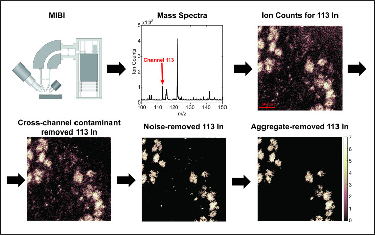
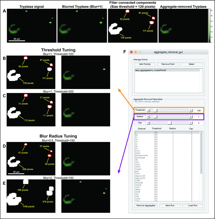
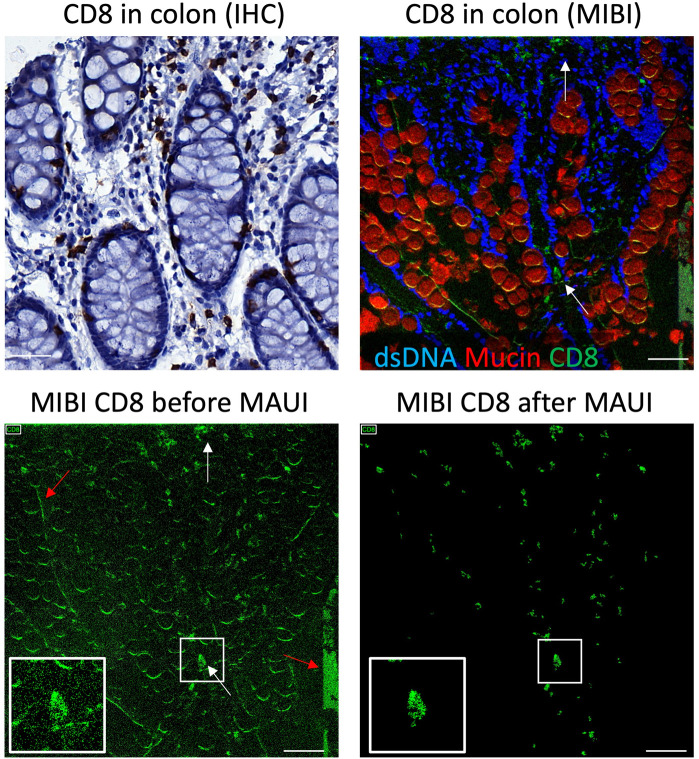
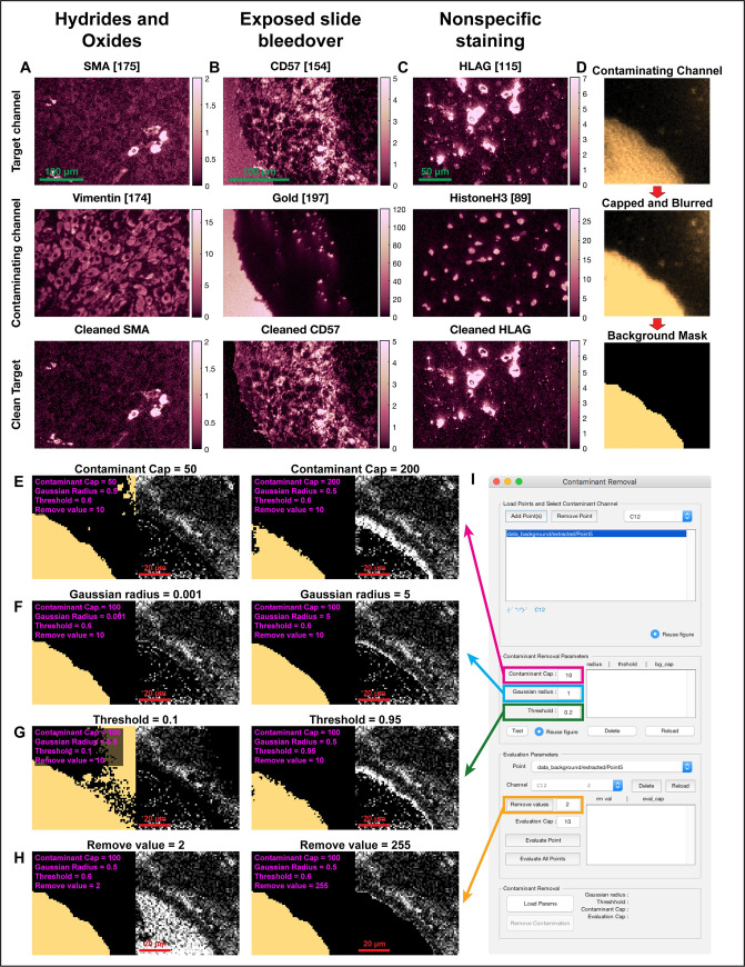
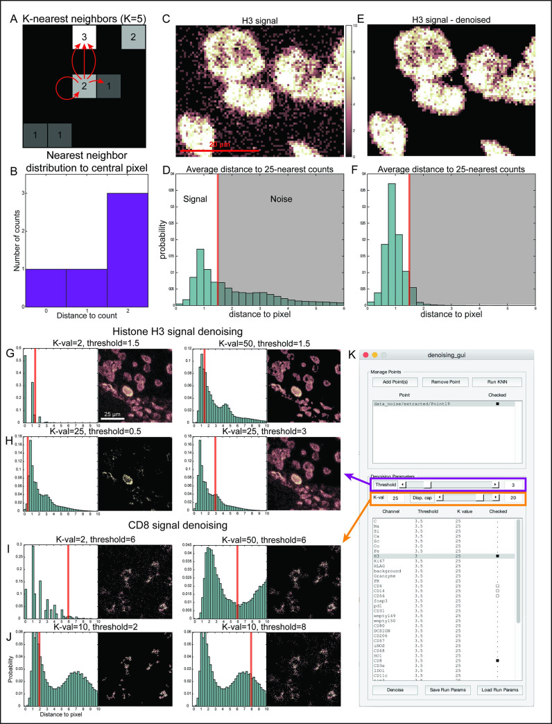
Mass Based Imaging (MBI) technologies such as Multiplexed Ion Beam Imaging by time of flight (MIBI-TOF) and Imaging Mass Cytometry (IMC) allow for the simultaneous measurement of the expression levels of 40 or more proteins in biological tissue, providing insight into cellular phenotypes and organization in situ. Imaging artifacts, resulting from the sample, assay or instrumentation complicate downstream analyses and require correction by domain experts. Here, we present MBI Analysis User Interface (MAUI), a series of graphical user interfaces that facilitate this data pre-processing, including the removal of channel crosstalk, noise and antibody aggregates. Our software streamlines these steps and accelerates processing by enabling real-time and interactive parameter tuning across multiple images.

摘要

基于质量的成像(MBI)技术,如飞行时间的多重离子束成像(MIBI-TOF)和成像质谱细胞术(IMC),允许同时测量生物组织中 40 种或更多蛋白质的表达水平,从而深入了解细胞表型和原位组织。由样本、检测或仪器引起的成像伪影会使下游分析变得复杂,需要领域专家进行校正。在这里,我们提出了 MBI 分析用户界面(MAUI),它是一系列图形用户界面,可促进数据预处理,包括去除通道串扰、噪声和抗体聚集。我们的软件通过在多个图像中实时和交互式地调整参数来简化这些步骤并加速处理。

https://cdn.ncbi.nlm.nih.gov/pmc/blobs/6c70/8084329/85efaf91d72a/pcbi.1008887.g005.jpg
https://cdn.ncbi.nlm.nih.gov/pmc/blobs/6c70/8084329/9391c0653bdb/pcbi.1008887.g001.jpg
https://cdn.ncbi.nlm.nih.gov/pmc/blobs/6c70/8084329/9aec03da92ee/pcbi.1008887.g002.jpg
https://cdn.ncbi.nlm.nih.gov/pmc/blobs/6c70/8084329/8d69414ddc1b/pcbi.1008887.g003.jpg
https://cdn.ncbi.nlm.nih.gov/pmc/blobs/6c70/8084329/900090f43d72/pcbi.1008887.g004.jpg
https://cdn.ncbi.nlm.nih.gov/pmc/blobs/6c70/8084329/85efaf91d72a/pcbi.1008887.g005.jpg

相似文献

1
MAUI (MBI Analysis User Interface)-An image processing pipeline for Multiplexed Mass Based Imaging.MAUI(MBI 分析用户界面)——一种用于多重基于质量的成像的图像处理流水线。
PLoS Comput Biol. 2021 Apr 19;17(4):e1008887. doi: 10.1371/journal.pcbi.1008887. eCollection 2021 Apr.
2
Cytokit: a single-cell analysis toolkit for high dimensional fluorescent microscopy imaging.细胞分析工具包:用于高维荧光显微镜成像的单细胞分析工具包。
BMC Bioinformatics. 2019 Sep 2;20(1):448. doi: 10.1186/s12859-019-3055-3.
3
GRAPE: a graphical pipeline environment for image analysis in adaptive magnetic resonance imaging.GRAPE:用于自适应磁共振成像中图像分析的图形化管道环境。
Int J Comput Assist Radiol Surg. 2017 Mar;12(3):449-457. doi: 10.1007/s11548-016-1495-z. Epub 2016 Oct 28.
4
Rapid Setup of Tissue Microarray and Tiled Area Imaging on the Multiplexed Ion Beam Imaging Microscope using the Tile/SED/Array Interface.利用 Tile/SED/Array 接口在多重离子束成像显微镜上快速构建组织微阵列和平铺区域成像。
J Vis Exp. 2023 Sep 15(199). doi: 10.3791/65615.
5
CyteGuide: Visual Guidance for Hierarchical Single-Cell Analysis.CyteGuide:用于层次化单细胞分析的可视化指导
IEEE Trans Vis Comput Graph. 2018 Jan;24(1):739-748. doi: 10.1109/TVCG.2017.2744318. Epub 2017 Aug 29.
6
Deep cell phenotyping and spatial analysis of multiplexed imaging with TRACERx-PHLEX.利用 TRACERx-PHLEX 进行深度细胞表型分析和多重成像的空间分析。
Nat Commun. 2024 Jun 15;15(1):5135. doi: 10.1038/s41467-024-48870-5.
7
An end-to-end workflow for multiplexed image processing and analysis.用于多重图像处理和分析的端到端工作流程。
Nat Protoc. 2023 Nov;18(11):3565-3613. doi: 10.1038/s41596-023-00881-0. Epub 2023 Oct 10.
8
Carving: scalable interactive segmentation of neural volume electron microscopy images.Carving:神经体积电子显微镜图像的可扩展交互式分割
Med Image Comput Comput Assist Interv. 2011;14(Pt 1):653-60. doi: 10.1007/978-3-642-23623-5_82.
9
Virtual MRI: a PC-based simulation of a clinical MR scanner.虚拟磁共振成像:基于个人计算机的临床磁共振扫描仪模拟。
Acad Radiol. 2005 Jan;12(1):85-96. doi: 10.1016/j.acra.2004.09.011.
10
Medical workstations for applied imaging and graphics research.用于应用成像和图形研究的医学工作站。
Comput Med Imaging Graph. 1994 Nov-Dec;18(6):403-11. doi: 10.1016/0895-6111(94)90077-9.

引用本文的文献

1
From surfing to diving into the tumor microenvironment through multiparametric imaging mass cytometry.从通过多参数成像质谱流式细胞术进行表面探索到深入肿瘤微环境。
Front Immunol. 2025 Apr 11;16:1544844. doi: 10.3389/fimmu.2025.1544844. eCollection 2025.
2
High-dimensional imaging using combinatorial channel multiplexing and deep learning.使用组合通道复用和深度学习的高维成像。
Nat Biotechnol. 2025 Mar 25. doi: 10.1038/s41587-025-02585-0.
3
Integrated workflow for analysis of immune enriched spatial proteomic data with IMmuneCite.

本文引用的文献

1
The Society for Immunotherapy of Cancer statement on best practices for multiplex immunohistochemistry (IHC) and immunofluorescence (IF) staining and validation.癌症免疫治疗学会关于多重免疫组织化学(IHC)和免疫荧光(IF)染色及验证最佳实践的声明。
J Immunother Cancer. 2020 May;8(1). doi: 10.1136/jitc-2019-000155.
2
Overview of multiplex immunohistochemistry/immunofluorescence techniques in the era of cancer immunotherapy.癌症免疫治疗时代的多重免疫组化/免疫荧光技术概述。
Cancer Commun (Lond). 2020 Apr;40(4):135-153. doi: 10.1002/cac2.12023. Epub 2020 Apr 17.
3
The single-cell pathology landscape of breast cancer.
使用IMmuneCite分析免疫富集空间蛋白质组学数据的集成工作流程。
Sci Rep. 2025 Mar 19;15(1):9394. doi: 10.1038/s41598-025-93060-y.
4
Addressing persistent challenges in digital image analysis of cancer tissue: resources developed from a hackathon.应对癌症组织数字图像分析中的持续挑战:源自黑客马拉松的资源
Mol Oncol. 2025 Jun;19(6):1565-1581. doi: 10.1002/1878-0261.13783. Epub 2025 Feb 10.
5
ImmuNet: a segmentation-free machine learning pipeline for immune landscape phenotyping in tumors by multiplex imaging.ImmuNet:一种用于通过多重成像对肿瘤免疫微环境进行表型分析的无分割机器学习流程。
Biol Methods Protoc. 2024 Dec 20;10(1):bpae094. doi: 10.1093/biomethods/bpae094. eCollection 2025.
6
Multiplexed image analysis: what have we achieved and where are we headed?多重图像分析:我们取得了哪些成果,又将走向何方?
Nat Methods. 2024 Dec;21(12):2212-2215. doi: 10.1038/s41592-024-02539-5.
7
PENGUIN: A rapid and efficient image preprocessing tool for multiplexed spatial proteomics.企鹅:一种用于多重空间蛋白质组学的快速高效图像预处理工具。
Comput Struct Biotechnol J. 2024 Oct 31;23:3920-3928. doi: 10.1016/j.csbj.2024.10.048. eCollection 2024 Dec.
8
A Hitchhiker's guide to high-dimensional tissue imaging with multiplexed ion beam imaging.高维组织离子束成像的多重离子束成像入门指南。
Methods Cell Biol. 2024;186:213-231. doi: 10.1016/bs.mcb.2024.02.018. Epub 2024 Mar 10.
9
Spatially resolved immune exhaustion within the alloreactive microenvironment predicts liver transplant rejection.在同种异体反应性微环境中,空间分辨的免疫衰竭可预测肝移植排斥。
Sci Adv. 2024 Apr 12;10(15):eadm8841. doi: 10.1126/sciadv.adm8841.
10
MAPS: pathologist-level cell type annotation from tissue images through machine learning.MAPS:通过机器学习对组织图像进行病理学家级别的细胞类型注释。
Nat Commun. 2024 Jan 2;15(1):28. doi: 10.1038/s41467-023-44188-w.
乳腺癌的单细胞病理学图谱。
Nature. 2020 Feb;578(7796):615-620. doi: 10.1038/s41586-019-1876-x. Epub 2020 Jan 20.
4
Immune monitoring using mass cytometry and related high-dimensional imaging approaches.使用质谱流式细胞术和相关的高维成像方法进行免疫监测。
Nat Rev Rheumatol. 2020 Feb;16(2):87-99. doi: 10.1038/s41584-019-0338-z. Epub 2019 Dec 31.
5
MIBI-TOF: A multiplexed imaging platform relates cellular phenotypes and tissue structure.MIBI-TOF:一种多重成像平台可关联细胞表型和组织结构。
Sci Adv. 2019 Oct 9;5(10):eaax5851. doi: 10.1126/sciadv.aax5851. eCollection 2019 Oct.
6
Immuno-SABER enables highly multiplexed and amplified protein imaging in tissues.免疫 SABER 技术可实现组织中高度多重化和放大的蛋白质成像。
Nat Biotechnol. 2019 Sep;37(9):1080-1090. doi: 10.1038/s41587-019-0207-y. Epub 2019 Aug 19.
7
Molecular, spatial, and functional single-cell profiling of the hypothalamic preoptic region.对下丘脑前区的分子、空间和功能单细胞进行分析。
Science. 2018 Nov 16;362(6416). doi: 10.1126/science.aau5324. Epub 2018 Nov 1.
8
Identification of spatially associated subpopulations by combining scRNAseq and sequential fluorescence in situ hybridization data.通过整合单细胞RNA测序和连续荧光原位杂交数据来鉴定空间相关亚群。
Nat Biotechnol. 2018 Oct 29. doi: 10.1038/nbt.4260.
9
A Structured Tumor-Immune Microenvironment in Triple Negative Breast Cancer Revealed by Multiplexed Ion Beam Imaging.多重离子束成像揭示三阴性乳腺癌中的结构化肿瘤免疫微环境。
Cell. 2018 Sep 6;174(6):1373-1387.e19. doi: 10.1016/j.cell.2018.08.039.
10
Deep Profiling of Mouse Splenic Architecture with CODEX Multiplexed Imaging.利用 CODEX 多重成像技术对小鼠脾脏结构进行深度剖析。
Cell. 2018 Aug 9;174(4):968-981.e15. doi: 10.1016/j.cell.2018.07.010. Epub 2018 Aug 2.