Department of Pathology, University of Veterinary Medicine Hannover, 30559 Hannover, Germany.
Department for Viral Zoonoses-One Health, Heinrich Pette Institute, Leibniz Institute for Experimental Virology, 20251 Hamburg, Germany.
Viruses. 2021 Apr 8;13(4):639. doi: 10.3390/v13040639.
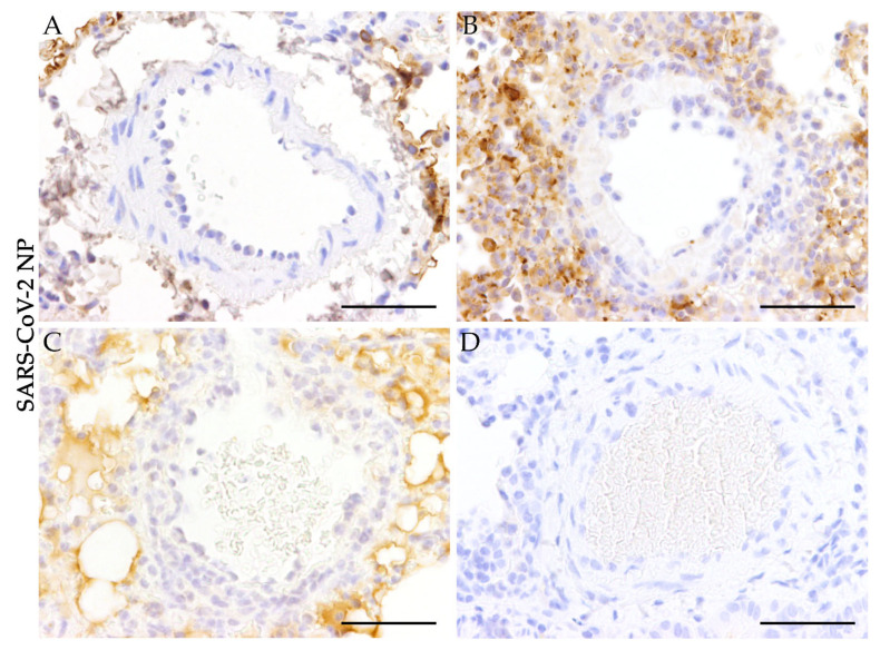
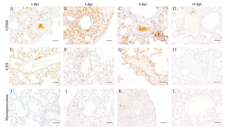
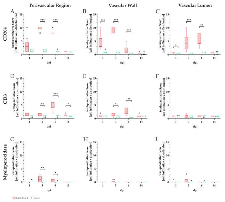
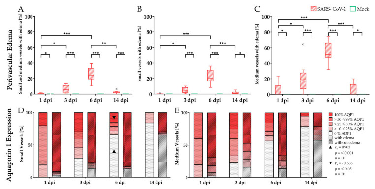
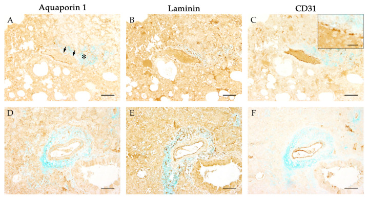
Vascular changes represent a characteristic feature of severe acute respiratory syndrome coronavirus-2 (SARS-CoV-2) infection leading to a breakdown of the vascular barrier and subsequent edema formation. The aim of this study was to provide a detailed characterization of the vascular alterations during SARS-CoV-2 infection and to evaluate the impaired vascular integrity. Groups of ten golden Syrian hamsters were infected intranasally with SARS-CoV-2 or phosphate-buffered saline (mock infection). Necropsies were performed at 1, 3, 6, and 14 days post-infection (dpi). Lung samples were investigated using hematoxylin and eosin, alcian blue, immunohistochemistry targeting aquaporin 1, CD3, CD204, CD31, laminin, myeloperoxidase, SARS-CoV-2 nucleoprotein, and transmission electron microscopy. SARS-CoV-2 infected animals showed endothelial hypertrophy, endothelialitis, and vasculitis. Inflammation mainly consisted of macrophages and lower numbers of T-lymphocytes and neutrophils/heterophils infiltrating the vascular walls as well as the perivascular region at 3 and 6 dpi. Affected vessels showed edema formation in association with loss of aquaporin 1 on endothelial cells. In addition, an ultrastructural investigation revealed disruption of the endothelium. Summarized, the presented findings indicate that loss of aquaporin 1 entails the loss of intercellular junctions resulting in paracellular leakage of edema as a key pathogenic mechanism in SARS-CoV-2 triggered pulmonary lesions.
血管改变是严重急性呼吸综合征冠状病毒 2 型(SARS-CoV-2)感染的特征性表现,导致血管屏障破坏和随后的水肿形成。本研究旨在详细描述 SARS-CoV-2 感染期间的血管改变,并评估受损的血管完整性。十组金黄地鼠经鼻腔感染 SARS-CoV-2 或磷酸盐缓冲盐水(模拟感染)。感染后 1、3、6 和 14 天进行尸检。使用苏木精和伊红、阿尔辛蓝、针对水通道蛋白 1、CD3、CD204、CD31、层粘连蛋白、髓过氧化物酶、SARS-CoV-2 核衣壳蛋白的免疫组织化学和透射电子显微镜检查肺组织样本。SARS-CoV-2 感染动物表现出血管内皮细胞肥大、内皮炎和血管炎。炎症主要由巨噬细胞和较少数量的 T 淋巴细胞和嗜中性粒细胞/异嗜性粒细胞浸润血管壁以及 3 和 6 dpi 的血管周围区域组成。受影响的血管伴有水肿形成,同时内皮细胞上的水通道蛋白 1 丢失。此外,超微结构研究显示内皮破坏。综上所述,这些发现表明水通道蛋白 1 的丢失导致细胞间连接的丢失,从而导致细胞旁渗漏水肿,这是 SARS-CoV-2 引发的肺损伤的关键发病机制。