UCCS BioFrontiers Center, University of Colorado at Colorado Springs, 1420 Austin Bluffs Parkway, Colorado Springs, CO 80918, USA.
George Mason University, 4400 University Drive, Fairfax, VA 22030, USA.
Gigascience. 2021 May 5;10(5). doi: 10.1093/gigascience/giab032.
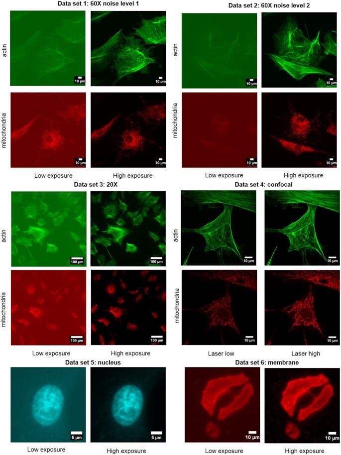
Fluorescence microscopy is an important technique in many areas of biological research. Two factors that limit the usefulness and performance of fluorescence microscopy are photobleaching of fluorescent probes during imaging and, when imaging live cells, phototoxicity caused by light exposure. Recently developed methods in machine learning are able to greatly improve the signal-to-noise ratio of acquired images. This allows researchers to record images with much shorter exposure times, which in turn minimizes photobleaching and phototoxicity by reducing the dose of light reaching the sample.
To use deep learning methods, a large amount of data is needed to train the underlying convolutional neural network. One way to do this involves use of pairs of fluorescence microscopy images acquired with long and short exposure times. We provide high-quality datasets that can be used to train and evaluate deep learning methods under development.
The availability of high-quality data is vital for training convolutional neural networks that are used in current machine learning approaches.
荧光显微镜是许多生物学研究领域的重要技术。在对荧光探针进行成像过程中,有两个因素限制了荧光显微镜的实用性和性能,即荧光探针的光漂白,以及当对活细胞进行成像时,由光暴露引起的光毒性。最近开发的机器学习方法能够极大地提高所获取图像的信噪比。这使得研究人员能够以更短的曝光时间记录图像,从而通过减少到达样品的光剂量来最小化光漂白和光毒性。
为了使用深度学习方法,需要大量的数据来训练基础卷积神经网络。一种方法是使用长曝光时间和短曝光时间采集的荧光显微镜图像对。我们提供高质量的数据集,可用于训练和评估正在开发的深度学习方法。
高质量数据的可用性对于训练在当前机器学习方法中使用的卷积神经网络至关重要。