• 文献检索
  • 文档翻译
  • 深度研究
  • 学术资讯
  • Suppr Zotero 插件Zotero 插件
  • 邀请有礼
  • 套餐&价格
  • 历史记录
应用&插件
Suppr Zotero 插件Zotero 插件浏览器插件Mac 客户端Windows 客户端微信小程序
定价
高级版会员购买积分包购买API积分包
服务
文献检索文档翻译深度研究API 文档MCP 服务
关于我们
关于 Suppr公司介绍联系我们用户协议隐私条款
关注我们

Suppr 超能文献

核心技术专利:CN118964589B侵权必究
粤ICP备2023148730 号-1Suppr @ 2026

文献检索

告别复杂PubMed语法,用中文像聊天一样搜索,搜遍4000万医学文献。AI智能推荐,让科研检索更轻松。

立即免费搜索

文件翻译

保留排版,准确专业,支持PDF/Word/PPT等文件格式,支持 12+语言互译。

免费翻译文档

深度研究

AI帮你快速写综述,25分钟生成高质量综述,智能提取关键信息,辅助科研写作。

立即免费体验

COVID-19 患者随访评估中的计算机断层扫描图像处理分析。

Computed Tomography Image Processing Analysis in COVID-19 Patient Follow-Up Assessment.

机构信息

Postgraduate Department, Instituto Tecnológico Superior de Lerdo, 35150 Lerdo DGO, Mexico.

Medical Familiar Unit, Instituto de Seguridad y Servicios Sociales de Los Trabajadores del Estado, 27268 Torreón COAH, Mexico.

出版信息

J Healthc Eng. 2021 Apr 29;2021:8869372. doi: 10.1155/2021/8869372. eCollection 2021.

DOI:10.1155/2021/8869372
PMID:33968356
原文链接:https://pmc.ncbi.nlm.nih.gov/articles/PMC8083830/
Abstract

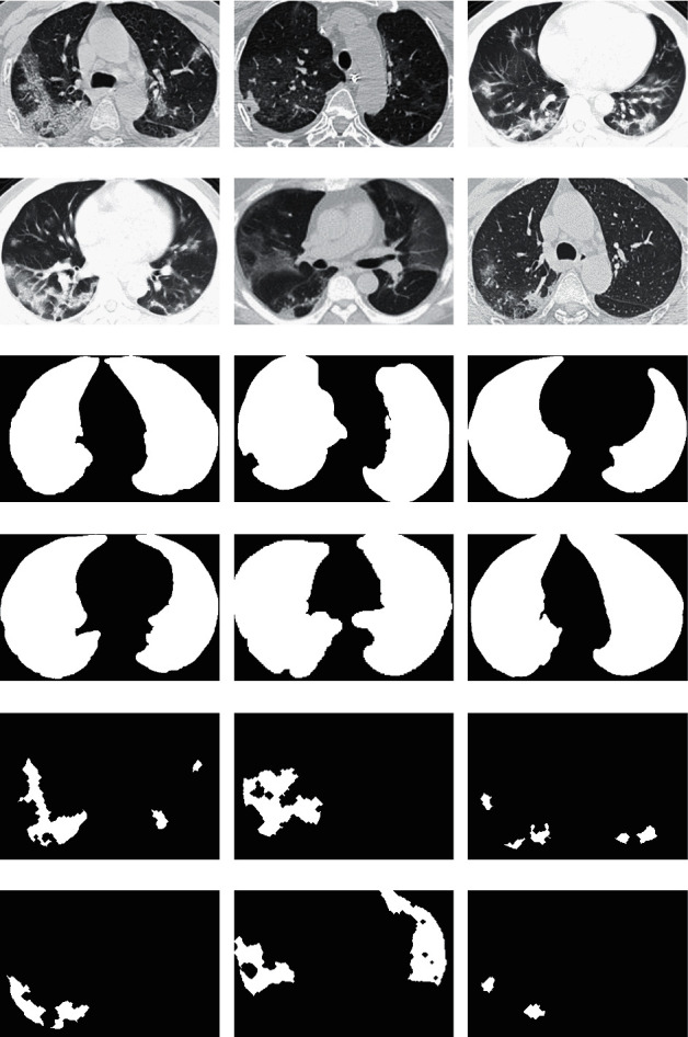
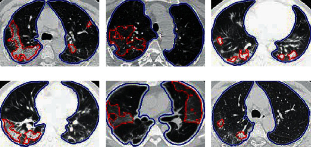
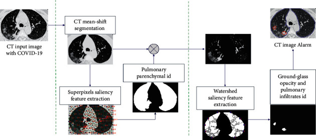
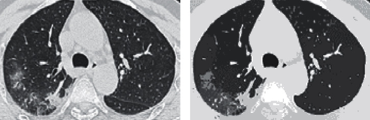
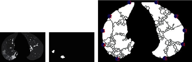
The rapid worldwide spread of the COVID-19 pandemic has infected patients around the world in a short space of time. Chest computed tomography (CT) images of patients who are infected with COVID-19 can offer early diagnosis and efficient forecast monitoring at a low cost. The diagnosis of COVID-19 on CT in an automated way can speed up many tasks and the application of medical treatments. This can help complement reverse transcription-polymerase chain reaction (RT-PCR) diagnosis. The aim of this work is to develop a system that automatically identifies ground-glass opacity (GGO) and pulmonary infiltrates (PIs) on CT images from patients with COVID-19. The purpose is to assess the disease progression during the patient's follow-up assessment and evaluation. We propose an efficient methodology that incorporates oversegmentation mean shift followed by superpixel-SLIC (simple linear iterative clustering) algorithm on CT images with COVID-19 for pulmonary parenchyma segmentation. To identify the pulmonary parenchyma, we described each superpixel cluster according to its position, grey intensity, second-order texture, and spatial-context-saliency features to classify by a tree random forest (TRF). Second, by applying the watershed segmentation to the mean-shift clusters, only pulmonary parenchyma segmentation-identified zones showed GGO and PI based on the description of each watershed cluster of its position, grey intensity, gradient entropy, second-order texture, Euclidean position to the border region of the PI zone, and global saliency features, after using TRF. Our classification results for pulmonary parenchyma identification on CT images with COVID-19 had a precision of over 92% and recall of over 92% on twofold cross validation. For GGO, the PI identification showed 96% precision and 96% recall on twofold cross validation.

摘要

COVID-19 大流行在全球范围内迅速蔓延,在短时间内感染了世界各地的患者。感染 COVID-19 的患者的胸部计算机断层扫描(CT)图像可以提供早期诊断和高效的预测监测,成本低廉。以自动化方式对 CT 上的 COVID-19 进行诊断可以加快许多任务和医疗处理的应用。这有助于补充逆转录-聚合酶链反应(RT-PCR)诊断。这项工作的目的是开发一种系统,该系统可以自动识别 COVID-19 患者 CT 图像上的磨玻璃密度(GGO)和肺部浸润(PI)。目的是在患者的随访评估和评估期间评估疾病进展。我们提出了一种有效的方法,该方法将 oversegmentation mean shift 与 COVID-19 患者 CT 图像上的 superpixel-SLIC(简单线性迭代聚类)算法结合使用,用于肺部实质分割。为了识别肺部实质,我们根据每个超像素簇的位置、灰度强度、二阶纹理和空间上下文显著性特征来描述每个超像素簇,然后通过树随机森林(TRF)进行分类。其次,通过将分水岭分割应用于均值移位簇,仅在根据每个分水岭簇的位置、灰度强度、梯度熵、二阶纹理、PI 区域边界区域的欧几里得位置和全局显著性特征对其进行描述后,才能识别出肺部实质分割识别的区域中的 GGO 和 PI。我们对 COVID-19 患者 CT 图像上的肺部实质识别的分类结果在两次交叉验证中,精度超过 92%,召回率超过 92%。对于 GGO,PI 识别在两次交叉验证中的准确率为 96%,召回率为 96%。

https://cdn.ncbi.nlm.nih.gov/pmc/blobs/d495/8083830/77fbb06b430e/JHE2021-8869372.006.jpg
https://cdn.ncbi.nlm.nih.gov/pmc/blobs/d495/8083830/161c520d6592/JHE2021-8869372.001.jpg
https://cdn.ncbi.nlm.nih.gov/pmc/blobs/d495/8083830/a07bf7e52b90/JHE2021-8869372.002.jpg
https://cdn.ncbi.nlm.nih.gov/pmc/blobs/d495/8083830/3102e2eb0491/JHE2021-8869372.003.jpg
https://cdn.ncbi.nlm.nih.gov/pmc/blobs/d495/8083830/22c0aec6e256/JHE2021-8869372.004.jpg
https://cdn.ncbi.nlm.nih.gov/pmc/blobs/d495/8083830/1e6ea726be83/JHE2021-8869372.005.jpg
https://cdn.ncbi.nlm.nih.gov/pmc/blobs/d495/8083830/77fbb06b430e/JHE2021-8869372.006.jpg

相似文献

1
Computed Tomography Image Processing Analysis in COVID-19 Patient Follow-Up Assessment.COVID-19 患者随访评估中的计算机断层扫描图像处理分析。
J Healthc Eng. 2021 Apr 29;2021:8869372. doi: 10.1155/2021/8869372. eCollection 2021.
2
COVID-19 pneumonia: computer-aided quantification of healthy lung parenchyma, emphysema, ground glass and consolidation on chest computed tomography (CT).COVID-19 肺炎:胸部计算机断层扫描(CT)上健康肺实质、肺气肿、磨玻璃影和实变的计算机辅助定量。
Radiol Med. 2021 Apr;126(4):553-560. doi: 10.1007/s11547-020-01305-9. Epub 2020 Nov 18.
3
Novel COVID-19 Diagnosis Delivery App Using Computed Tomography Images Analyzed with Saliency-Preprocessing and Deep Learning.基于显著性预处理和深度学习分析的新型 COVID-19 诊断交付应用的计算机断层扫描图像。
Tomography. 2022 Jun 20;8(3):1618-1630. doi: 10.3390/tomography8030134.
4
Deep Learning Algorithm for COVID-19 Classification Using Chest X-Ray Images.基于胸部 X 光图像的 COVID-19 分类深度学习算法。
Comput Math Methods Med. 2021 Nov 9;2021:9269173. doi: 10.1155/2021/9269173. eCollection 2021.
5
JCS: An Explainable COVID-19 Diagnosis System by Joint Classification and Segmentation.JCS:基于联合分类与分割的 COVID-19 可解释诊断系统。
IEEE Trans Image Process. 2021;30:3113-3126. doi: 10.1109/TIP.2021.3058783. Epub 2021 Feb 24.
6
Clinical and laboratory data, radiological structured report findings and quantitative evaluation of lung involvement on baseline chest CT in COVID-19 patients to predict prognosis.对 COVID-19 患者基线胸部 CT 上的临床和实验室数据、放射学结构化报告结果以及肺部受累的定量评估,以预测其预后。
Radiol Med. 2021 Jan;126(1):29-39. doi: 10.1007/s11547-020-01293-w. Epub 2020 Oct 12.
7
Value of minimum intensity projections for chest CT in COVID-19 patients.COVID-19 患者胸部 CT 最小密度投影的价值。
Eur J Radiol. 2021 Feb;135:109478. doi: 10.1016/j.ejrad.2020.109478. Epub 2020 Dec 14.
8
Deep-learning algorithms for the interpretation of chest radiographs to aid in the triage of COVID-19 patients: A multicenter retrospective study.深度学习算法在解读胸部 X 光片中对 COVID-19 患者分诊的辅助作用:一项多中心回顾性研究。
PLoS One. 2020 Nov 24;15(11):e0242759. doi: 10.1371/journal.pone.0242759. eCollection 2020.
9
Classification of COVID-19 Chest CT Images Based on Ensemble Deep Learning.基于集成深度学习的 COVID-19 胸部 CT 图像分类。
J Healthc Eng. 2021 Apr 20;2021:5528441. doi: 10.1155/2021/5528441. eCollection 2021.
10
SOM-LWL method for identification of COVID-19 on chest X-rays.基于 SOM-LWL 算法的胸部 X 光片 COVID-19 识别方法。
PLoS One. 2021 Feb 24;16(2):e0247176. doi: 10.1371/journal.pone.0247176. eCollection 2021.

引用本文的文献

1
Challenges issues and future recommendations of deep learning techniques for SARS-CoV-2 detection utilising X-ray and CT images: a comprehensive review.利用X射线和CT图像进行SARS-CoV-2检测的深度学习技术面临的挑战、问题及未来建议:全面综述
PeerJ Comput Sci. 2024 Dec 24;10:e2517. doi: 10.7717/peerj-cs.2517. eCollection 2024.
2
Classification of Severity of Lung Parenchyma Using Saliency and Discrete Cosine Transform Energy in Computed Tomography of Patients With COVID-19.利用显著性和离散余弦变换能量对新冠肺炎患者计算机断层扫描中的肺实质严重程度进行分类
Int J Telemed Appl. 2025 Jan 6;2025:4420410. doi: 10.1155/ijta/4420410. eCollection 2025.
3

本文引用的文献

1
RETRACTED ARTICLE: Deep learning system to screen coronavirus disease 2019 pneumonia.撤稿文章:用于筛查2019冠状病毒病肺炎的深度学习系统。
Appl Intell (Dordr). 2023;53(4):4874. doi: 10.1007/s10489-020-01714-3. Epub 2020 Apr 22.
2
Leveraging Data Science to Combat COVID-19: A Comprehensive Review.利用数据科学抗击新冠疫情:全面综述
IEEE Trans Artif Intell. 2020 Sep 2;1(1):85-103. doi: 10.1109/TAI.2020.3020521. eCollection 2020 Aug.
3
Imaging Profile of the COVID-19 Infection: Radiologic Findings and Literature Review.新型冠状病毒肺炎感染的影像学表现:放射学发现与文献综述
Novel Deep CNNs Explore Regions, Boundaries, and Residual Learning for COVID-19 Infection Analysis in Lung CT.
新型深度卷积神经网络探索肺部 CT 中 COVID-19 感染分析的区域、边界和残差学习。
Tomography. 2024 Aug 3;10(8):1205-1221. doi: 10.3390/tomography10080091.
4
Artificially-generated consolidations and balanced augmentation increase performance of U-net for lung parenchyma segmentation on MR images.人工生成的强化和平衡增强提高了 U-net 在 MR 图像上对肺实质分割的性能。
PLoS One. 2023 May 9;18(5):e0285378. doi: 10.1371/journal.pone.0285378. eCollection 2023.
5
COVID-19 Detection by Means of ECG, Voice, and X-ray Computerized Systems: A Review.通过心电图、语音和X射线计算机系统检测COVID-19:综述
Bioengineering (Basel). 2023 Feb 3;10(2):198. doi: 10.3390/bioengineering10020198.
6
Application of Machine Learning and Deep Learning Techniques for COVID-19 Screening Using Radiological Imaging: A Comprehensive Review.使用放射成像的机器学习和深度学习技术在COVID-19筛查中的应用:综述
SN Comput Sci. 2023;4(1):65. doi: 10.1007/s42979-022-01464-8. Epub 2022 Nov 24.
7
Recommender System for the Efficient Treatment of COVID-19 Using a Convolutional Neural Network Model and Image Similarity.基于卷积神经网络模型和图像相似度的新冠高效治疗推荐系统
Diagnostics (Basel). 2022 Nov 5;12(11):2700. doi: 10.3390/diagnostics12112700.
8
Efficient COVID-19 super pixel segmentation algorithm using MCFO-based SLIC.基于基于多宇宙萤火虫优化算法的简单线性迭代聚类的高效新冠病毒超像素分割算法
J Ambient Intell Humaniz Comput. 2023;14(7):9217-9232. doi: 10.1007/s12652-022-04425-4. Epub 2022 Oct 21.
9
Novel COVID-19 Diagnosis Delivery App Using Computed Tomography Images Analyzed with Saliency-Preprocessing and Deep Learning.基于显著性预处理和深度学习分析的新型 COVID-19 诊断交付应用的计算机断层扫描图像。
Tomography. 2022 Jun 20;8(3):1618-1630. doi: 10.3390/tomography8030134.
10
Optimal Deep-Learning-Enabled Intelligent Decision Support System for SARS-CoV-2 Classification.用于 SARS-CoV-2 分类的最优深度学习智能决策支持系统。
J Healthc Eng. 2022 Jan 25;2022:4130674. doi: 10.1155/2022/4130674. eCollection 2022.
Radiol Cardiothorac Imaging. 2020 Feb 13;2(1):e200034. doi: 10.1148/ryct.2020200034. eCollection 2020 Feb.
4
Large-scale screening to distinguish between COVID-19 and community-acquired pneumonia using infection size-aware classification.利用感染大小感知分类进行大规模筛选,以区分 COVID-19 和社区获得性肺炎。
Phys Med Biol. 2021 Mar 17;66(6):065031. doi: 10.1088/1361-6560/abe838.
5
Deep Learning Enables Accurate Diagnosis of Novel Coronavirus (COVID-19) With CT Images.深度学习利用 CT 图像准确诊断新型冠状病毒(COVID-19)。
IEEE/ACM Trans Comput Biol Bioinform. 2021 Nov-Dec;18(6):2775-2780. doi: 10.1109/TCBB.2021.3065361. Epub 2021 Dec 8.
6
A deep learning algorithm using CT images to screen for Corona virus disease (COVID-19).利用 CT 图像进行冠状病毒病(COVID-19)筛查的深度学习算法。
Eur Radiol. 2021 Aug;31(8):6096-6104. doi: 10.1007/s00330-021-07715-1. Epub 2021 Feb 24.
7
JCS: An Explainable COVID-19 Diagnosis System by Joint Classification and Segmentation.JCS:基于联合分类与分割的 COVID-19 可解释诊断系统。
IEEE Trans Image Process. 2021;30:3113-3126. doi: 10.1109/TIP.2021.3058783. Epub 2021 Feb 24.
8
COVID-Net: a tailored deep convolutional neural network design for detection of COVID-19 cases from chest X-ray images.COVID-Net:一种针对胸部 X 光图像中 COVID-19 病例检测的定制化深度卷积神经网络设计。
Sci Rep. 2020 Nov 11;10(1):19549. doi: 10.1038/s41598-020-76550-z.
9
Deep learning-based model for detecting 2019 novel coronavirus pneumonia on high-resolution computed tomography.基于深度学习的高分辨率计算机断层扫描 2019 年新型冠状病毒肺炎检测模型。
Sci Rep. 2020 Nov 5;10(1):19196. doi: 10.1038/s41598-020-76282-0.
10
Multi-task deep learning based CT imaging analysis for COVID-19 pneumonia: Classification and segmentation.基于多任务深度学习的 COVID-19 肺炎 CT 成像分析:分类与分割。
Comput Biol Med. 2020 Nov;126:104037. doi: 10.1016/j.compbiomed.2020.104037. Epub 2020 Oct 8.