Gouveia-Freitas Kaylene, Bastos-Leite António J
Faculty of Medicine, University of Porto, Alameda do Professor Hernâni Monteiro, 4200-319, Porto, Portugal.
Neuroradiology. 2021 Oct;63(10):1581-1597. doi: 10.1007/s00234-021-02718-7. Epub 2021 May 21.
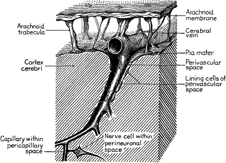
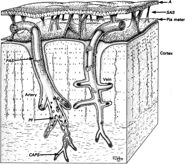
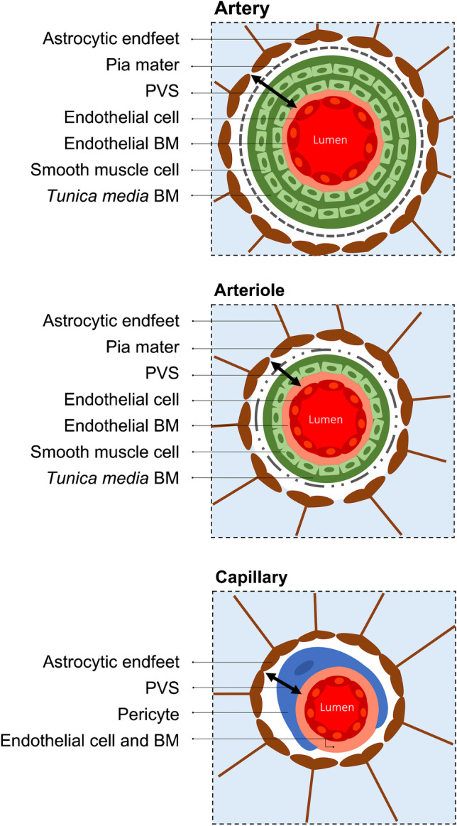
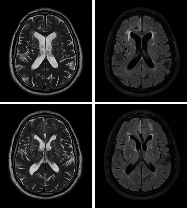
Perivascular spaces (PVS) of the brain, often called Virchow-Robin spaces, comprise fluid, cells and connective tissue, and are externally limited by astrocytic endfeet. PVS are involved in clearing brain waste and belong to the "glymphatic" system and/or the "intramural periarterial drainage" pathway through the basement membranes of the arteries. Related brain waste clearance systems include the blood-brain barrier, scavenger cells, cerebrospinal fluid, perineural lymphatic drainage pathways and the newly characterised meningeal lymphatic vessels. Any functional abnormality of PVS or related clearance systems might lead to accumulation of brain waste. It has been postulated that PVS enlargement can be secondary to accumulation of β-amyloid. Lack of integrity of the vascular wall, microbleeds, cerebral amyloid angiopathy (CAA) and enlarged PVS often occur in the preclinical stages of Alzheimer's disease, preceding substantial brain atrophy. PVS enlargement in the form of état criblé at the basal ganglia has also been considered to reflect focal atrophy, most probably secondary to ischaemic injury, based upon both pathological and imaging arguments. In addition, distinct topographic patterns of enlarged PVS are related to different types of microangiopathy: CAA is linked to enlarged juxtacortical PVS, whereas subjects with vascular risk factors tend to have enlarged PVS in the basal ganglia. Therefore, enlarged PVS are progressively being regarded as a marker of neurodegenerative and cerebrovascular pathology. The present review addresses the evolving concept of PVS and brain waste clearance systems, the potential relevance of their dysfunction to neurodegenerative and cerebrovascular pathology, and potential therapeutic approaches of interest.
脑周血管间隙(PVS),通常称为魏尔啸-罗宾间隙,由液体、细胞和结缔组织组成,其外部由星形胶质细胞终足界定。PVS参与清除脑内废物,属于“类淋巴”系统和/或通过动脉基底膜的“壁内动脉周围引流”途径。相关的脑内废物清除系统包括血脑屏障、吞噬细胞、脑脊液、神经周围淋巴引流途径以及新发现的脑膜淋巴管。PVS或相关清除系统的任何功能异常都可能导致脑内废物积聚。据推测,PVS扩大可能继发于β-淀粉样蛋白的积聚。血管壁完整性缺失、微出血、脑淀粉样血管病(CAA)和PVS扩大常发生在阿尔茨海默病临床前期,早于明显的脑萎缩。基于病理和影像学证据,基底节呈筛状的PVS扩大也被认为反映了局灶性萎缩,最可能继发于缺血性损伤。此外,PVS扩大的不同地形模式与不同类型的微血管病变有关:CAA与皮质旁PVS扩大有关,而有血管危险因素的受试者基底节PVS往往扩大。因此,PVS扩大正逐渐被视为神经退行性和脑血管病理的标志物。本综述阐述了PVS和脑内废物清除系统的不断演变的概念、其功能障碍与神经退行性和脑血管病理的潜在相关性以及潜在的治疗方法。