• 文献检索
  • 文档翻译
  • 深度研究
  • 学术资讯
  • Suppr Zotero 插件Zotero 插件
  • 邀请有礼
  • 套餐&价格
  • 历史记录
应用&插件
Suppr Zotero 插件Zotero 插件浏览器插件Mac 客户端Windows 客户端微信小程序
定价
高级版会员购买积分包购买API积分包
服务
文献检索文档翻译深度研究API 文档MCP 服务
关于我们
关于 Suppr公司介绍联系我们用户协议隐私条款
关注我们

Suppr 超能文献

核心技术专利:CN118964589B侵权必究
粤ICP备2023148730 号-1Suppr @ 2026

文献检索

告别复杂PubMed语法,用中文像聊天一样搜索,搜遍4000万医学文献。AI智能推荐,让科研检索更轻松。

立即免费搜索

文件翻译

保留排版,准确专业,支持PDF/Word/PPT等文件格式,支持 12+语言互译。

免费翻译文档

深度研究

AI帮你快速写综述,25分钟生成高质量综述,智能提取关键信息,辅助科研写作。

立即免费体验

青光眼可疑患者视乳头深层区域血管密度损失及其与筛板特征的关联

Vessel Density Loss of the Deep Peripapillary Area in Glaucoma Suspects and Its Association with Features of the Lamina Cribrosa.

作者信息

Jeon Soo-Ji, Park Hae-Young Lopilly, Park Chan-Kee

机构信息

Apgujeong St. Mary's Eye Center, 859 Eonju-ro, Gangnam-gu, Seoul 06023, Korea.

Department of Ophthalmology, Seoul St. Mary's Hospital, College of Medicine, The Catholic University of Korea, 222 Banpo-daero, Seocho-gu, Seoul 06591, Korea.

出版信息

J Clin Med. 2021 May 28;10(11):2373. doi: 10.3390/jcm10112373.

DOI:10.3390/jcm10112373
PMID:34071218
原文链接:https://pmc.ncbi.nlm.nih.gov/articles/PMC8197842/
Abstract

PURPOSE

To investigate the association of decreased vessel density (VD) in the deep peripapillary region and structural features of the lamina cribrosa (LC).

MATERIALS AND METHODS

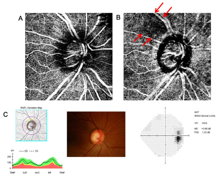
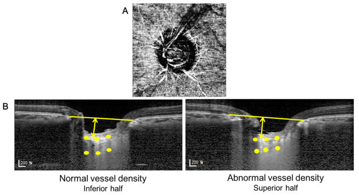
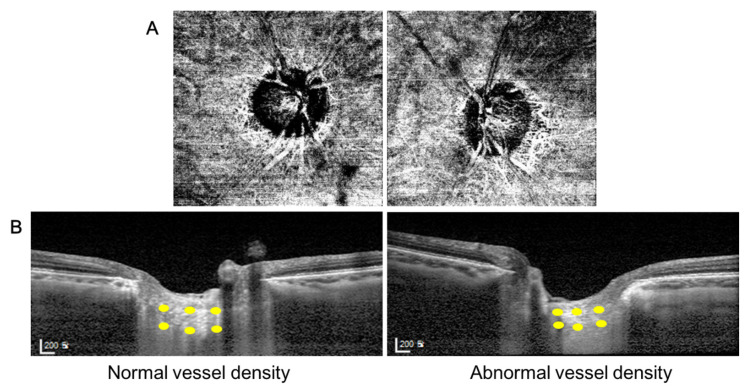
70 eyes of glaucoma suspects with enlarged cup-to-disc ratio were scanned and 51 eyes with adequate image quality were included in this study. All subjects had localized VD defects in the deep layer but intact VD in the superficial layer around the peripapillary region using optical coherence tomography angiography (OCTA). Only single-hemizone OCTA results from one eye of each subject had to fulfill the distinctive feature mentioned above to perform inter-eye and inter-hemizone comparisons. The thickness and depth of the LC, and prelaminar thickness were measured using enhanced depth imaging OCT (EDI-OCT). Paired t-tests were performed to evaluate differences in measurements of the LC and prelaminar thickness within each individual. -values lower than 0.05 was considered to be statistically significant.

RESULTS

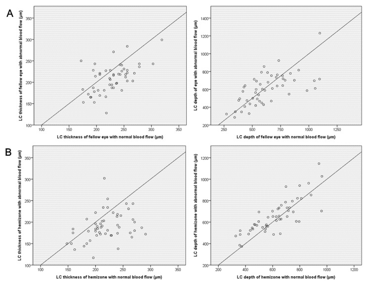
Eyes with deep VD defects in the peripapillary region in OCTA had thinner LC than the fellow eyes. The hemizone with the deep VD defects in the peripapillary region had a thinner LC and a deeper depth of LC than the other hemizone in the same eye. According to logistic regression analysis, a thin LC was a significant factor associated with deep VD defect in the peripapillary region.

CONCLUSIONS

Glaucoma suspect eyes with deep VD defects in the peripapillary area exhibited structural differences in the LC. The structural changes of the LC was associated with the vessel density in the deep peripapillary layer at the stage of suspected glaucoma.

摘要

目的

研究视乳头周围深层血管密度(VD)降低与筛板(LC)结构特征之间的关联。

材料与方法

对70例杯盘比增大的青光眼可疑患者的眼睛进行扫描,本研究纳入了51例图像质量合格的眼睛。所有受试者使用光学相干断层扫描血管造影(OCTA)检查发现视乳头周围区域深层存在VD缺陷,但表层VD正常。每个受试者仅一只眼睛的单半区OCTA结果须满足上述独特特征,以便进行眼间和半区间比较。使用增强深度成像OCT(EDI-OCT)测量LC的厚度、深度以及板层前厚度。采用配对t检验评估每个个体LC测量值和板层前厚度的差异。P值低于0.05被认为具有统计学意义。

结果

OCTA显示视乳头周围区域存在深层VD缺陷的眼睛,其LC比对侧眼更薄。视乳头周围区域存在深层VD缺陷的半区,其LC比同一只眼睛的另一个半区更薄,且LC深度更深。根据逻辑回归分析,薄的LC是视乳头周围区域深层VD缺陷的一个重要相关因素。

结论

视乳头周围区域存在深层VD缺陷的青光眼可疑患者的眼睛,其LC表现出结构差异。在青光眼可疑阶段,LC的结构变化与视乳头周围深层的血管密度相关。

https://cdn.ncbi.nlm.nih.gov/pmc/blobs/dc49/8197842/765fdcdfcded/jcm-10-02373-g004.jpg
https://cdn.ncbi.nlm.nih.gov/pmc/blobs/dc49/8197842/7619451e6fc6/jcm-10-02373-g001.jpg
https://cdn.ncbi.nlm.nih.gov/pmc/blobs/dc49/8197842/e79336267abf/jcm-10-02373-g002.jpg
https://cdn.ncbi.nlm.nih.gov/pmc/blobs/dc49/8197842/ea9399cba8c9/jcm-10-02373-g003.jpg
https://cdn.ncbi.nlm.nih.gov/pmc/blobs/dc49/8197842/765fdcdfcded/jcm-10-02373-g004.jpg

相似文献

1
Vessel Density Loss of the Deep Peripapillary Area in Glaucoma Suspects and Its Association with Features of the Lamina Cribrosa.青光眼可疑患者视乳头深层区域血管密度损失及其与筛板特征的关联
J Clin Med. 2021 May 28;10(11):2373. doi: 10.3390/jcm10112373.
2
Microvascular Changes in Peripapillary and Optic Nerve Head Tissues After Trabeculectomy in Primary Open-Angle Glaucoma.原发性开角型青光眼小梁切除术后周边视网膜和视盘组织的微血管变化。
Invest Ophthalmol Vis Sci. 2018 Sep 4;59(11):4614-4621. doi: 10.1167/iovs.18-25038.
3
Correlation of Visual Field With Peripapillary Vessel Density Through Optical Coherence Tomography Angiography in Normal-Tension Glaucoma.正常眼压性青光眼中通过光学相干断层扫描血管造影术评估视野与视乳头周围血管密度的相关性
Transl Vis Sci Technol. 2020 Dec 17;9(13):26. doi: 10.1167/tvst.9.13.26. eCollection 2020 Dec.
4
Alterations of the Lamina Cribrosa Are Associated with Peripapillary Retinoschisis in Glaucoma and Pachychoroid Spectrum Disease.神经纤维层筛板改变与青光眼和脉络膜增厚谱疾病的视盘周围视网膜劈裂有关。
Ophthalmology. 2016 Oct;123(10):2066-76. doi: 10.1016/j.ophtha.2016.06.033. Epub 2016 Aug 6.
5
Optical coherence tomography angiography of the peripapillary region and macula in normal, primary open angle glaucoma, pseudoexfoliation glaucoma and ocular hypertension eyes.正常眼、原发性开角型青光眼、剥脱性青光眼和高眼压症患者眼的视乳头周围区域和黄斑的光学相干断层扫描血管造影
Int J Ophthalmol. 2020 May 18;13(5):744-754. doi: 10.18240/ijo.2020.05.08. eCollection 2020.
6
OCTA vessel density changes in the macular zone in glaucomatous eyes.青光眼患者眼部黄斑区光学相干断层扫描血管密度的变化
Graefes Arch Clin Exp Ophthalmol. 2018 Aug;256(8):1499-1508. doi: 10.1007/s00417-018-3965-1. Epub 2018 Apr 10.
7
Deep Retinal Layer Microvasculature Dropout Detected by the Optical Coherence Tomography Angiography in Glaucoma.光学相干断层扫描血管造影术检测青光眼患者视网膜深层微血管缺失
Ophthalmology. 2016 Dec;123(12):2509-2518. doi: 10.1016/j.ophtha.2016.09.002. Epub 2016 Oct 18.
8
Optical Coherence Tomography Angiography of the Peripapillary Retina in Primary Angle-Closure Glaucoma.原发性闭角型青光眼视乳头周围视网膜的光学相干断层扫描血管造影
Am J Ophthalmol. 2017 Oct;182:194-200. doi: 10.1016/j.ajo.2017.07.024. Epub 2017 Aug 7.
9
Quantitative analysis of neural tissues around the optic disc after panretinal photocoagulation in patients with diabetic retinopathy.糖尿病视网膜病变患者全视网膜光凝术后视盘周围神经组织的定量分析。
PLoS One. 2017 Oct 17;12(10):e0186229. doi: 10.1371/journal.pone.0186229. eCollection 2017.
10
Optical Coherence Tomography Angiography Macular and Peripapillary Vessel Perfusion Density in Healthy Subjects, Glaucoma Suspects, and Glaucoma Patients.光学相干断层扫描血管造影术检测健康受试者、青光眼疑似患者和青光眼患者黄斑及视乳头周围血管灌注密度
Invest Ophthalmol Vis Sci. 2017 Nov 1;58(13):5713-5722. doi: 10.1167/iovs.17-22865.

引用本文的文献

1
Features Associated with Visible Lamina Cribrosa Pores in Individuals of African Ancestry with Glaucoma: Primary Open-Angle African Ancestry Glaucoma Genetics (POAAGG) Study.非洲裔青光眼患者中与可见筛板孔相关的特征:原发性开角型非洲裔青光眼遗传学(POAAGG)研究。
Vision (Basel). 2024 Apr 18;8(2):24. doi: 10.3390/vision8020024.
2
Association of choroidal blood flow with autonomic dysfunction in patients with normal tension glaucoma.正常眼压性青光眼患者脉络膜血流与自主神经功能障碍的相关性。
Sci Rep. 2022 Mar 24;12(1):5136. doi: 10.1038/s41598-022-09162-4.
3
Updates in Clinical and Translational Glaucoma Research.

本文引用的文献

1
Optical Coherence Tomography Angiography in Glaucoma.光学相干断层扫描血管造影术在青光眼中的应用。
J Glaucoma. 2020 Apr;29(4):312-321. doi: 10.1097/IJG.0000000000001463.
2
Association of Retinal Blood Flow with Progression of Visual Field in Glaucoma.视网膜血流与青光眼视野进展的关系。
Sci Rep. 2019 Nov 14;9(1):16813. doi: 10.1038/s41598-019-53354-4.
3
Projection-Resolved Optical Coherence Tomography Angiography of the Peripapillary Retina in Glaucoma.青光眼周边视网膜的层析光学相干断层扫描血管造影术。
临床与转化性青光眼研究进展
J Clin Med. 2021 Dec 31;11(1):221. doi: 10.3390/jcm11010221.
Am J Ophthalmol. 2019 Nov;207:99-109. doi: 10.1016/j.ajo.2019.05.024. Epub 2019 Jun 3.
4
A Review of OCT Angiography in Glaucoma.青光眼的光学相干断层扫描血管造影综述
Semin Ophthalmol. 2019;34(4):279-286. doi: 10.1080/08820538.2019.1620807. Epub 2019 Jun 3.
5
Quantitative Analysis of Retinal and Choroidal Vascular Parameters in Patients With Low Tension Glaucoma.低眼压性青光眼患者视网膜和脉络膜血管参数的定量分析。
J Glaucoma. 2019 Jun;28(6):557-562. doi: 10.1097/IJG.0000000000001242.
6
Effect of Macular Vascular Density on Central Visual Function and Macular Structure in Glaucoma Patients.青光眼患者黄斑血管密度对中心视觉功能和黄斑结构的影响。
Sci Rep. 2018 Oct 30;8(1):16009. doi: 10.1038/s41598-018-34417-4.
7
Extracellular matrix scaffolding in angiogenesis and capillary homeostasis.细胞外基质支架在血管生成和毛细血管稳态中的作用。
Semin Cell Dev Biol. 2019 May;89:147-156. doi: 10.1016/j.semcdb.2018.08.007. Epub 2018 Sep 5.
8
Features of the Choroidal Microvasculature in Peripapillary Atrophy Are Associated With Visual Field Damage in Myopic Patients.周边性视神经萎缩的脉络膜微血管特征与近视患者的视野损害有关。
Am J Ophthalmol. 2018 Aug;192:206-216. doi: 10.1016/j.ajo.2018.05.027. Epub 2018 Jun 5.
9
OCTA vessel density changes in the macular zone in glaucomatous eyes.青光眼患者眼部黄斑区光学相干断层扫描血管密度的变化
Graefes Arch Clin Exp Ophthalmol. 2018 Aug;256(8):1499-1508. doi: 10.1007/s00417-018-3965-1. Epub 2018 Apr 10.
10
Retinal vasculature in glaucoma: a review.青光眼的视网膜血管系统:综述
BMJ Open Ophthalmol. 2017 Jul 11;1(1):e000032. doi: 10.1136/bmjophth-2016-000032. eCollection 2017.