Patrick G. Johnston Centre for Cancer Research, Queen's University, Belfast, UK.
National Physical Laboratory, London, UK.
Radiat Oncol. 2021 Jun 12;16(1):104. doi: 10.1186/s13014-021-01829-y.
The recent implementation of MR-Linacs has highlighted theranostic opportunities of contrast agents in both imaging and radiotherapy. There is a lack of data exploring the potential of superparamagnetic iron oxide nanoparticles (SPIONs) as radiosensitisers. Through preclinical 225 kVp exposures, this study aimed to characterise the uptake and radiobiological effects of SPIONs in tumour cell models in vitro and to provide proof-of-principle application in a xenograft tumour model.
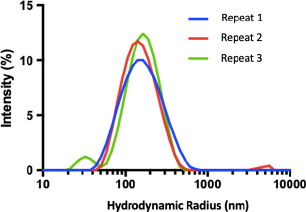
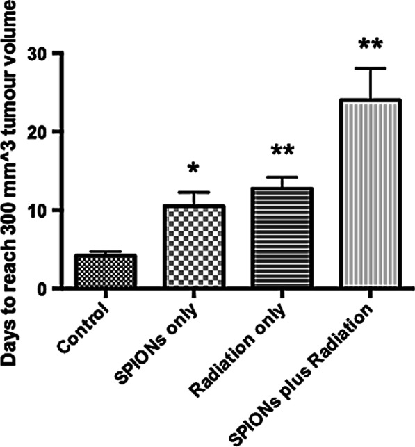
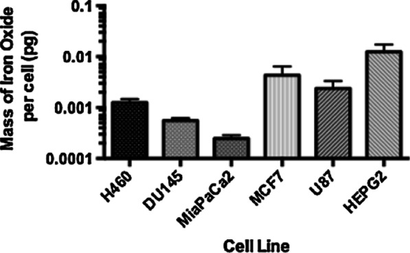
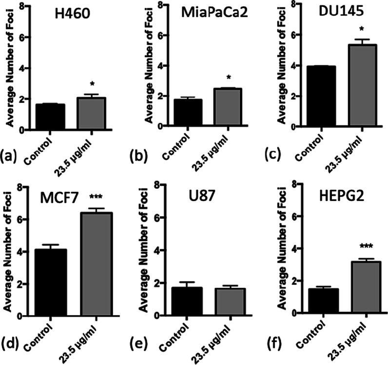
SPIONs were also characterised to determine their hydrodynamic radius using dynamic light scattering and uptake was measured using ICP-MS in 6 cancer cell lines; H460, MiaPaCa2, DU145, MCF7, U87 and HEPG2. The impact of SPIONs on radiobiological response was determined by measuring DNA damage using 53BP1 immunofluorescence and cell survival. Sensitisation Enhancement Ratios (SERs) were compared with the predicted Dose Enhancement Ratios (DEFs) based on physical absorption estimations. In vivo efficacy was demonstrated using a subcutaneous H460 xenograft tumour model in SCID mice by following intra-tumoural injection of SPIONs.
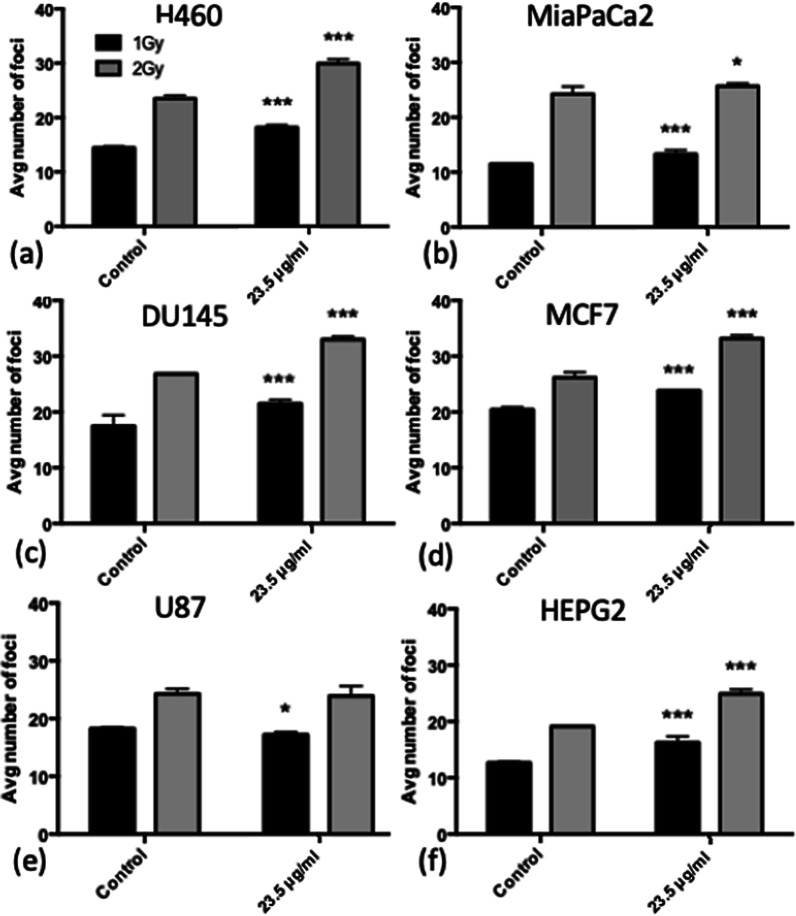
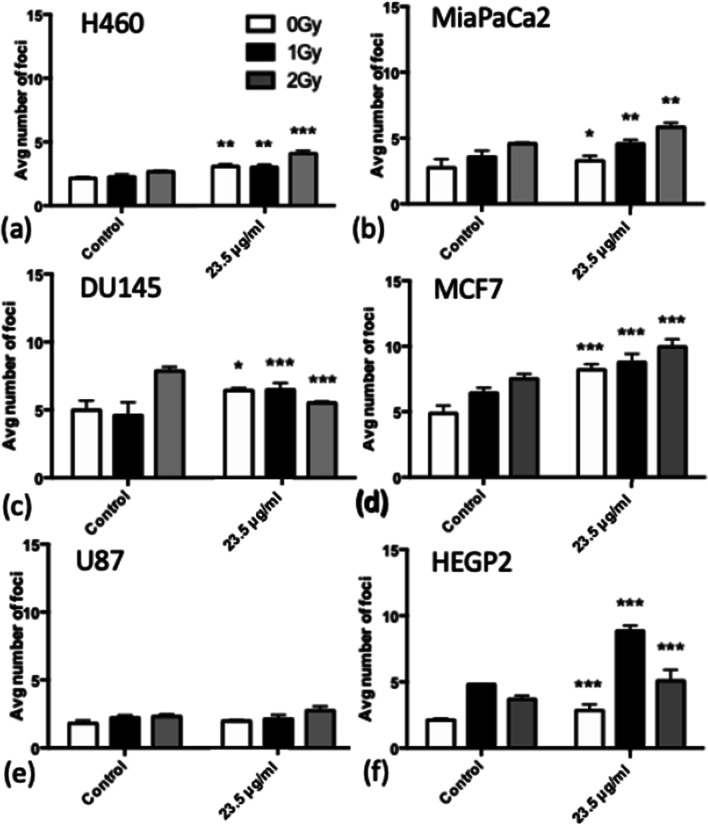
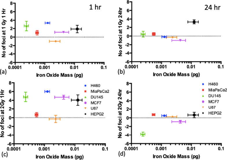
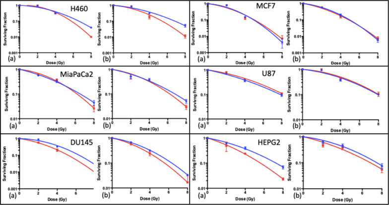
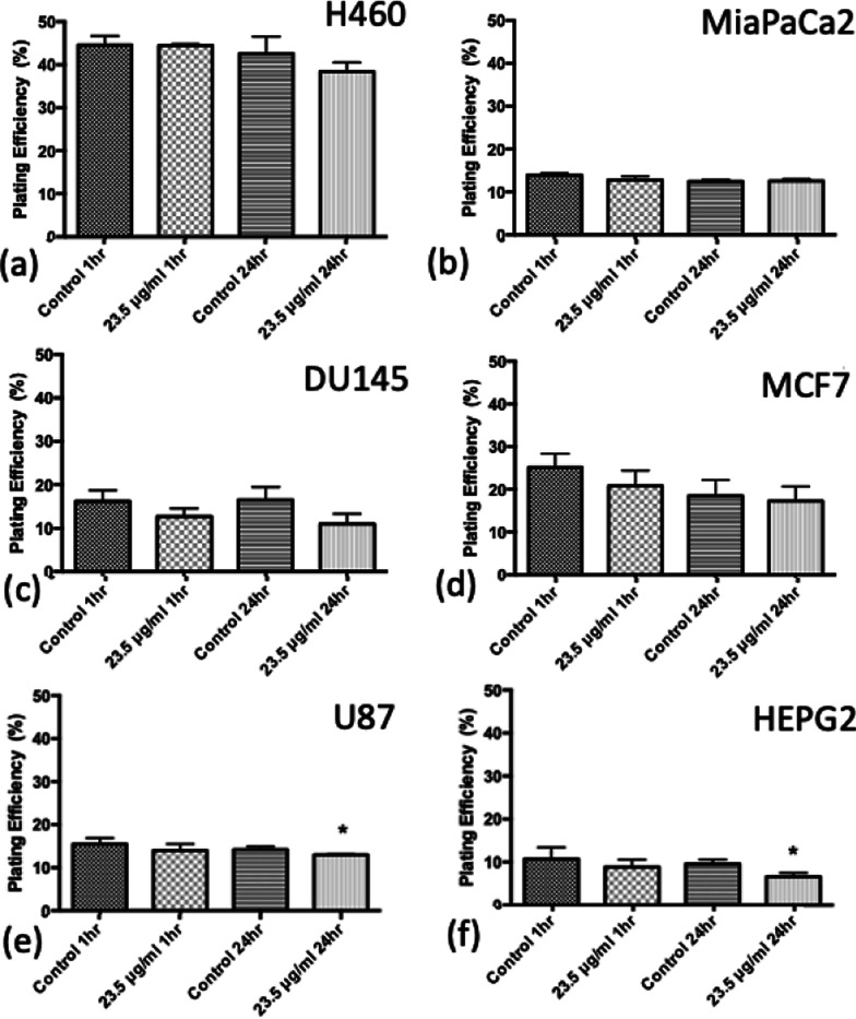
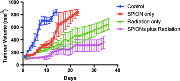
The hydrodynamic radius was found to be between 110 and 130 nm, with evidence of being monodisperse in nature. SPIONs significantly increased DNA damage in all cell lines with the exception of U87 cells at a dose of 1 Gy, 1 h post-irradiation. Levels of DNA damage correlated with the cell survival, in which all cell lines except U87 cells showed an increased sensitivity (P < 0.05) in the linear quadratic curve fit for 1 h exposure to 23.5 μg/ml SPIONs. There was also a 30.1% increase in the number of DNA damage foci found for HEPG2 cells at 2 Gy. No strong correlation was found between SPION uptake and DNA damage at any dose, yet the biological consequences of SPIONs on radiosensitisation were found to be much greater, with SERs up to 1.28 ± 0.03, compared with predicted physical dose enhancement levels of 1.0001. In vivo, intra-tumoural injection of SPIONs combined with radiation showed significant tumour growth delay compared to animals treated with radiation or SPIONs alone (P < 0.05).
SPIONs showed radiosensitising effects in 5 out of 6 cancer cell lines. No correlation was found between the cell-specific uptake of SPIONs into the cells and DNA damage levels. The in vivo study found a significant decrease in the tumour growth rate.
最近实施的磁共振直线加速器(MR-Linacs)突出了对比剂在成像和放射治疗中的治疗诊断机会。缺乏探索超顺磁氧化铁纳米颗粒(SPIONs)作为放射增敏剂的潜力的数据。通过临床前 225 kVp 照射,本研究旨在描述 SPIONs 在体外肿瘤细胞模型中的摄取和放射生物学效应,并提供在异种移植肿瘤模型中的原理验证应用。
还对 SPIONs 进行了特征描述,以使用动态光散射确定其水动力半径,并使用 ICP-MS 在 6 种癌细胞系(H460、MiaPaCa2、DU145、MCF7、U87 和 HEPG2)中测量摄取量。使用 53BP1 免疫荧光测量 DNA 损伤来确定 SPIONs 对放射生物学反应的影响,并测量细胞存活。比较了基于物理吸收估算的 SPIONs 的敏感性增强比(SER)与预测的剂量增强比(DEF)。通过在 SCID 小鼠的皮下 H460 异种移植肿瘤模型中进行肿瘤内注射 SPIONs,证明了体内疗效。
发现水动力半径在 110 至 130nm 之间,具有单分散性的证据。SPIONs 在除 U87 细胞外的所有细胞系中均在 1Gy 辐射后 1 小时显着增加了 DNA 损伤。DNA 损伤水平与细胞存活相关,其中除 U87 细胞外的所有细胞系在 1 小时暴露于 23.5μg/ml SPIONs 的线性二次曲线拟合中显示出更高的敏感性(P<0.05)。在 2Gy 时,HEPG2 细胞的 DNA 损伤焦点数量也增加了 30.1%。在任何剂量下,都没有发现 SPION 摄取与 DNA 损伤之间的强相关性,但 SPIONs 对放射增敏的生物学后果被发现要大得多,SER 高达 1.28±0.03,而预测的物理剂量增强水平为 1.0001。在体内,与单独接受放射治疗或 SPIONs 治疗的动物相比,肿瘤内注射 SPIONs 联合放射治疗可显著延缓肿瘤生长(P<0.05)。
SPIONs 在 6 种癌细胞系中的 5 种中表现出放射增敏作用。没有发现 SPIONs 进入细胞的细胞特异性摄取与 DNA 损伤水平之间存在相关性。体内研究发现肿瘤生长速度显着降低。