• 文献检索
  • 文档翻译
  • 深度研究
  • 学术资讯
  • Suppr Zotero 插件Zotero 插件
  • 邀请有礼
  • 套餐&价格
  • 历史记录
应用&插件
Suppr Zotero 插件Zotero 插件浏览器插件Mac 客户端Windows 客户端微信小程序
定价
高级版会员购买积分包购买API积分包
服务
文献检索文档翻译深度研究API 文档MCP 服务
关于我们
关于 Suppr公司介绍联系我们用户协议隐私条款
关注我们

Suppr 超能文献

核心技术专利:CN118964589B侵权必究
粤ICP备2023148730 号-1Suppr @ 2026

文献检索

告别复杂PubMed语法,用中文像聊天一样搜索,搜遍4000万医学文献。AI智能推荐,让科研检索更轻松。

立即免费搜索

文件翻译

保留排版,准确专业,支持PDF/Word/PPT等文件格式,支持 12+语言互译。

免费翻译文档

深度研究

AI帮你快速写综述,25分钟生成高质量综述,智能提取关键信息,辅助科研写作。

立即免费体验

伴有或不伴有视网膜分支血管阻塞的正常眼压性青光眼患者的视盘血管密度

Optic Disc Vascular Density in Normal-Tension Glaucoma Eyes with or without Branch Retinal Vessel Occlusion.

作者信息

Baek Jiwon, Jeon Soo-Ji, Kim Jin-Ho, Park Chan-Kee, Park Hae-Young L

机构信息

Department of Ophthalmology, Bucheon St. Mary's Hospital, College of Medicine, The Catholic University of Korea, Bucheon 14647, Korea.

Department of Ophthalmology, Eunpyeong St. Mary's Hospital, College of Medicine, The Catholic University of Korea, Seoul 03312, Korea.

出版信息

J Clin Med. 2021 Jun 10;10(12):2574. doi: 10.3390/jcm10122574.

DOI:10.3390/jcm10122574
PMID:34200846
原文链接:https://pmc.ncbi.nlm.nih.gov/articles/PMC8230548/
Abstract

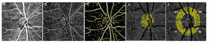
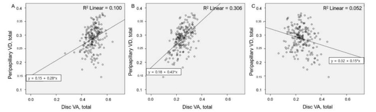
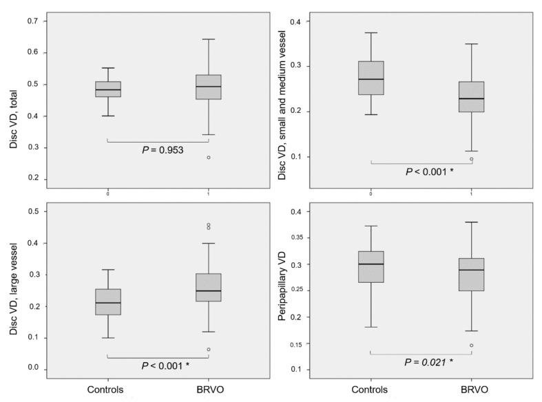
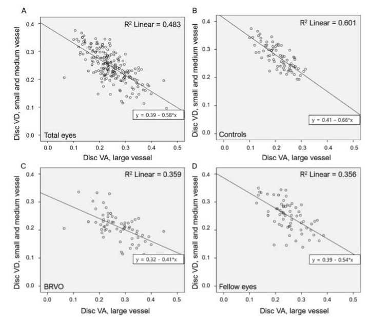
We analyzed the vascular densities (VDs) of the optic disc areas in eyes with normal-tension glaucoma (NTG) according to their branch retinal vessel occlusion (BRVO) status. The VDs of the optic discs and peripapillary areas of 68 NTG patients with BRVO (BRVO group; BRVO eyes and fellow eyes) and 37 patients with NTG alone (control eyes) were measured on angiographic images obtained via swept-source optical coherence tomography angiography. VDs were compared among groups and correlations were assessed. The VD of the optic disc large vessel was the highest in BRVO eyes, followed by the fellow eyes and controls (all < 0.05). Conversely, small and medium vessel VD was in the opposite order (all < 0.05). Large vessel VD was negatively correlated with small and medium vessel VD (r = -0.697, < 0.001). Peripapillary VD was lower in the BRVO eyes than in the control and fellow eyes ( < 0.001 and = 0.861, respectively). In conclusion, significant changes in the distribution of VDs for optic disc larger vessel and small and medium vessels were observed in both eyes of NTG patients with BRVO, compared to NTG patients without BRVO.

摘要

我们根据视网膜分支血管阻塞(BRVO)状态,分析了正常眼压性青光眼(NTG)患者视盘区域的血管密度(VDs)。通过扫频光学相干断层扫描血管造影获得的血管造影图像,测量了68例患有BRVO的NTG患者(BRVO组;BRVO眼和对侧眼)和37例单纯NTG患者(对照眼)的视盘和视乳头周围区域的VDs。比较了各组之间的VDs,并评估了相关性。视盘大血管的VD在BRVO眼中最高,其次是对侧眼和对照眼(均P<0.05)。相反,中小血管VD的顺序则相反(均P<0.05)。大血管VD与中小血管VD呈负相关(r=-0.697,P<0.001)。BRVO眼的视乳头周围VD低于对照眼和对侧眼(分别为P<0.001和P=0.861)。总之,与无BRVO的NTG患者相比,患有BRVO的NTG患者双眼视盘大血管以及中小血管的VD分布均有显著变化。

https://cdn.ncbi.nlm.nih.gov/pmc/blobs/345d/8230548/a0addd4bd9c2/jcm-10-02574-g004.jpg
https://cdn.ncbi.nlm.nih.gov/pmc/blobs/345d/8230548/89d8c7f25ddb/jcm-10-02574-g001.jpg
https://cdn.ncbi.nlm.nih.gov/pmc/blobs/345d/8230548/233c72b6c5a8/jcm-10-02574-g002.jpg
https://cdn.ncbi.nlm.nih.gov/pmc/blobs/345d/8230548/0fd43edfee3f/jcm-10-02574-g003.jpg
https://cdn.ncbi.nlm.nih.gov/pmc/blobs/345d/8230548/a0addd4bd9c2/jcm-10-02574-g004.jpg

相似文献

1
Optic Disc Vascular Density in Normal-Tension Glaucoma Eyes with or without Branch Retinal Vessel Occlusion.伴有或不伴有视网膜分支血管阻塞的正常眼压性青光眼患者的视盘血管密度
J Clin Med. 2021 Jun 10;10(12):2574. doi: 10.3390/jcm10122574.
2
Evaluation of microvascular network with optical coherence tomography angiography (OCTA) in branch retinal vein occlusion (BRVO).应用光相干断层扫描血管造影术(OCTA)评估分支视网膜静脉阻塞(BRVO)的微血管网络。
BMC Ophthalmol. 2020 Apr 19;20(1):154. doi: 10.1186/s12886-020-01405-0.
3
Quantitative Assessment Parameters of Peripapillary Regions with Branch Retinal Vein Occlusion by Using Optical Coherence Tomography Angiography.利用光相干断层扫描血管造影术对分支型视网膜静脉阻塞的视盘周围区域进行定量评估参数。
Biomed Res Int. 2022 Nov 3;2022:9281630. doi: 10.1155/2022/9281630. eCollection 2022.
4
Optical Coherence Tomography Angiography of Optic Disc in Eyes With Primary Open-angle Glaucoma and Normal-tension Glaucoma.原发性开角型青光眼和正常眼压性青光眼患者视盘的光学相干断层扫描血管造影。
J Glaucoma. 2019 Mar;28(3):243-251. doi: 10.1097/IJG.0000000000001184.
5
Comparison of lamina cribrosa properties and the peripapillary vessel density between branch retinal vein occlusion and normal-tension glaucoma.比较分支型视网膜静脉阻塞和正常眼压型青光眼的视盘筛板特性和视盘周围血管密度。
PLoS One. 2020 Oct 2;15(10):e0240109. doi: 10.1371/journal.pone.0240109. eCollection 2020.
6
Correlation of Visual Field With Peripapillary Vessel Density Through Optical Coherence Tomography Angiography in Normal-Tension Glaucoma.正常眼压性青光眼中通过光学相干断层扫描血管造影术评估视野与视乳头周围血管密度的相关性
Transl Vis Sci Technol. 2020 Dec 17;9(13):26. doi: 10.1167/tvst.9.13.26. eCollection 2020 Dec.
7
Quantification of retinal microvasculature and neurodegeneration changes in branch retinal vein occlusion after resolution of cystoid macular edema on optical coherence tomography angiography.在光学相干断层扫描血管造影检查发现并解决了囊样黄斑水肿后,对视网膜分支静脉阻塞的视网膜微血管和神经退行性变变化进行定量分析。
Indian J Ophthalmol. 2019 Nov;67(11):1864-1869. doi: 10.4103/ijo.IJO_1554_18.
8
Does Glaucoma Share Common Pathogenesis with Branch Retinal Vein Occlusion?青光眼与视网膜分支静脉阻塞是否具有共同的发病机制?
PLoS One. 2016 Jun 15;11(6):e0156966. doi: 10.1371/journal.pone.0156966. eCollection 2016.
9
Disc hemorrhages in patients with both normal tension glaucoma and branch retinal vein occlusion in different eyes.双眼分别患有正常眼压性青光眼和视网膜分支静脉阻塞的患者的视盘出血。
Korean J Ophthalmol. 2007 Dec;21(4):222-7. doi: 10.3341/kjo.2007.21.4.222.
10
Evaluation of the radial peripapillary capillary density in unilateral branch retinal vein occlusion and the unaffected fellow eyes.单侧视网膜分支静脉阻塞及其未受影响的对侧眼的视盘周围视网膜毛细血管密度评估。
Ther Adv Ophthalmol. 2022 Apr 17;14:25158414221090092. doi: 10.1177/25158414221090092. eCollection 2022 Jan-Dec.

引用本文的文献

1
Updates in Clinical and Translational Glaucoma Research.临床与转化性青光眼研究进展
J Clin Med. 2021 Dec 31;11(1):221. doi: 10.3390/jcm11010221.

本文引用的文献

1
Comparison of lamina cribrosa properties and the peripapillary vessel density between branch retinal vein occlusion and normal-tension glaucoma.比较分支型视网膜静脉阻塞和正常眼压型青光眼的视盘筛板特性和视盘周围血管密度。
PLoS One. 2020 Oct 2;15(10):e0240109. doi: 10.1371/journal.pone.0240109. eCollection 2020.
2
Association Between Parapapillary Choroidal Vessel Density Measured With Optical Coherence Tomography Angiography and Future Visual Field Progression in Patients With Glaucoma.光学相干断层扫描血管造影测量的脉络膜旁血管密度与青光眼患者未来视野进展的相关性。
JAMA Ophthalmol. 2019 Jun 1;137(6):681-688. doi: 10.1001/jamaophthalmol.2019.0422.
3
Changes in Peripapillary Microvasculature and Retinal Thickness in the Fellow Eyes of Patients With Unilateral Retinal Vein Occlusion: An OCTA Study.
单侧视网膜静脉阻塞患者对侧眼的视盘周围微血管和视网膜厚度的变化:OCTA 研究。
Invest Ophthalmol Vis Sci. 2019 Feb 1;60(2):823-829. doi: 10.1167/iovs.18-26288.
4
Diagnostic performance of optical coherence tomography angiography in glaucoma: a systematic review and meta-analysis.光学相干断层扫描血管造影术在青光眼诊断中的性能:系统评价和荟萃分析。
Br J Ophthalmol. 2019 Nov;103(11):1677-1684. doi: 10.1136/bjophthalmol-2018-313461. Epub 2019 Feb 6.
5
Optical Coherence Tomography Angiography of Optic Disc in Eyes With Primary Open-angle Glaucoma and Normal-tension Glaucoma.原发性开角型青光眼和正常眼压性青光眼患者视盘的光学相干断层扫描血管造影。
J Glaucoma. 2019 Mar;28(3):243-251. doi: 10.1097/IJG.0000000000001184.
6
Primary angle closure and primary angle closure glaucoma in retinal vein occlusion.视网膜静脉阻塞中的原发性房角关闭和原发性房角关闭性青光眼。
Acta Ophthalmol. 2019 May;97(3):e364-e372. doi: 10.1111/aos.13879. Epub 2018 Sep 21.
7
Macular and Optic Nerve Head Vessel Density and Progressive Retinal Nerve Fiber Layer Loss in Glaucoma.青光眼的黄斑和视神经头血管密度与进行性视网膜神经纤维层丢失。
Ophthalmology. 2018 Nov;125(11):1720-1728. doi: 10.1016/j.ophtha.2018.05.006. Epub 2018 Jun 12.
8
Clinical Features of Japanese Patients with Central Retinal Vein Occlusion Complicated by Normal-Tension Glaucoma: A Retrospective Study.中心性视网膜静脉阻塞合并正常眼压性青光眼的日本患者的临床特征:一项回顾性研究。
Ophthalmologica. 2017;237(3):173-179. doi: 10.1159/000459636. Epub 2017 Mar 11.
9
Glaucoma Progression in the Unaffected Fellow Eye of Glaucoma Patients Who Developed Unilateral Branch Retinal Vein Occlusion.发生单侧视网膜分支静脉阻塞的青光眼患者未受影响的对侧眼中青光眼的进展情况。
Am J Ophthalmol. 2017 Mar;175:194-200. doi: 10.1016/j.ajo.2016.10.009. Epub 2016 Oct 25.
10
Relationship between Optical Coherence Tomography Angiography Vessel Density and Severity of Visual Field Loss in Glaucoma.光学相干断层扫描血管造影血管密度与青光眼视野缺损严重程度之间的关系
Ophthalmology. 2016 Dec;123(12):2498-2508. doi: 10.1016/j.ophtha.2016.08.041. Epub 2016 Oct 7.