Jang Hyun-Jong, Song In-Hye, Lee Sung-Hak
Catholic Big Data Integration Center, Department of Physiology, College of Medicine, The Catholic University of Korea, Seoul 06591, Korea.
Department of Hospital Pathology, Seoul St. Mary's Hospital, College of Medicine, The Catholic University of Korea, Seoul 06591, Korea.
Cancers (Basel). 2021 Jul 29;13(15):3811. doi: 10.3390/cancers13153811.
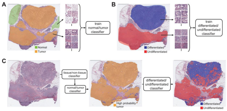
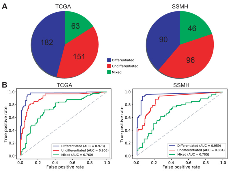
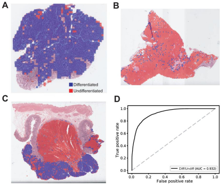
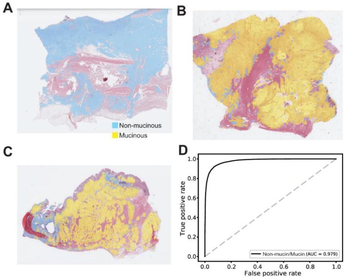
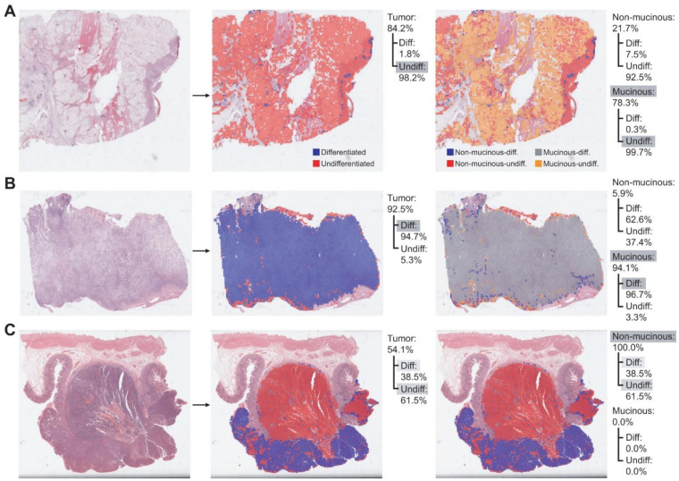
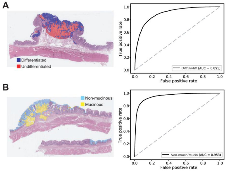
Histomorphologic types of gastric cancer (GC) have significant prognostic values that should be considered during treatment planning. Because the thorough quantitative review of a tissue slide is a laborious task for pathologists, deep learning (DL) can be a useful tool to support pathologic workflow. In the present study, a fully automated approach was applied to distinguish differentiated/undifferentiated and non-mucinous/mucinous tumor types in GC tissue whole-slide images from The Cancer Genome Atlas (TCGA) stomach adenocarcinoma dataset (TCGA-STAD). By classifying small patches of tissue images into differentiated/undifferentiated and non-mucinous/mucinous tumor tissues, the relative proportion of GC tissue subtypes can be easily quantified. Furthermore, the distribution of different tissue subtypes can be clearly visualized. The patch-level areas under the curves for the receiver operating characteristic curves for the differentiated/undifferentiated and non-mucinous/mucinous classifiers were 0.932 and 0.979, respectively. We also validated the classifiers on our own GC datasets and confirmed that the generalizability of the classifiers is excellent. The results indicate that the DL-based tissue classifier could be a useful tool for the quantitative analysis of cancer tissue slides. By combining DL-based classifiers for various molecular and morphologic variations in tissue slides, the heterogeneity of tumor tissues can be unveiled more efficiently.
胃癌(GC)的组织形态学类型具有重要的预后价值,在制定治疗方案时应予以考虑。由于对组织切片进行全面的定量评估对病理学家来说是一项艰巨的任务,深度学习(DL)可以成为支持病理工作流程的有用工具。在本研究中,我们应用了一种全自动方法,从癌症基因组图谱(TCGA)胃腺癌数据集(TCGA-STAD)的GC组织全切片图像中区分分化型/未分化型以及非黏液型/黏液型肿瘤类型。通过将组织图像的小斑块分类为分化型/未分化型和非黏液型/黏液型肿瘤组织,可以轻松量化GC组织亚型的相对比例。此外,不同组织亚型的分布可以清晰地可视化。分化型/未分化型和非黏液型/黏液型分类器的受试者操作特征曲线的曲线下面积分别为0.932和0.979。我们还在自己的GC数据集上对分类器进行了验证,并确认分类器的泛化能力非常出色。结果表明,基于DL的组织分类器可以成为癌症组织切片定量分析的有用工具。通过结合基于DL的分类器对组织切片中的各种分子和形态学变异进行分析,可以更有效地揭示肿瘤组织的异质性。