Department of Anatomy and Neurosciences, Amsterdam UMC, Vrije Universiteit Amsterdam, O|2 building 13W09, De Boelelaan 1108, 1081 HV, Amsterdam, The Netherlands.
Brain Tumor Center Amsterdam, Cancer Center Amsterdam, Amsterdam UMC, Vrije Universiteit Amsterdam, Amsterdam, The Netherlands.
Sci Rep. 2021 Sep 23;11(1):18990. doi: 10.1038/s41598-021-97818-y.
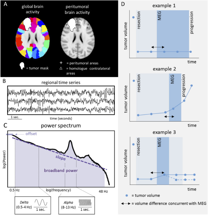
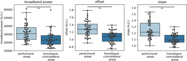
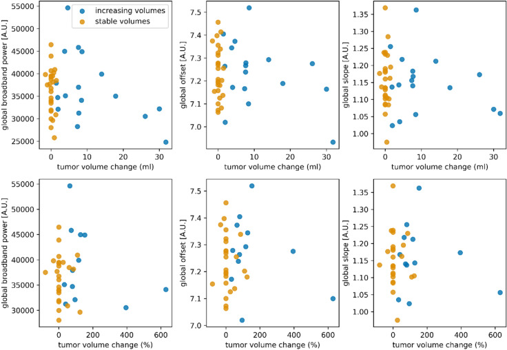
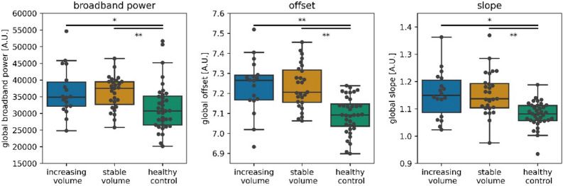
Non-invasively measured brain activity is related to progression-free survival in glioma patients, suggesting its potential as a marker of glioma progression. We therefore assessed the relationship between brain activity and increasing tumor volumes on routine clinical magnetic resonance imaging (MRI) in glioma patients. Postoperative magnetoencephalography (MEG) was recorded in 45 diffuse glioma patients. Brain activity was estimated using three measures (absolute broadband power, offset and slope) calculated at three spatial levels: global average, averaged across the peritumoral areas, and averaged across the homologues of these peritumoral areas in the contralateral hemisphere. Tumors were segmented on MRI. Changes in tumor volume between the two scans surrounding the MEG were calculated and correlated with brain activity. Brain activity was compared between patient groups classified into having increasing or stable tumor volume. Results show that brain activity was significantly increased in the tumor hemisphere in general, and in peritumoral regions specifically. However, none of the measures and spatial levels of brain activity correlated with changes in tumor volume, nor did they differ between patients with increasing versus stable tumor volumes. Longitudinal studies in more homogeneous subgroups of glioma patients are necessary to further explore the clinical potential of non-invasively measured brain activity.
非侵入性测量的大脑活动与胶质瘤患者的无进展生存期相关,表明其可能是胶质瘤进展的标志物。因此,我们评估了胶质瘤患者常规临床磁共振成像(MRI)上肿瘤体积增加与大脑活动之间的关系。对 45 名弥漫性胶质瘤患者进行了术后脑磁图(MEG)记录。使用三种测量方法(绝对宽带功率、偏移和斜率)在三个空间水平上估计大脑活动:全球平均值、肿瘤周围区域的平均值和对侧半球这些肿瘤周围区域的平均值。在 MRI 上对肿瘤进行分割。计算 MEG 前后两次扫描之间肿瘤体积的变化,并与大脑活动相关联。将具有增加或稳定肿瘤体积的患者组进行比较。结果表明,总的来说,肿瘤半球的大脑活动增加,肿瘤周围区域的大脑活动增加更为明显。然而,没有任何措施和大脑活动的空间水平与肿瘤体积的变化相关,也没有在肿瘤体积增加和稳定的患者之间存在差异。在更同质的胶质瘤患者亚组中进行纵向研究,以进一步探索非侵入性测量大脑活动的临床潜力。