Oulefki Adel, Agaian Sos, Trongtirakul Thaweesak, Benbelkacem Samir, Aouam Djamel, Zenati-Henda Nadia, Abdelli Mohamed-Lamine
Centre de Développement des Technologies Avancées (CDTA), PO. Box 17 Baba Hassen, Algiers 16081, Algeria.
Dept. of Computer Science, College of Staten Island, New York, 2800 Victory Blvd Staten Island, New York 10314, USA.
Biomed Signal Process Control. 2022 Mar;73:103371. doi: 10.1016/j.bspc.2021.103371. Epub 2021 Nov 24.
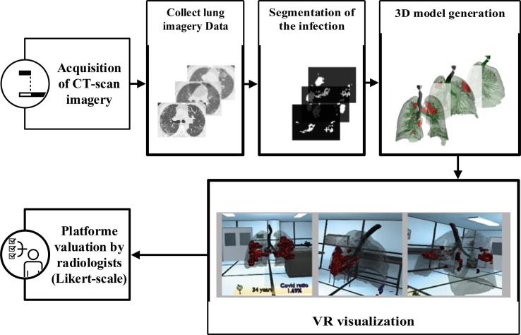
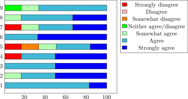
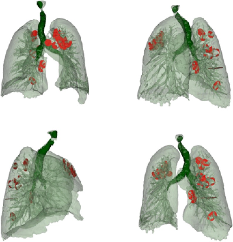
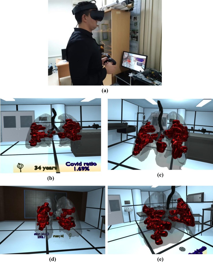
Coronavirus disease (COVID-19) is a severe infectious disease that causes respiratory illness and has had devastating medical and economic consequences globally. Therefore, early, and precise diagnosis is critical to control disease progression and management. Compared to the very popular RT-PCR (reverse-transcription polymerase chain reaction) method, chest CT imaging is a more consistent, sensible, and fast approach for identifying and managing infected COVID-19 patients, specifically in the epidemic area. CT images use computational methods to combine 2D X-ray images and transform them into 3D images. One major drawback of CT scans in diagnosing COVID-19 is creating false-negative effects, especially early infection. This article aims to combine novel CT imaging tools and Virtual Reality (VR) technology and generate an automatize system for accurately screening COVID-19 disease and navigating 3D visualizations of medical scenes. The key benefits of this system are a) it offers stereoscopic depth perception, b) give better insights and comprehension into the overall imaging data, c) it allows doctors to visualize the 3D models, manipulate them, study the inside 3D data, and do several kinds of measurements, and finally d) it has the capacity of real-time interactivity and accurately visualizes dynamic 3D volumetric data. The tool provides novel visualizations for medical practitioners to identify and analyze the change in the shape of COVID-19 infectious. The second objective of this work is to generate, the first time, the CT African patient COVID-19 scan datasets containing 224 patients positive for an infection and 70 regular patients CT-scan images. Computer simulations demonstrate that the proposed method's effectiveness comparing with state-of-the-art baselines methods. The results have also been evaluated with medical professionals. The developed system could be used for medical education professional training and a telehealth VR platform.
冠状病毒病(COVID-19)是一种严重的传染病,可导致呼吸系统疾病,并在全球范围内造成了毁灭性的医学和经济后果。因此,早期准确诊断对于控制疾病进展和管理至关重要。与非常流行的逆转录聚合酶链反应(RT-PCR)方法相比,胸部CT成像对于识别和管理感染COVID-19的患者,特别是在疫区,是一种更一致、更灵敏且快速的方法。CT图像使用计算方法将二维X射线图像组合并转换为三维图像。CT扫描在诊断COVID-19时的一个主要缺点是会产生假阴性结果,尤其是在早期感染时。本文旨在结合新型CT成像工具和虚拟现实(VR)技术,生成一个用于准确筛查COVID-19疾病并浏览医学场景三维可视化的自动化系统。该系统的主要优点包括:a)它提供立体深度感知;b)能更好地洞察和理解整体成像数据;c)允许医生可视化三维模型、进行操作、研究内部三维数据并进行多种测量;最后d)它具有实时交互能力,并能准确可视化动态三维容积数据。该工具为医学从业者提供了新颖的可视化手段,以识别和分析COVID-19感染形状的变化。这项工作的第二个目标是首次生成包含224例感染阳性患者和70例正常患者CT扫描图像的非洲患者COVID-19 CT扫描数据集。计算机模拟证明了所提方法与现有最先进基线方法相比的有效性。结果也经过了医学专业人员的评估。所开发的系统可用于医学教育专业培训和远程医疗VR平台。