• 文献检索
  • 文档翻译
  • 深度研究
  • 学术资讯
  • Suppr Zotero 插件Zotero 插件
  • 邀请有礼
  • 套餐&价格
  • 历史记录
应用&插件
Suppr Zotero 插件Zotero 插件浏览器插件Mac 客户端Windows 客户端微信小程序
定价
高级版会员购买积分包购买API积分包
服务
文献检索文档翻译深度研究API 文档MCP 服务
关于我们
关于 Suppr公司介绍联系我们用户协议隐私条款
关注我们

Suppr 超能文献

核心技术专利:CN118964589B侵权必究
粤ICP备2023148730 号-1Suppr @ 2026

文献检索

告别复杂PubMed语法,用中文像聊天一样搜索,搜遍4000万医学文献。AI智能推荐,让科研检索更轻松。

立即免费搜索

文件翻译

保留排版,准确专业,支持PDF/Word/PPT等文件格式,支持 12+语言互译。

免费翻译文档

深度研究

AI帮你快速写综述,25分钟生成高质量综述,智能提取关键信息,辅助科研写作。

立即免费体验

细胞内结构的红外纳米成像和层析成像。

Infrared nanoscopy and tomography of intracellular structures.

机构信息

Freie Universität Berlin, Department of Physics, Experimental Molecular Biophysics, Arnimallee 14, 14195, Berlin, Germany.

Freie Universität Berlin, Department of Physics, Experimental Biophysics and Space Sciences, Arnimallee 14, 14195, Berlin, Germany.

出版信息

Commun Biol. 2021 Nov 30;4(1):1341. doi: 10.1038/s42003-021-02876-7.

DOI:10.1038/s42003-021-02876-7
PMID:34848821
原文链接:https://pmc.ncbi.nlm.nih.gov/articles/PMC8633277/
Abstract

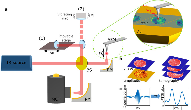
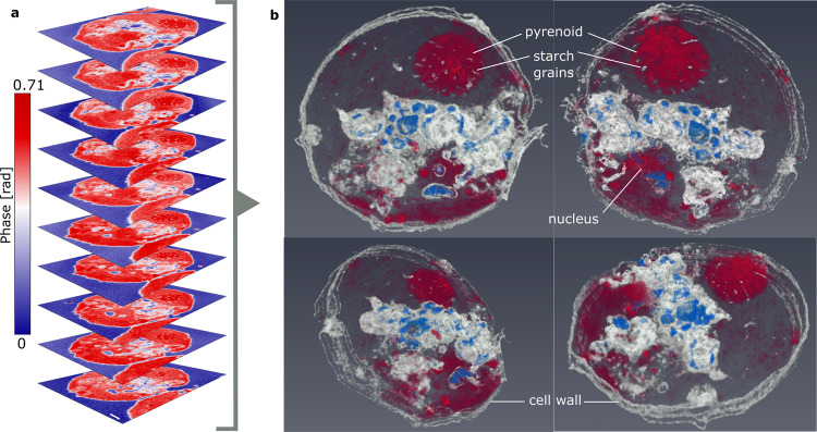
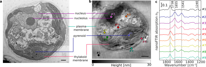
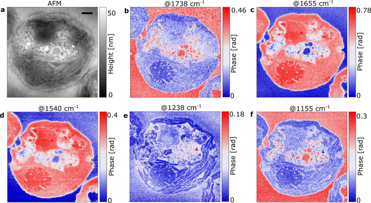
Although techniques such as fluorescence-based super-resolution imaging or confocal microscopy simultaneously gather both morphological and chemical data, these techniques often rely on the use of localized and chemically specific markers. To eliminate this flaw, we have developed a method of examining cellular cross sections using the imaging power of scattering-type scanning near-field optical microscopy and Fourier-transform infrared spectroscopy at a spatial resolution far beyond the diffraction limit. Herewith, nanoscale surface and volumetric chemical imaging is performed using the intrinsic contrast generated by the characteristic absorption of mid-infrared radiation by the covalent bonds. We employ infrared nanoscopy to study the subcellular structures of eukaryotic (Chlamydomonas reinhardtii) and prokaryotic (Escherichia coli) species, revealing chemically distinct regions within each cell such as the microtubular structure of the flagellum. Serial 100 nm-thick cellular cross-sections were compiled into a tomogram yielding a three-dimensional infrared image of subcellular structure distribution at 20 nm resolution. The presented methodology is able to image biological samples complementing current fluorescence nanoscopy but at less interference due to the low energy of infrared radiation and the absence of labeling.

摘要

尽管荧光超分辨率成像或共聚焦显微镜等技术可以同时收集形态和化学数据,但这些技术通常依赖于使用局部和化学特异性标记物。为了消除这一缺陷,我们开发了一种使用散射型近场光学显微镜和傅里叶变换红外光谱的成像能力来检查细胞切片的方法,其空间分辨率远远超过衍射极限。在此,使用共价键对中红外辐射的特征吸收产生的固有对比度来进行纳米级表面和体积化学成像。我们采用红外纳米显微镜研究真核(莱茵衣藻)和原核(大肠杆菌)生物的亚细胞结构,揭示每个细胞内化学性质不同的区域,如鞭毛的微管结构。100nm 厚的细胞连续切片被汇编成一个体素,生成亚细胞结构分布的三维红外图像,分辨率为 20nm。所提出的方法能够对生物样本进行成像,补充当前的荧光纳米显微镜,但由于红外辐射能量低且无需标记,因此干扰较小。

https://cdn.ncbi.nlm.nih.gov/pmc/blobs/9cb0/8633277/4933c6574b27/42003_2021_2876_Fig5_HTML.jpg
https://cdn.ncbi.nlm.nih.gov/pmc/blobs/9cb0/8633277/9b0d8835100c/42003_2021_2876_Fig1_HTML.jpg
https://cdn.ncbi.nlm.nih.gov/pmc/blobs/9cb0/8633277/feef7112dc2a/42003_2021_2876_Fig2_HTML.jpg
https://cdn.ncbi.nlm.nih.gov/pmc/blobs/9cb0/8633277/14289d6ec8d6/42003_2021_2876_Fig3_HTML.jpg
https://cdn.ncbi.nlm.nih.gov/pmc/blobs/9cb0/8633277/755aa1123956/42003_2021_2876_Fig4_HTML.jpg
https://cdn.ncbi.nlm.nih.gov/pmc/blobs/9cb0/8633277/4933c6574b27/42003_2021_2876_Fig5_HTML.jpg

相似文献

1
Infrared nanoscopy and tomography of intracellular structures.细胞内结构的红外纳米成像和层析成像。
Commun Biol. 2021 Nov 30;4(1):1341. doi: 10.1038/s42003-021-02876-7.
2
Super-resolution mid-infrared spectro-microscopy of biological applications through tapping mode and peak force tapping mode atomic force microscope.通过敲击模式和峰值力敲击模式原子力显微镜对生物应用的超分辨率中红外光谱显微镜。
Adv Drug Deliv Rev. 2022 Jan;180:114080. doi: 10.1016/j.addr.2021.114080. Epub 2021 Dec 11.
3
Infrared-spectroscopic, dynamic near-field microscopy of living cells and nanoparticles in water.水中活细胞和纳米粒子的红外光谱、动态近场显微镜。
Sci Rep. 2021 Nov 8;11(1):21860. doi: 10.1038/s41598-021-01425-w.
4
Nanoscale infrared absorption spectroscopy of individual nanoparticles enabled by scattering-type near-field microscopy.利用散射型近场显微镜实现单个纳米颗粒的纳米级红外吸收光谱学。
ACS Nano. 2011 Aug 23;5(8):6494-9. doi: 10.1021/nn2017638. Epub 2011 Jul 27.
5
Atomic force microscope infrared spectroscopy of griseofulvin nanocrystals.灰黄霉素纳米晶体的原子力显微镜红外光谱。
Anal Chem. 2013 Dec 3;85(23):11449-55. doi: 10.1021/ac4025889. Epub 2013 Nov 12.
6
Fluorescently Guided Optical Photothermal Infrared Microspectroscopy for Protein-Specific Bioimaging at Subcellular Level.荧光引导光热红外显微光谱法在亚细胞水平的蛋白质特异性生物成像。
J Med Chem. 2023 Feb 23;66(4):2542-2549. doi: 10.1021/acs.jmedchem.2c01359. Epub 2023 Jan 4.
7
Nanoscale Optical Microscopy and Spectroscopy Using Near-Field Probes.使用近场探针的纳米级光学显微镜和光谱学。
Annu Rev Chem Biomol Eng. 2018 Jun 7;9:365-387. doi: 10.1146/annurev-chembioeng-060817-084150. Epub 2018 Mar 29.
8
Sub-100 nm IR spectromicroscopy of living cells.活细胞的亚100纳米红外光谱显微镜技术
Opt Lett. 2008 Jul 15;33(14):1611-3. doi: 10.1364/ol.33.001611.
9
High-resolution Fourier-transform infrared chemical imaging with multiple synchrotron beams.高分辨率傅里叶变换红外化学成像与多同步辐射光束。
Nat Methods. 2011 May;8(5):413-6. doi: 10.1038/nmeth.1585. Epub 2011 Mar 20.
10
Chemically resolved imaging of biological cells and thin films by infrared scanning near-field optical microscopy.通过红外扫描近场光学显微镜对生物细胞和薄膜进行化学分辨成像。
Biophys J. 2003 Oct;85(4):2705-10. doi: 10.1016/S0006-3495(03)74693-1.

引用本文的文献

1
Nanoscale Mechanical Manipulation of Ultrathin SiN Membranes Enabling Infrared Near-Field Microscopy of Liquid-Immersed samples.超薄氮化硅膜的纳米级机械操纵实现液浸样品的红外近场显微镜观察
Small. 2024 Nov;20(47):e2402568. doi: 10.1002/smll.202402568. Epub 2024 Aug 15.
2
Nanosecond Transient IR Spectroscopy of Halorhodopsin in Living Cells.活细胞中嗜盐视紫红质的纳秒瞬态红外光谱
J Am Chem Soc. 2024 Jul 17;146(28):19118-19127. doi: 10.1021/jacs.4c03891. Epub 2024 Jul 1.
3
Infrared nanoimaging of neuronal ultrastructure and nanoparticle interaction with cells.

本文引用的文献

1
Spectroscopic investigations under whole-cell conditions provide new insight into the metal hydride chemistry of [FeFe]-hydrogenase.全细胞条件下的光谱研究为[FeFe]-氢化酶的金属氢化物化学提供了新的见解。
Chem Sci. 2020 Apr 14;11(18):4608-4617. doi: 10.1039/d0sc00512f.
2
Atomic-resolution protein structure determination by cryo-EM.利用冷冻电镜技术进行原子分辨率的蛋白质结构测定。
Nature. 2020 Nov;587(7832):157-161. doi: 10.1038/s41586-020-2833-4. Epub 2020 Oct 21.
3
Infrared Scattering-Type Scanning Near-Field Optical Microscopy of Biomembranes in Water.
神经元超微结构的红外纳米成像及纳米颗粒与细胞的相互作用。
Nanoscale. 2024 Mar 21;16(12):6190-6198. doi: 10.1039/d3nr04948e.
4
Molecular design and synthesis of methoxy-substitued spiropyrans with photomodulated NIR-fluorescence.甲氧取代螺吡喃的光调控近红外荧光分子设计与合成。
Photochem Photobiol Sci. 2023 Nov;22(11):2651-2673. doi: 10.1007/s43630-023-00479-1. Epub 2023 Sep 21.
5
Visualization of Nanocarriers and Drugs in Cells and Tissue.纳米载体和药物在细胞和组织中的可视化。
Handb Exp Pharmacol. 2024;284:153-189. doi: 10.1007/164_2023_684.
6
Label-free nanoscale mapping of intracellular organelle chemistry.无标记纳米尺度细胞内细胞器化学物质成像
Commun Biol. 2023 May 31;6(1):583. doi: 10.1038/s42003-023-04943-7.
7
Toward the Next Frontiers of Vibrational Bioimaging.迈向振动生物成像的新前沿。
Chem Biomed Imaging. 2023 Mar 28;1(1):3-17. doi: 10.1021/cbmi.3c00004. eCollection 2023 Apr 24.
8
Scattering-type Scanning Near-Field Optical Microscopy of Polymer-Coated Gold Nanoparticles.聚合物包覆金纳米粒子的散射型扫描近场光学显微镜
ACS Omega. 2022 Mar 24;7(13):11353-11362. doi: 10.1021/acsomega.2c00410. eCollection 2022 Apr 5.
水生物膜的红外散射型扫描近场光学显微镜观察。
J Phys Chem Lett. 2020 Oct 1;11(19):8183-8188. doi: 10.1021/acs.jpclett.0c01769. Epub 2020 Sep 15.
4
FTIR Nanospectroscopy Shows Molecular Structures of Plant Biominerals and Cell Walls.傅里叶变换红外纳米光谱技术揭示植物生物矿质和细胞壁的分子结构。
Anal Chem. 2020 Oct 20;92(20):13694-13701. doi: 10.1021/acs.analchem.0c00271. Epub 2020 Sep 28.
5
Subsurface chemical nanoidentification by nano-FTIR spectroscopy.通过纳米傅里叶变换红外光谱法进行地下化学纳米识别。
Nat Commun. 2020 Jul 3;11(1):3359. doi: 10.1038/s41467-020-17034-6.
6
Infrared and Raman chemical imaging and spectroscopy at the nanoscale.纳米尺度的红外和拉曼化学成像与光谱学。
Chem Soc Rev. 2020 Jun 7;49(11):3315-3347. doi: 10.1039/c8cs00916c. Epub 2020 May 19.
7
In Liquid Infrared Scattering Scanning Near-Field Optical Microscopy for Chemical and Biological Nanoimaging.在液体红外散射扫描近场光学显微镜中进行化学和生物纳米成像。
Nano Lett. 2020 Jun 10;20(6):4497-4504. doi: 10.1021/acs.nanolett.0c01291. Epub 2020 May 7.
8
Visibility of subsurface nanostructures in scattering-type scanning near-field optical microscopy imaging.散射型扫描近场光学显微镜成像中地下纳米结构的可见性
Opt Express. 2020 Mar 2;28(5):6696-6707. doi: 10.1364/OE.386713.
9
Deep learning a boon for biophotonics?深度学习对生物光子学是福音吗?
J Biophotonics. 2020 Jun;13(6):e201960186. doi: 10.1002/jbio.201960186. Epub 2020 Mar 30.
10
A Series of Fortunate Events: Introducing Chlamydomonas as a Reference Organism.幸运事件系列:介绍衣藻作为参考生物。
Plant Cell. 2019 Aug;31(8):1682-1707. doi: 10.1105/tpc.18.00952. Epub 2019 Jun 12.