• 文献检索
  • 文档翻译
  • 深度研究
  • 学术资讯
  • Suppr Zotero 插件Zotero 插件
  • 邀请有礼
  • 套餐&价格
  • 历史记录
应用&插件
Suppr Zotero 插件Zotero 插件浏览器插件Mac 客户端Windows 客户端微信小程序
定价
高级版会员购买积分包购买API积分包
服务
文献检索文档翻译深度研究API 文档MCP 服务
关于我们
关于 Suppr公司介绍联系我们用户协议隐私条款
关注我们

Suppr 超能文献

核心技术专利:CN118964589B侵权必究
粤ICP备2023148730 号-1Suppr @ 2026

文献检索

告别复杂PubMed语法,用中文像聊天一样搜索,搜遍4000万医学文献。AI智能推荐,让科研检索更轻松。

立即免费搜索

文件翻译

保留排版,准确专业,支持PDF/Word/PPT等文件格式,支持 12+语言互译。

免费翻译文档

深度研究

AI帮你快速写综述,25分钟生成高质量综述,智能提取关键信息,辅助科研写作。

立即免费体验

基于机器学习的骨肉瘤动态对比增强磁共振成像影像组学列线图用于评估新辅助化疗疗效

Machine Learning-Based Radiomics Nomogram With Dynamic Contrast-Enhanced MRI of the Osteosarcoma for Evaluation of Efficacy of Neoadjuvant Chemotherapy.

作者信息

Zhang Lu, Ge Yinghui, Gao Qiuru, Zhao Fei, Cheng Tianming, Li Hailiang, Xia Yuwei

机构信息

Department of Medical Imaging, People's Hospital of Zhengzhou University Henan Provincial People's Hospital, Zhengzhou, China.

Department of Orthopedics, People's Hospital of Zhengzhou University Henan Provincial People's Hospital, Zhengzhou, China.

出版信息

Front Oncol. 2021 Nov 15;11:758921. doi: 10.3389/fonc.2021.758921. eCollection 2021.

DOI:10.3389/fonc.2021.758921
PMID:34868973
原文链接:https://pmc.ncbi.nlm.nih.gov/articles/PMC8634262/
Abstract

OBJECTIVES

This study aims to evaluate the value of machine learning-based dynamic contrast-enhanced MRI (DCE-MRI) radiomics nomogram in prediction treatment response of neoadjuvant chemotherapy (NAC) in patients with osteosarcoma.

METHODS

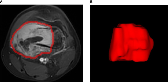
A total of 102 patients with osteosarcoma and who underwent NAC were enrolled in this study. All patients received a DCE-MRI scan before NAC. The Response Evaluation Criteria in Solid Tumors was used as the standard to evaluate the NAC response with complete remission and partial remission in the effective group, stable disease, and progressive disease in the ineffective group. The following semi-quantitative parameters of DCE-MRI were calculated: early dynamic enhancement wash-in slope (Slope), time to peak (TTP), and enhancement rate (R). The acquired data is randomly divided into 70% for training and 30% for testing. Variance threshold, univariate feature selection, and least absolute shrinkage and selection operator were used to select the optimal features. Three classifiers (K-nearest neighbor, KNN; support vector machine, SVM; and logistic regression, LR) were implemented for model establishment. The performance of different classifiers and conventional semi-quantitative parameters was evaluated by confusion matrix and receiver operating characteristic curves. Furthermore, clinically relevant risk factors including age, tumor size and site, pathological fracture, and surgical staging were collected to evaluate their predictive values for the efficacy of NAC. The selected clinical features and imaging features were combined to establish the model and the nomogram, and then the predictive efficacy was evaluated.

RESULTS

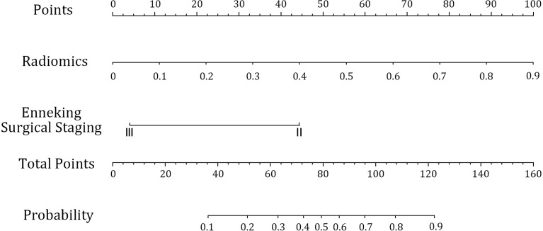
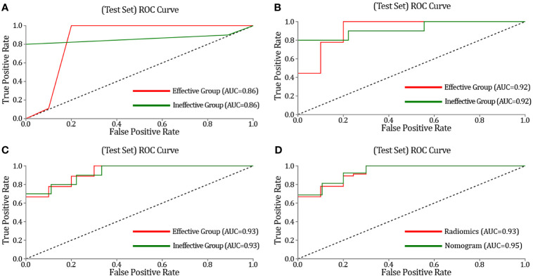
The clinical relevance risk factor analysis demonstrates that only surgical stage was an independent predictor of NAC. A total of seven radiomic features were selected, and three machine learning models (KNN, SVM, and LR) were established based on such features. The prediction accuracy (ACC) of these three models was 0.89, 0.84, and 0.84, respectively. The area under the subject curve (AUC) of these three models was 0.86, 0.92, and 0.93, respectively. As for Slope, TTP, and R parameters, the prediction ACC was 0.91, 0.89, and 0.81, respectively, while the AUC was 0.87, 0.85, and 0.83, respectively. In both the training and testing sets, the ACC and AUC of the combined model were higher than those of the radiomics models (ACC = 0.91 and AUC = 0.95), which indicate an outstanding performance of our proposed model.

CONCLUSIONS

The radiomics nomogram demonstrates satisfactory predictive results for the treatment response of patients with osteosarcoma before NAC. This finding may provide a new decision basis to improve the treatment plan.

摘要

目的

本研究旨在评估基于机器学习的动态对比增强磁共振成像(DCE-MRI)影像组学列线图在预测骨肉瘤患者新辅助化疗(NAC)治疗反应中的价值。

方法

本研究共纳入102例接受NAC的骨肉瘤患者。所有患者在NAC前均接受了DCE-MRI扫描。实体瘤疗效评价标准被用作评估NAC反应的标准,有效组为完全缓解和部分缓解,无效组为疾病稳定和疾病进展。计算DCE-MRI的以下半定量参数:早期动态增强流入斜率(Slope)、达峰时间(TTP)和增强率(R)。将获取的数据随机分为70%用于训练,30%用于测试。采用方差阈值、单变量特征选择和最小绝对收缩和选择算子来选择最佳特征。实施三种分类器(K近邻,KNN;支持向量机,SVM;逻辑回归,LR)进行模型建立。通过混淆矩阵和受试者工作特征曲线评估不同分类器和传统半定量参数的性能。此外,收集包括年龄、肿瘤大小和部位、病理性骨折和手术分期等临床相关危险因素,以评估它们对NAC疗效的预测价值。将选定的临床特征和影像特征相结合建立模型和列线图,然后评估预测疗效。

结果

临床相关性危险因素分析表明,只有手术分期是NAC的独立预测因素。共选择了七个影像组学特征,并基于这些特征建立了三种机器学习模型(KNN、SVM和LR)。这三种模型的预测准确率(ACC)分别为0.89、0.84和0.84。这三种模型的受试者曲线下面积(AUC)分别为0.86、0.92和0.93。至于Slope、TTP和R参数,预测ACC分别为0.91、0.89和0.81,而AUC分别为0.87、0.85和0.83。在训练集和测试集中,联合模型的ACC和AUC均高于影像组学模型(ACC = 0.91,AUC = 0.95),这表明我们提出的模型具有出色的性能。

结论

影像组学列线图对骨肉瘤患者NAC前的治疗反应显示出令人满意的预测结果。这一发现可能为改进治疗方案提供新的决策依据。

https://cdn.ncbi.nlm.nih.gov/pmc/blobs/6d3d/8634262/e024792466da/fonc-11-758921-g007.jpg
https://cdn.ncbi.nlm.nih.gov/pmc/blobs/6d3d/8634262/8173a1b94701/fonc-11-758921-g001.jpg
https://cdn.ncbi.nlm.nih.gov/pmc/blobs/6d3d/8634262/b74d5d75f9c7/fonc-11-758921-g002.jpg
https://cdn.ncbi.nlm.nih.gov/pmc/blobs/6d3d/8634262/0811dda27917/fonc-11-758921-g003.jpg
https://cdn.ncbi.nlm.nih.gov/pmc/blobs/6d3d/8634262/688c77ac345a/fonc-11-758921-g004.jpg
https://cdn.ncbi.nlm.nih.gov/pmc/blobs/6d3d/8634262/4b284ed15893/fonc-11-758921-g005.jpg
https://cdn.ncbi.nlm.nih.gov/pmc/blobs/6d3d/8634262/13a644d905a5/fonc-11-758921-g006.jpg
https://cdn.ncbi.nlm.nih.gov/pmc/blobs/6d3d/8634262/e024792466da/fonc-11-758921-g007.jpg

相似文献

1
Machine Learning-Based Radiomics Nomogram With Dynamic Contrast-Enhanced MRI of the Osteosarcoma for Evaluation of Efficacy of Neoadjuvant Chemotherapy.基于机器学习的骨肉瘤动态对比增强磁共振成像影像组学列线图用于评估新辅助化疗疗效
Front Oncol. 2021 Nov 15;11:758921. doi: 10.3389/fonc.2021.758921. eCollection 2021.
2
Evaluation of the neoadjuvant chemotherapy response in osteosarcoma using the MRI DWI-based machine learning radiomics nomogram.使用基于MRI扩散加权成像的机器学习影像组学列线图评估骨肉瘤新辅助化疗反应
Front Oncol. 2024 Mar 21;14:1345576. doi: 10.3389/fonc.2024.1345576. eCollection 2024.
3
MRI-based radiomics signature for pretreatment prediction of pathological response to neoadjuvant chemotherapy in osteosarcoma: a multicenter study.基于 MRI 的放射组学特征预测骨肉瘤新辅助化疗的病理反应:一项多中心研究。
Eur Radiol. 2021 Oct;31(10):7913-7924. doi: 10.1007/s00330-021-07748-6. Epub 2021 Mar 30.
4
Evaluation of Multiparametric MRI Radiomics-Based Nomogram in Prediction of Response to Neoadjuvant Chemotherapy in Breast Cancer: A Two-Center study.基于多参数 MRI 放射组学的列线图预测乳腺癌新辅助化疗反应的评价:一项多中心研究。
Clin Breast Cancer. 2023 Aug;23(6):e331-e344. doi: 10.1016/j.clbc.2023.05.010. Epub 2023 May 27.
5
Machine learning model based on enhanced CT radiomics for the preoperative prediction of lymphovascular invasion in esophageal squamous cell carcinoma.基于增强CT影像组学的机器学习模型用于术前预测食管鳞状细胞癌的淋巴管侵犯
Front Oncol. 2024 Feb 23;14:1308317. doi: 10.3389/fonc.2024.1308317. eCollection 2024.
6
[Preoperative prediction of HER-2 expression status in breast cancer based on MRI radiomics model].基于MRI影像组学模型的乳腺癌HER-2表达状态术前预测
Zhonghua Zhong Liu Za Zhi. 2024 May 23;46(5):428-437. doi: 10.3760/cma.j.cn112152-20230816-00086.
7
Automated prediction of the neoadjuvant chemotherapy response in osteosarcoma with deep learning and an MRI-based radiomics nomogram.深度学习和基于 MRI 的放射组学列线图预测骨肉瘤新辅助化疗反应。
Eur Radiol. 2022 Sep;32(9):6196-6206. doi: 10.1007/s00330-022-08735-1. Epub 2022 Apr 2.
8
Multi-modal radiomics model to predict treatment response to neoadjuvant chemotherapy for locally advanced rectal cancer.多模态放射组学模型预测局部晚期直肠癌新辅助化疗的治疗反应。
World J Gastroenterol. 2020 May 21;26(19):2388-2402. doi: 10.3748/wjg.v26.i19.2388.
9
Delta Radiomics Based on MRI for Predicting Axillary Lymph Node Pathologic Complete Response After Neoadjuvant Chemotherapy in Breast Cancer Patients.基于MRI的Delta放射组学预测乳腺癌患者新辅助化疗后腋窝淋巴结病理完全缓解
Acad Radiol. 2025 Jan;32(1):37-49. doi: 10.1016/j.acra.2024.07.052. Epub 2024 Sep 13.
10
Deep learning radiomic analysis of DCE-MRI combined with clinical characteristics predicts pathological complete response to neoadjuvant chemotherapy in breast cancer.DCE-MRI的深度学习影像组学分析结合临床特征可预测乳腺癌新辅助化疗的病理完全缓解。
Front Oncol. 2023 Jan 5;12:1041142. doi: 10.3389/fonc.2022.1041142. eCollection 2022.

引用本文的文献

1
Integrating radiomics, artificial intelligence, and molecular signatures in bone and soft tissue tumors: advances in diagnosis and prognostication.整合放射组学、人工智能和分子特征于骨与软组织肿瘤:诊断与预后的进展
Front Oncol. 2025 Aug 18;15:1613133. doi: 10.3389/fonc.2025.1613133. eCollection 2025.
2
Artificial Intelligence in Primary Malignant Bone Tumor Imaging: A Narrative Review.原发性恶性骨肿瘤成像中的人工智能:一项叙述性综述。
Diagnostics (Basel). 2025 Jul 4;15(13):1714. doi: 10.3390/diagnostics15131714.
3
UK guidelines for the management of bone sarcomas.

本文引用的文献

1
Radiological Assessment and Outcome of Local Disease Progression after Neoadjuvant Chemotherapy in Children and Adolescents with Localized Osteosarcoma.局限性骨肉瘤患儿及青少年新辅助化疗后局部疾病进展的放射学评估及预后
J Clin Med. 2020 Dec 17;9(12):4070. doi: 10.3390/jcm9124070.
2
Cardiac SPECT radiomic features repeatability and reproducibility: A multi-scanner phantom study.心脏 SPECT 影像组学特征的可重复性:一项多扫描仪 phantom 研究。
J Nucl Cardiol. 2021 Dec;28(6):2730-2744. doi: 10.1007/s12350-020-02109-0. Epub 2020 Apr 24.
3
Radiomics Analysis of Dynamic Contrast-Enhanced Magnetic Resonance Imaging for the Prediction of Sentinel Lymph Node Metastasis in Breast Cancer.
英国骨肉瘤管理指南。
Br J Cancer. 2025 Jan;132(1):32-48. doi: 10.1038/s41416-024-02868-4. Epub 2024 Nov 16.
4
Contrast-enhanced CT radiomics combined with multiple machine learning algorithms for preoperative identification of lymph node metastasis in pancreatic ductal adenocarcinoma.对比增强CT影像组学联合多种机器学习算法用于术前鉴别胰腺导管腺癌淋巴结转移
Front Oncol. 2024 Sep 13;14:1342317. doi: 10.3389/fonc.2024.1342317. eCollection 2024.
5
Preoperative prediction of high-grade osteosarcoma response to neoadjuvant therapy based on a plain CT radiomics model: A dual-center study.基于平扫CT影像组学模型的高级别骨肉瘤对新辅助治疗反应的术前预测:一项双中心研究
J Bone Oncol. 2024 Jun 8;47:100614. doi: 10.1016/j.jbo.2024.100614. eCollection 2024 Aug.
6
Relative Wash-In Rate in Dynamic Contrast-Enhanced Magnetic Resonance Imaging as a New Prognostic Biomarker for Event-Free Survival in 82 Patients with Osteosarcoma: A Multicenter Study.动态对比增强磁共振成像中的相对洗入率作为82例骨肉瘤患者无事件生存期的新预后生物标志物:一项多中心研究
Cancers (Basel). 2024 May 21;16(11):1954. doi: 10.3390/cancers16111954.
7
CT and MRI radiomics of bone and soft-tissue sarcomas: an updated systematic review of reproducibility and validation strategies.骨与软组织肉瘤的CT和MRI影像组学:关于可重复性和验证策略的最新系统综述
Insights Imaging. 2024 Feb 27;15(1):54. doi: 10.1186/s13244-024-01614-x.
8
Deep Learning Approaches to Osteosarcoma Diagnosis and Classification: A Comparative Methodological Approach.骨肉瘤诊断与分类的深度学习方法:一种比较性的方法学途径
Cancers (Basel). 2023 Apr 13;15(8):2290. doi: 10.3390/cancers15082290.
9
Cancer-Specific Survival after Limb Salvage versus Amputation in Children and Adolescents with Osteosarcoma: A Population-Based Analysis with Propensity Score Matching.骨肉瘤患儿及青少年保肢与截肢术后的癌症特异性生存率:一项基于倾向评分匹配的人群分析
J Oncol. 2023 Apr 8;2023:8635829. doi: 10.1155/2023/8635829. eCollection 2023.
10
3D vs. 2D MRI radiomics in skeletal Ewing sarcoma: Feature reproducibility and preliminary machine learning analysis on neoadjuvant chemotherapy response prediction.骨骼尤文肉瘤中3D与2D MRI影像组学:特征可重复性及新辅助化疗反应预测的初步机器学习分析
Front Oncol. 2022 Dec 2;12:1016123. doi: 10.3389/fonc.2022.1016123. eCollection 2022.
动态对比增强磁共振成像的影像组学分析用于预测乳腺癌前哨淋巴结转移
Front Oncol. 2019 Sep 30;9:980. doi: 10.3389/fonc.2019.00980. eCollection 2019.
4
Clinical evaluation of neoadjuvant chemotherapy for osteosarcoma.骨肉瘤新辅助化疗的临床评估
J BUON. 2019 May-Jun;24(3):1181-1185.
5
Gray-level discretization impacts reproducible MRI radiomics texture features.灰度离散化影响可重现的 MRI 放射组学纹理特征。
PLoS One. 2019 Mar 7;14(3):e0213459. doi: 10.1371/journal.pone.0213459. eCollection 2019.
6
T -based MRI Delta-radiomics improve response prediction in soft-tissue sarcomas treated by neoadjuvant chemotherapy.基于 T1 加权 MRI 的 Delta 放射组学可改善新辅助化疗治疗的软组织肉瘤的反应预测。
J Magn Reson Imaging. 2019 Aug;50(2):497-510. doi: 10.1002/jmri.26589. Epub 2018 Dec 19.
7
Bone sarcomas: ESMO-PaedCan-EURACAN Clinical Practice Guidelines for diagnosis, treatment and follow-up.骨肉瘤:ESMO-儿童癌症-欧洲骨肉瘤协作组诊断、治疗及随访临床实践指南
Ann Oncol. 2018 Oct 1;29(Suppl 4):iv79-iv95. doi: 10.1093/annonc/mdy310.
8
Differentiation between vasogenic edema and infiltrative tumor in patients with high-grade gliomas using texture patch-based analysis.基于纹理块分析鉴别高级别胶质瘤患者血管源性水肿与浸润性肿瘤
J Magn Reson Imaging. 2018 Jan 3. doi: 10.1002/jmri.25939.
9
Radiomics and radiogenomics in lung cancer: A review for the clinician.肺癌的影像组学和放射组学:临床医生的综述。
Lung Cancer. 2018 Jan;115:34-41. doi: 10.1016/j.lungcan.2017.10.015. Epub 2017 Nov 8.
10
Current and future therapeutic approaches for osteosarcoma.骨肉瘤的当前及未来治疗方法
Expert Rev Anticancer Ther. 2018 Jan;18(1):39-50. doi: 10.1080/14737140.2018.1413939. Epub 2017 Dec 14.