Brawner D L, Cutler J E
Infect Immun. 1986 Jan;51(1):327-36. doi: 10.1128/iai.51.1.327-336.1986.
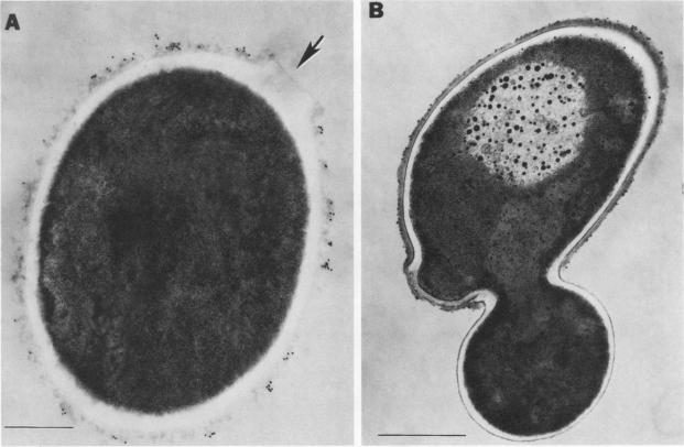
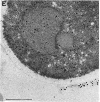
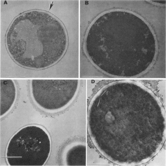
Variability in the expression of two different cell surface carbohydrate determinants was examined with two agglutinating immunoglobulin M monoclonal antibodies (H9 and C6) and immunoelectron microscopy during growth of three strains of Candida albicans. A single strain of Candida parapsilosis did not express either antigen at any time during growth. Antigens were detected on the surface of C. albicans by agglutination tests with either H9 or C6 over a 48-h growth period. The difference in specificities of the monoclonal antibodies was demonstrated by Ouchterlony double-diffusion tests with solubilized antigens and by variabilities in the reactivity of the agglutinins among yeast strains. The antigenic determinants were isolated by specific immunoprecipitation and protease digestion and characterized by methods including high-pressure liquid chromatography, gas-liquid chromatography, and mass spectroscopy with both chemical and electron ionization. These determinants both contain mannose and glucose. In the case of antigen H9, an additional carbohydrate was detected with gas chromatography and mass spectroscopy. The location of antigens on individual cells was determined by indirect labeling of the determinants, first reacting cells with H9 or C6 followed by goat anti-mouse antibody conjugated with 20-nm colloidal gold particles. Transmission electron microscopy was used to examine cells. The antigens that were reactive with the monoclonal antibodies were associated with a flocculent surface layer. Expression of this layer and expression of the antigens is a dynamic process which is growth phase and strain dependent. The antigens were not expressed on very young cells and disappeared from the cell surface of most C. albicans strains with age. The use of monoclonal antibody to cell surface determinants may allow characterization of cell surface antigens of C. albicans and be helpful in establishing receptors which mediate adherence.
在三株白色念珠菌生长过程中,使用两种凝集免疫球蛋白M单克隆抗体(H9和C6)以及免疫电子显微镜检查了两种不同细胞表面碳水化合物决定簇的表达情况。近平滑念珠菌的一个菌株在生长过程中的任何时候都不表达这两种抗原。在48小时的生长期间,通过用H9或C6进行凝集试验,在白色念珠菌表面检测到了抗原。通过用溶解抗原进行的Ouchterlony双扩散试验以及酵母菌株间凝集素反应性的变化,证明了单克隆抗体特异性的差异。通过特异性免疫沉淀和蛋白酶消化分离抗原决定簇,并通过包括高压液相色谱、气液色谱以及化学电离和电子电离质谱在内的方法进行表征。这些决定簇都含有甘露糖和葡萄糖。就抗原H9而言,通过气相色谱和质谱检测到了一种额外的碳水化合物。通过对决定簇进行间接标记来确定单个细胞上抗原的位置,首先使细胞与H9或C6反应,然后与结合了20纳米胶体金颗粒的山羊抗小鼠抗体反应。使用透射电子显微镜检查细胞。与单克隆抗体反应的抗原与絮状表面层相关。该层的表达和抗原的表达是一个动态过程,依赖于生长阶段和菌株。抗原在非常年轻的细胞上不表达,并且随着年龄增长在大多数白色念珠菌菌株细胞表面消失。使用针对细胞表面决定簇的单克隆抗体可能有助于表征白色念珠菌的细胞表面抗原,并有助于建立介导黏附的受体。