• 文献检索
  • 文档翻译
  • 深度研究
  • 学术资讯
  • Suppr Zotero 插件Zotero 插件
  • 邀请有礼
  • 套餐&价格
  • 历史记录
应用&插件
Suppr Zotero 插件Zotero 插件浏览器插件Mac 客户端Windows 客户端微信小程序
定价
高级版会员购买积分包购买API积分包
服务
文献检索文档翻译深度研究API 文档MCP 服务
关于我们
关于 Suppr公司介绍联系我们用户协议隐私条款
关注我们

Suppr 超能文献

核心技术专利:CN118964589B侵权必究
粤ICP备2023148730 号-1Suppr @ 2026

文献检索

告别复杂PubMed语法,用中文像聊天一样搜索,搜遍4000万医学文献。AI智能推荐,让科研检索更轻松。

立即免费搜索

文件翻译

保留排版,准确专业,支持PDF/Word/PPT等文件格式,支持 12+语言互译。

免费翻译文档

深度研究

AI帮你快速写综述,25分钟生成高质量综述,智能提取关键信息,辅助科研写作。

立即免费体验

一个可扩展的信息学框架,用于通过数字病理标本、计算机成像工具和先进挖掘能力增强中央癌症登记系统。

An Expandable Informatics Framework for Enhancing Central Cancer Registries with Digital Pathology Specimens, Computational Imaging Tools, and Advanced Mining Capabilities.

作者信息

Foran David J, Durbin Eric B, Chen Wenjin, Sadimin Evita, Sharma Ashish, Banerjee Imon, Kurc Tahsin, Li Nan, Stroup Antoinette M, Harris Gerald, Gu Annie, Schymura Maria, Gupta Rajarsi, Bremer Erich, Balsamo Joseph, DiPrima Tammy, Wang Feiqiao, Abousamra Shahira, Samaras Dimitris, Hands Isaac, Ward Kevin, Saltz Joel H

机构信息

Center for Biomedical Informatics, Rutgers Cancer Institute of New Jersey, New Brunswick, NJ, USA.

Department of Pathology and Laboratory Medicine, Rutgers-Robert Wood Johnson Medical School, Piscataway, NJ, USA.

出版信息

J Pathol Inform. 2022 Jan 5;13:5. doi: 10.4103/jpi.jpi_31_21. eCollection 2022.

DOI:10.4103/jpi.jpi_31_21
PMID:35136672
原文链接:https://pmc.ncbi.nlm.nih.gov/articles/PMC8794027/
Abstract

BACKGROUND

Population-based state cancer registries are an authoritative source for cancer statistics in the United States. They routinely collect a variety of data, including patient demographics, primary tumor site, stage at diagnosis, first course of treatment, and survival, on every cancer case that is reported across all U.S. states and territories. The goal of our project is to enrich NCI's Surveillance, Epidemiology, and End Results (SEER) registry data with high-quality population-based biospecimen data in the form of digital pathology, machine-learning-based classifications, and quantitative histopathology imaging feature sets (referred to here as ).

MATERIALS AND METHODS

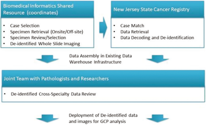
As part of the project, the underlying informatics infrastructure was designed, tested, and implemented through close collaboration with several participating SEER registries to ensure consistency with registry processes, computational scalability, and ability to support creation of population cohorts that span multiple sites. Utilizing computational imaging algorithms and methods to both generate indices and search for matches makes it possible to reduce inter- and intra-observer inconsistencies and to improve the objectivity with which large image repositories are interrogated.

RESULTS

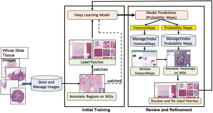
Our team has created and continues to expand a well-curated repository of high-quality digitized pathology images corresponding to subjects whose data are routinely collected by the collaborating registries. Our team has systematically deployed and tested key, visual analytic methods to facilitate automated creation of population cohorts for epidemiological studies and tools to support visualization of feature clusters and evaluation of whole-slide images. As part of these efforts, we are developing and optimizing advanced search and matching algorithms to facilitate automated, content-based retrieval of digitized specimens based on their underlying image features and staining characteristics.

CONCLUSION

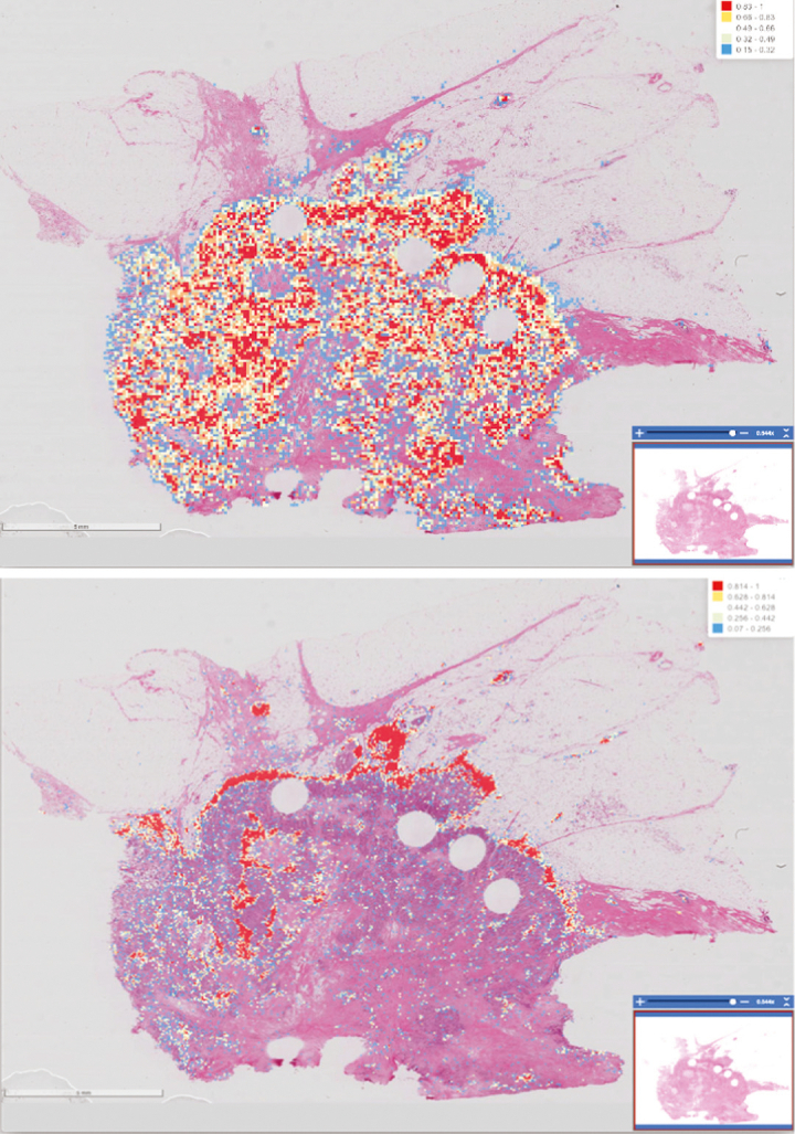
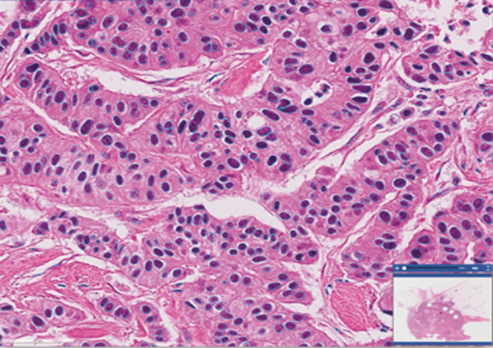
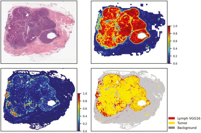
To meet the challenges of this project, we established the analytic pipelines, methods, and workflows to support the expansion and management of a growing repository of high-quality digitized pathology and information-rich, population cohorts containing objective imaging and clinical attributes to facilitate studies that seek to discriminate among different subtypes of disease, stratify patient populations, and perform comparisons of tumor characteristics within and across patient cohorts. We have also successfully developed a suite of tools based on a deep-learning method to perform quantitative characterizations of tumor regions, assess infiltrating lymphocyte distributions, and generate objective nuclear feature measurements. As part of these efforts, our team has implemented reliable methods that enable investigators to systematically search through large repositories to automatically retrieve digitized pathology specimens and correlated clinical data based on their computational signatures.

摘要

背景

基于人群的州癌症登记处是美国癌症统计数据的权威来源。它们定期收集各类数据,包括患者人口统计学信息、原发肿瘤部位、诊断时的分期、初始治疗方案以及生存情况等,涉及美国所有州和领地上报的每一例癌症病例。我们项目的目标是以数字病理学、基于机器学习的分类以及定量组织病理学成像特征集(此处称为[未提及具体名称])的形式,用高质量的基于人群的生物样本数据丰富美国国立癌症研究所(NCI)的监测、流行病学和最终结果(SEER)登记处数据。

材料与方法

作为项目的一部分,通过与多个参与的SEER登记处密切合作,设计、测试并实施了基础信息学基础设施,以确保与登记处流程一致、具备计算可扩展性以及支持创建跨多个地点的人群队列的能力。利用计算成像算法和方法来生成索引并寻找匹配项,能够减少观察者之间和观察者内部的不一致性,并提高查询大型图像存储库的客观性。

结果

我们的团队已经创建并持续扩充一个精心整理的高质量数字化病理图像存储库,这些图像对应的数据由合作登记处常规收集。我们的团队系统地部署并测试了关键的视觉分析方法,以促进为流行病学研究自动创建人群队列,以及支持特征聚类可视化和全切片图像评估的工具。作为这些努力的一部分,我们正在开发和优化先进的搜索和匹配算法,以便基于数字化标本的基础图像特征和染色特性,促进基于内容的自动检索。

结论

为应对该项目的挑战,我们建立了分析管道、方法和工作流程,以支持扩充和管理不断增长的高质量数字化病理学存储库以及包含客观成像和临床属性的信息丰富的人群队列,从而便于开展旨在区分疾病不同亚型、对患者群体进行分层以及在患者队列内部和之间比较肿瘤特征的研究。我们还成功开发了一套基于深度学习方法的工具,用于对肿瘤区域进行定量表征、评估浸润淋巴细胞分布并生成客观的核特征测量值。作为这些努力的一部分,我们的团队实施了可靠的方法,使研究人员能够系统地搜索大型存储库,以根据其计算特征自动检索数字化病理标本和相关临床数据。

https://cdn.ncbi.nlm.nih.gov/pmc/blobs/b6c1/8794027/88cb64da24d6/gr7.jpg
https://cdn.ncbi.nlm.nih.gov/pmc/blobs/b6c1/8794027/5cad238a44f4/gr1.jpg
https://cdn.ncbi.nlm.nih.gov/pmc/blobs/b6c1/8794027/aa0bfb8f2288/gr2.jpg
https://cdn.ncbi.nlm.nih.gov/pmc/blobs/b6c1/8794027/88e8df72e1cd/gr3.jpg
https://cdn.ncbi.nlm.nih.gov/pmc/blobs/b6c1/8794027/56fdb50bab04/gr4.jpg
https://cdn.ncbi.nlm.nih.gov/pmc/blobs/b6c1/8794027/363241a07db5/gr5.jpg
https://cdn.ncbi.nlm.nih.gov/pmc/blobs/b6c1/8794027/fba79ec46948/gr6.jpg
https://cdn.ncbi.nlm.nih.gov/pmc/blobs/b6c1/8794027/88cb64da24d6/gr7.jpg

相似文献

1
An Expandable Informatics Framework for Enhancing Central Cancer Registries with Digital Pathology Specimens, Computational Imaging Tools, and Advanced Mining Capabilities.一个可扩展的信息学框架,用于通过数字病理标本、计算机成像工具和先进挖掘能力增强中央癌症登记系统。
J Pathol Inform. 2022 Jan 5;13:5. doi: 10.4103/jpi.jpi_31_21. eCollection 2022.
2
An Intelligent Search & Retrieval System (IRIS) and Clinical and Research Repository for Decision Support Based on Machine Learning and Joint Kernel-based Supervised Hashing.一种智能搜索与检索系统(IRIS)以及基于机器学习和联合核监督哈希的用于决策支持的临床与研究知识库。
Cancer Inform. 2024 Feb 4;23:11769351231223806. doi: 10.1177/11769351231223806. eCollection 2024.
3
A data model and database for high-resolution pathology analytical image informatics.用于高分辨率病理学分析图像信息学的数据模型与数据库。
J Pathol Inform. 2011;2:32. doi: 10.4103/2153-3539.83192. Epub 2011 Jul 26.
4
Image analysis and machine learning in digital pathology: Challenges and opportunities.数字病理学中的图像分析与机器学习:挑战与机遇
Med Image Anal. 2016 Oct;33:170-175. doi: 10.1016/j.media.2016.06.037. Epub 2016 Jul 4.
5
Content-based histopathology image retrieval using CometCloud.基于内容的组织病理学图像检索 CometCloud 方法。
BMC Bioinformatics. 2014 Aug 26;15(1):287. doi: 10.1186/1471-2105-15-287.
6
A high-performance spatial database based approach for pathology imaging algorithm evaluation.一种基于高性能空间数据库的病理学成像算法评估方法。
J Pathol Inform. 2013 Mar 14;4:5. doi: 10.4103/2153-3539.108543. Print 2013.
7
The future of Cochrane Neonatal.考克兰新生儿协作网的未来。
Early Hum Dev. 2020 Nov;150:105191. doi: 10.1016/j.earlhumdev.2020.105191. Epub 2020 Sep 12.
8
Critical Care Network in the State of Qatar.卡塔尔国重症监护网络。
Qatar Med J. 2019 Nov 7;2019(2):2. doi: 10.5339/qmj.2019.qccc.2. eCollection 2019.
9
Developing a low-cost, open-source, locally manufactured workstation and computational pipeline for automated histopathology evaluation using deep learning.开发一种低成本、开源、本地制造的工作站和计算管道,用于使用深度学习进行自动化组织病理学评估。
EBioMedicine. 2024 Sep;107:105276. doi: 10.1016/j.ebiom.2024.105276. Epub 2024 Aug 27.
10
The NCI All Ireland Cancer Conference.美国国家癌症研究所全爱尔兰癌症会议。
Oncologist. 1999;4(4):275-277.

引用本文的文献

1
An Intelligent Search & Retrieval System (IRIS) and Clinical and Research Repository for Decision Support Based on Machine Learning and Joint Kernel-based Supervised Hashing.一种智能搜索与检索系统(IRIS)以及基于机器学习和联合核监督哈希的用于决策支持的临床与研究知识库。
Cancer Inform. 2024 Feb 4;23:11769351231223806. doi: 10.1177/11769351231223806. eCollection 2024.
2
Biobanking in the digital pathology era.数字病理学时代的生物样本库。
Oncol Res. 2022 Aug 31;29(4):229-233. doi: 10.32604/or.2022.024892. eCollection 2021.

本文引用的文献

1
Robust Histopathology Image Analysis: to Label or to Synthesize?强大的组织病理学图像分析:标记还是合成?
Proc IEEE Comput Soc Conf Comput Vis Pattern Recognit. 2019 Jun;2019:8533-8542. doi: 10.1109/CVPR.2019.00873. Epub 2020 Jan 9.
2
A Digital Pathology Solution to Resolve the Tissue Floater Conundrum.一种解决组织漂浮物难题的数字病理学解决方案。
Arch Pathol Lab Med. 2021 Mar 1;145(3):359-364. doi: 10.5858/arpa.2020-0034-OA.
3
Deep learning in digital pathology image analysis: a survey.深度学习在数字病理学图像分析中的应用:综述。
Front Med. 2020 Aug;14(4):470-487. doi: 10.1007/s11684-020-0782-9. Epub 2020 Jul 29.
4
AI in Medical Imaging Informatics: Current Challenges and Future Directions.人工智能在医学影像信息学中的应用:当前的挑战与未来方向。
IEEE J Biomed Health Inform. 2020 Jul;24(7):1837-1857. doi: 10.1109/JBHI.2020.2991043.
5
Editorial: Computational Pathology.社论:计算病理学
Front Med (Lausanne). 2020 Jun 9;7:245. doi: 10.3389/fmed.2020.00245. eCollection 2020.
6
Dataset of segmented nuclei in hematoxylin and eosin stained histopathology images of ten cancer types.十类癌症的苏木精和伊红染色组织病理学图像分割细胞核数据集。
Sci Data. 2020 Jun 19;7(1):185. doi: 10.1038/s41597-020-0528-1.
7
PRISM: A Platform for Imaging in Precision Medicine.PRISM:精准医学成像平台。
JCO Clin Cancer Inform. 2020 Jun;4:491-499. doi: 10.1200/CCI.20.00001.
8
Report on computational assessment of Tumor Infiltrating Lymphocytes from the International Immuno-Oncology Biomarker Working Group.国际免疫肿瘤生物标志物工作组关于肿瘤浸润淋巴细胞的计算评估报告。
NPJ Breast Cancer. 2020 May 12;6:16. doi: 10.1038/s41523-020-0154-2. eCollection 2020.
9
Tumor-infiltrating lymphocytes are associated with poor prognosis in invasive lobular breast carcinoma.肿瘤浸润淋巴细胞与浸润性小叶乳腺癌不良预后相关。
Mod Pathol. 2020 Nov;33(11):2198-2207. doi: 10.1038/s41379-020-0561-9. Epub 2020 May 13.
10
Utilizing Automated Breast Cancer Detection to Identify Spatial Distributions of Tumor-Infiltrating Lymphocytes in Invasive Breast Cancer.利用自动化乳腺癌检测识别浸润性乳腺癌中肿瘤浸润淋巴细胞的空间分布。
Am J Pathol. 2020 Jul;190(7):1491-1504. doi: 10.1016/j.ajpath.2020.03.012. Epub 2020 Apr 8.