• 文献检索
  • 文档翻译
  • 深度研究
  • 学术资讯
  • Suppr Zotero 插件Zotero 插件
  • 邀请有礼
  • 套餐&价格
  • 历史记录
应用&插件
Suppr Zotero 插件Zotero 插件浏览器插件Mac 客户端Windows 客户端微信小程序
定价
高级版会员购买积分包购买API积分包
服务
文献检索文档翻译深度研究API 文档MCP 服务
关于我们
关于 Suppr公司介绍联系我们用户协议隐私条款
关注我们

Suppr 超能文献

核心技术专利:CN118964589B侵权必究
粤ICP备2023148730 号-1Suppr @ 2026

文献检索

告别复杂PubMed语法,用中文像聊天一样搜索,搜遍4000万医学文献。AI智能推荐,让科研检索更轻松。

立即免费搜索

文件翻译

保留排版,准确专业,支持PDF/Word/PPT等文件格式,支持 12+语言互译。

免费翻译文档

深度研究

AI帮你快速写综述,25分钟生成高质量综述,智能提取关键信息,辅助科研写作。

立即免费体验

十类癌症的苏木精和伊红染色组织病理学图像分割细胞核数据集。

Dataset of segmented nuclei in hematoxylin and eosin stained histopathology images of ten cancer types.

机构信息

Computer Science Department, 203C New Computer Science Building, Stony Brook University, Stony Brook, NY, 11794, USA.

Biomedical Informatics Department, HSC L3-045, Stony Brook Medicine, Stony Brook University, Stony Brook, NY, 11794, USA.

出版信息

Sci Data. 2020 Jun 19;7(1):185. doi: 10.1038/s41597-020-0528-1.

DOI:10.1038/s41597-020-0528-1
PMID:32561748
原文链接:https://pmc.ncbi.nlm.nih.gov/articles/PMC7305328/
Abstract

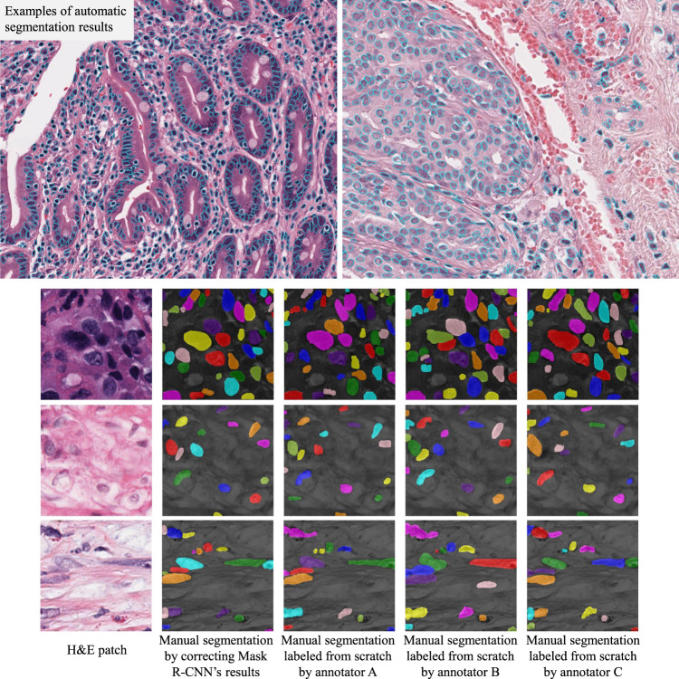
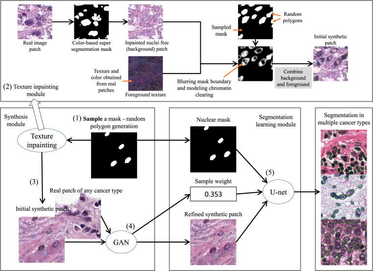
The distribution and appearance of nuclei are essential markers for the diagnosis and study of cancer. Despite the importance of nuclear morphology, there is a lack of large scale, accurate, publicly accessible nucleus segmentation data. To address this, we developed an analysis pipeline that segments nuclei in whole slide tissue images from multiple cancer types with a quality control process. We have generated nucleus segmentation results in 5,060 Whole Slide Tissue images from 10 cancer types in The Cancer Genome Atlas. One key component of our work is that we carried out a multi-level quality control process (WSI-level and image patch-level), to evaluate the quality of our segmentation results. The image patch-level quality control used manual segmentation ground truth data from 1,356 sampled image patches. The datasets we publish in this work consist of roughly 5 billion quality controlled nuclei from more than 5,060 TCGA WSIs from 10 different TCGA cancer types and 1,356 manually segmented TCGA image patches from the same 10 cancer types plus additional 4 cancer types.

摘要

细胞核的分布和形态是癌症诊断和研究的重要标志物。尽管细胞核形态学很重要,但缺乏大规模、准确、公开可获取的细胞核分割数据。针对这一问题,我们开发了一种分析流水线,可对来自多种癌症类型的全切片组织图像中的细胞核进行分割,并带有质量控制流程。我们已经在 10 种癌症类型的 5,060 张全切片组织图像中生成了细胞核分割结果。我们工作的一个关键组成部分是,我们执行了多级质量控制流程(全切片组织图像级和图像斑块级),以评估我们的分割结果的质量。图像斑块级质量控制使用了来自 1,356 个采样图像斑块的手动分割地面实况数据。我们在这项工作中发布的数据集包含来自 10 种不同 TCGA 癌症类型的 5,060 张 TCGA 全切片组织图像和 1,356 张来自相同 10 种癌症类型以及另外 4 种癌症类型的手动分割 TCGA 图像斑块的大约 50 亿个经过质量控制的细胞核。

https://cdn.ncbi.nlm.nih.gov/pmc/blobs/bbc7/7305328/94fbba794af7/41597_2020_528_Fig5_HTML.jpg
https://cdn.ncbi.nlm.nih.gov/pmc/blobs/bbc7/7305328/f4f10c52194c/41597_2020_528_Fig1_HTML.jpg
https://cdn.ncbi.nlm.nih.gov/pmc/blobs/bbc7/7305328/3b4d3064ffea/41597_2020_528_Fig2_HTML.jpg
https://cdn.ncbi.nlm.nih.gov/pmc/blobs/bbc7/7305328/2070ae5a22f6/41597_2020_528_Fig3_HTML.jpg
https://cdn.ncbi.nlm.nih.gov/pmc/blobs/bbc7/7305328/e5c661b9189d/41597_2020_528_Fig4_HTML.jpg
https://cdn.ncbi.nlm.nih.gov/pmc/blobs/bbc7/7305328/94fbba794af7/41597_2020_528_Fig5_HTML.jpg

相似文献

1
Dataset of segmented nuclei in hematoxylin and eosin stained histopathology images of ten cancer types.十类癌症的苏木精和伊红染色组织病理学图像分割细胞核数据集。
Sci Data. 2020 Jun 19;7(1):185. doi: 10.1038/s41597-020-0528-1.
2
Tumor grading model employing geometric analysis of histopathological images with characteristic nuclei dictionary.基于具有特征核字典的组织病理学图像的几何分析的肿瘤分级模型。
Comput Biol Med. 2022 Oct;149:106008. doi: 10.1016/j.compbiomed.2022.106008. Epub 2022 Aug 17.
3
Using cell nuclei features to detect colon cancer tissue in hematoxylin and eosin stained slides.利用细胞核特征在苏木精和伊红染色切片中检测结肠癌组织。
Cytometry A. 2017 Aug;91(8):785-793. doi: 10.1002/cyto.a.23175. Epub 2017 Jul 20.
4
Automated cell nuclear segmentation in color images of hematoxylin and eosin-stained breast biopsy.苏木精和伊红染色的乳腺活检彩色图像中的自动细胞核分割
Anal Quant Cytol Histol. 2003 Dec;25(6):321-31.
5
FEEDNet: a feature enhanced encoder-decoder LSTM network for nuclei instance segmentation for histopathological diagnosis.FEEDNet:一种用于组织病理学诊断的细胞核实例分割的特征增强编码器-解码器长短期记忆网络。
Phys Med Biol. 2022 Sep 28;67(19). doi: 10.1088/1361-6560/ac8594.
6
NuInsSeg: A fully annotated dataset for nuclei instance segmentation in H&E-stained histological images.NuInsSeg:H&E 染色组织学图像中细胞核实例分割的完全标注数据集。
Sci Data. 2024 Mar 14;11(1):295. doi: 10.1038/s41597-024-03117-2.
7
Detection of malignant melanoma in H&E-stained images using deep learning techniques.利用深度学习技术在 H&E 染色图像中检测恶性黑色素瘤。
Tissue Cell. 2021 Dec;73:101659. doi: 10.1016/j.tice.2021.101659. Epub 2021 Sep 29.
8
Nuclei segmentation of HE stained histopathological images based on feature global delivery connection network.基于特征全局传递连接网络的 HE 染色组织病理学图像细胞核分割。
PLoS One. 2022 Sep 15;17(9):e0273682. doi: 10.1371/journal.pone.0273682. eCollection 2022.
9
CellViT: Vision Transformers for precise cell segmentation and classification.CellViT:用于精确细胞分割和分类的视觉Transformer
Med Image Anal. 2024 May;94:103143. doi: 10.1016/j.media.2024.103143. Epub 2024 Mar 16.
10
HoLy-Net: Segmentation of histological images of diffuse large B-cell lymphoma.HoLy-Net:弥漫性大 B 细胞淋巴瘤组织学图像分割。
Comput Biol Med. 2024 Mar;170:107978. doi: 10.1016/j.compbiomed.2024.107978. Epub 2024 Jan 11.

引用本文的文献

1
Novel feature-based method for multi-modal biomedical image registration compared to intensity-based technique.与基于强度的技术相比,用于多模态生物医学图像配准的基于特征的新方法。
Sci Rep. 2025 Aug 1;15(1):28183. doi: 10.1038/s41598-025-12862-2.
2
Deep-learning based colorectal cancer pathological analysis with hyperspectral light field microscopy.基于深度学习的高光谱光场显微镜结直肠癌病理分析
iScience. 2025 Jun 25;28(8):112987. doi: 10.1016/j.isci.2025.112987. eCollection 2025 Aug 15.
3
Tumour nuclear size heterogeneity as a biomarker for post-radiotherapy outcomes in gynecological malignancies.

本文引用的文献

1
MoNuSAC2020: A Multi-Organ Nuclei Segmentation and Classification Challenge.MoNuSAC2020:多器官细胞核分割与分类挑战赛
IEEE Trans Med Imaging. 2021 Dec;40(12):3413-3423. doi: 10.1109/TMI.2021.3085712. Epub 2021 Nov 30.
2
Robust Histopathology Image Analysis: to Label or to Synthesize?强大的组织病理学图像分析:标记还是合成?
Proc IEEE Comput Soc Conf Comput Vis Pattern Recognit. 2019 Jun;2019:8533-8542. doi: 10.1109/CVPR.2019.00873. Epub 2020 Jan 9.
3
Methods for Segmentation and Classification of Digital Microscopy Tissue Images.
肿瘤核大小异质性作为妇科恶性肿瘤放疗后预后的生物标志物。
Phys Imaging Radiat Oncol. 2025 Jun 19;35:100793. doi: 10.1016/j.phro.2025.100793. eCollection 2025 Jul.
4
Nuclei segmentation and classification from histopathology images using federated learning for end-edge platform.使用联邦学习在端边缘平台上对组织病理学图像进行细胞核分割和分类。
PLoS One. 2025 Jul 10;20(7):e0322749. doi: 10.1371/journal.pone.0322749. eCollection 2025.
5
Nuclei-level prior knowledge constrained multiple instance learning for breast histopathology whole slide image classification.基于细胞核水平先验知识约束的多实例学习用于乳腺组织病理学全切片图像分类
iScience. 2024 Apr 26;27(6):109826. doi: 10.1016/j.isci.2024.109826. eCollection 2024 Jun 21.
6
NuInsSeg: A fully annotated dataset for nuclei instance segmentation in H&E-stained histological images.NuInsSeg:H&E 染色组织学图像中细胞核实例分割的完全标注数据集。
Sci Data. 2024 Mar 14;11(1):295. doi: 10.1038/s41597-024-03117-2.
7
Computational pathology: A survey review and the way forward.计算病理学:综述与未来发展方向
J Pathol Inform. 2024 Jan 14;15:100357. doi: 10.1016/j.jpi.2023.100357. eCollection 2024 Dec.
8
Attention De-sparsification Matters: Inducing diversity in digital pathology representation learning.注意力去稀疏化很重要:在数字病理学表示学习中引入多样性。
Med Image Anal. 2024 Apr;93:103070. doi: 10.1016/j.media.2023.103070. Epub 2023 Dec 28.
9
Preparing Data for Artificial Intelligence in Pathology with Clinical-Grade Performance.利用临床级性能为病理学中的人工智能准备数据。
Diagnostics (Basel). 2023 Oct 3;13(19):3115. doi: 10.3390/diagnostics13193115.
10
A Large-scale Synthetic Pathological Dataset for Deep Learning-enabled Segmentation of Breast Cancer.用于深度学习辅助乳腺癌分割的大规模合成病理数据集。
Sci Data. 2023 Apr 21;10(1):231. doi: 10.1038/s41597-023-02125-y.
数字显微镜组织图像的分割与分类方法
Front Bioeng Biotechnol. 2019 Apr 2;7:53. doi: 10.3389/fbioe.2019.00053. eCollection 2019.
4
Structured crowdsourcing enables convolutional segmentation of histology images.结构化众包可实现组织学图像的卷积分割。
Bioinformatics. 2019 Sep 15;35(18):3461-3467. doi: 10.1093/bioinformatics/btz083.
5
Segmentation of Nuclei in Histopathology Images by Deep Regression of the Distance Map.基于距离图深度回归的组织病理学图像细胞核分割。
IEEE Trans Med Imaging. 2019 Feb;38(2):448-459. doi: 10.1109/TMI.2018.2865709.
6
Sparse Autoencoder for Unsupervised Nucleus Detection and Representation in Histopathology Images.用于组织病理学图像中无监督细胞核检测与表征的稀疏自动编码器
Pattern Recognit. 2019 Feb;86:188-200. doi: 10.1016/j.patcog.2018.09.007. Epub 2018 Sep 13.
7
Mask R-CNN.Mask R-CNN。
IEEE Trans Pattern Anal Mach Intell. 2020 Feb;42(2):386-397. doi: 10.1109/TPAMI.2018.2844175. Epub 2018 Jun 5.
8
Center-Focusing Multi-task CNN with Injected Features for Classification of Glioma Nuclear Images.具有注入特征的中心聚焦多任务卷积神经网络用于胶质瘤细胞核图像分类
IEEE Winter Conf Appl Comput Vis. 2017 Mar;2017:834-841. doi: 10.1109/WACV.2017.98. Epub 2017 May 15.
9
PanCancer insights from The Cancer Genome Atlas: the pathologist's perspective.从癌症基因组图谱中获得的泛癌症洞察:病理学家视角。
J Pathol. 2018 Apr;244(5):512-524. doi: 10.1002/path.5028. Epub 2018 Feb 22.
10
A Containerized Software System for Generation, Management, and Exploration of Features from Whole Slide Tissue Images.一种用于从全切片组织图像生成、管理和探索特征的容器化软件系统。
Cancer Res. 2017 Nov 1;77(21):e79-e82. doi: 10.1158/0008-5472.CAN-17-0316.