Zhang Haijie, Yin Fu, Chen Menglin, Yang Liyang, Qi Anqi, Cui Weiwei, Yang Shanshan, Wen Ge
Department of Imaging, Nanfang Hospital, Southern Medical University, Guangzhou, China.
PET/CT Center, Department of Nuclear Medicine, First Affiliated Hospital of Shenzhen University, Shenzhen Second People's Hospital, Shenzhen, China.
Front Oncol. 2022 Jan 27;11:742547. doi: 10.3389/fonc.2021.742547. eCollection 2021.
Many patients experience recurrence of renal cell carcinoma (RCC) after radical and partial nephrectomy. Radiomics nomogram is a newly used noninvasive tool that could predict tumor phenotypes.
To investigate Radiomics Features (RFs) associated with progression-free survival (PFS) of RCC, assessing its incremental value over clinical factors, and to develop a visual nomogram in order to provide reference for individualized treatment.
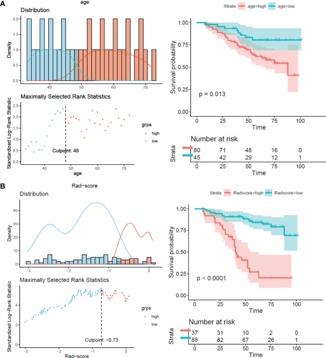
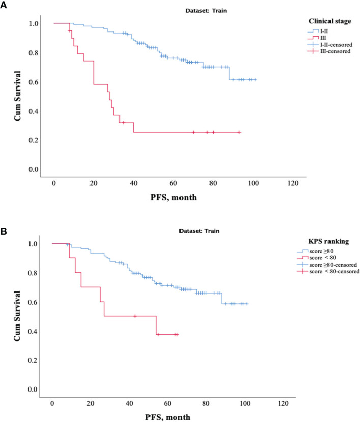
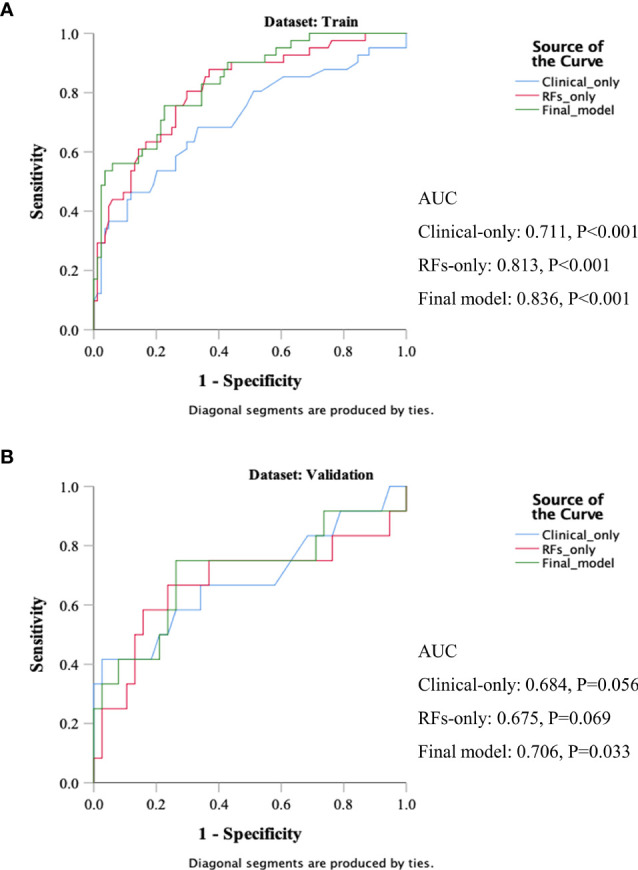
The RFs and clinicopathological data of 175 patients (125 in the training set and 50 in the validation set) with clear cell RCC (ccRCC) were retrospectively analyzed. In the training set, RFs were extracted from multiphase enhanced CT tumor volume and selected using the stability LASSO feature selection algorithm. A radiomics nomogram final model was developed that incorporated the RFs weighted sum and selected clinical predictors based on the multivariate Cox proportional hazard regression. The performances of a clinical variables-only model, RFs-only model, and the final model were compared by receiver operator characteristic (ROC) analysis and DeLong test. Nomogram performance was determined and validated with respect to its discrimination, calibration, reclassification, and clinical usefulness.
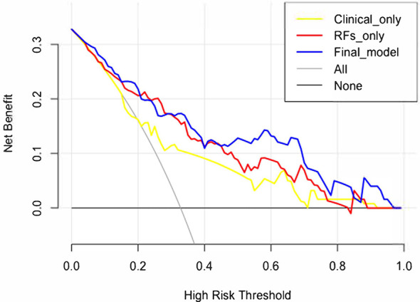
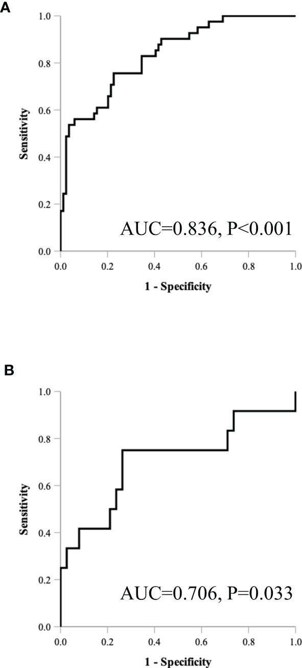
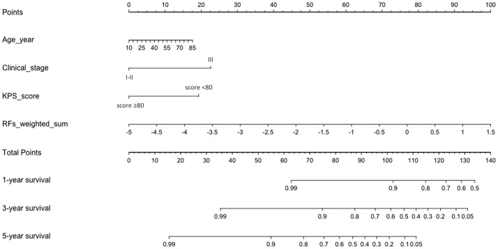
The radiomics nomogram included age, clinical stage, KPS score, and RFs weighted sum, which consisted of 6 selected RFs. The final model showed good discrimination, with a C-index of 0.836 and 0.706 in training and validation, and good calibration. In the training set, the C-index of the final model was significantly larger than the clinical-only model (DeLong test, = 0.008). From the clinical variables-only model to the final model, the reclassification of net reclassification improvement was 18.03%, and the integrated discrimination improvement was 19.08%. Decision curve analysis demonstrated the clinical usefulness of the radiomics nomogram.
The CT-based RF is an improvement factor for clinical variables-only model. The radiomics nomogram provides individualized risk assessment of postoperative PFS for patients with RCC.
许多患者在根治性肾切除术和部分肾切除术后会出现肾细胞癌(RCC)复发。放射组学列线图是一种新使用的无创工具,可预测肿瘤表型。
研究与RCC无进展生存期(PFS)相关的放射组学特征(RFs),评估其相对于临床因素的增量价值,并开发一种可视化列线图,为个体化治疗提供参考。
回顾性分析175例透明细胞肾细胞癌(ccRCC)患者(训练集125例,验证集50例)的RFs和临床病理数据。在训练集中,从多期增强CT肿瘤体积中提取RFs,并使用稳定性LASSO特征选择算法进行选择。开发了一个放射组学列线图最终模型,该模型结合了RFs加权和,并基于多变量Cox比例风险回归选择临床预测因子。通过受试者操作特征(ROC)分析和DeLong检验比较仅临床变量模型、仅RFs模型和最终模型的性能。确定列线图性能,并就其区分度、校准、重新分类和临床实用性进行验证。
放射组学列线图包括年龄、临床分期、KPS评分和RFs加权和,后者由6个选定的RFs组成。最终模型显示出良好的区分度,训练集和验证集的C指数分别为0.836和0.706,且校准良好。在训练集中,最终模型的C指数显著大于仅临床模型(DeLong检验,=0.008)。从仅临床变量模型到最终模型,净重新分类改善的重新分类为18.03%,综合区分改善为19.08%。决策曲线分析证明了放射组学列线图的临床实用性。
基于CT的RFs是仅临床变量模型的一个改善因素。放射组学列线图为RCC患者术后PFS提供了个体化风险评估。