Jekel Leon, Brim Waverly R, von Reppert Marc, Staib Lawrence, Cassinelli Petersen Gabriel, Merkaj Sara, Subramanian Harry, Zeevi Tal, Payabvash Seyedmehdi, Bousabarah Khaled, Lin MingDe, Cui Jin, Brackett Alexandria, Mahajan Amit, Omuro Antonio, Johnson Michele H, Chiang Veronica L, Malhotra Ajay, Scheffler Björn, Aboian Mariam S
Department of Radiology and Biomedical Imaging, Yale School of Medicine, 333 Cedar Street, P.O. Box 208042, New Haven, CT 06510, USA.
DKFZ Division of Translational Neurooncology at the WTZ, German Cancer Consortium, DKTK Partner Site, University Hospital Essen, 45147 Essen, Germany.
Cancers (Basel). 2022 Mar 8;14(6):1369. doi: 10.3390/cancers14061369.
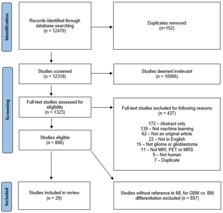
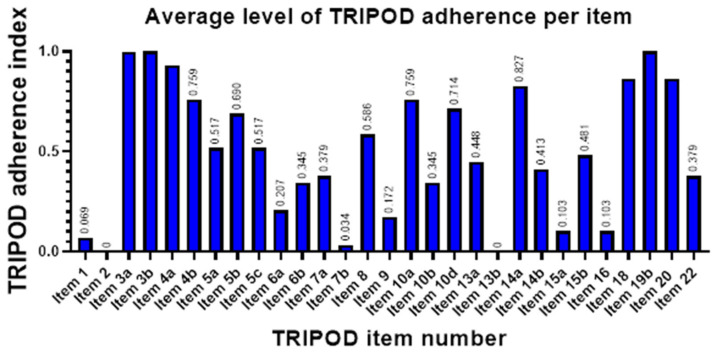
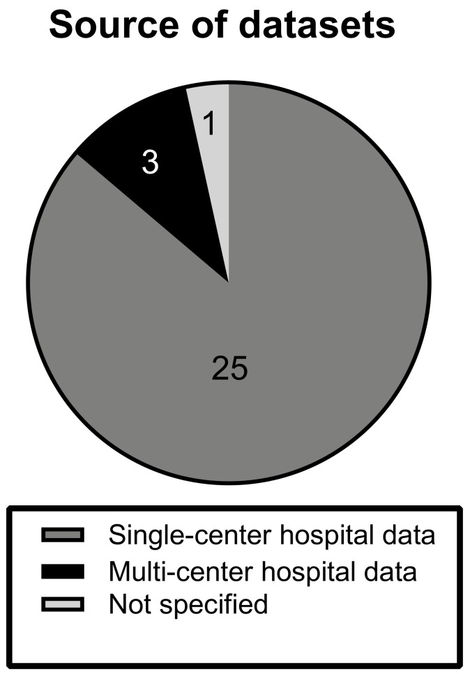
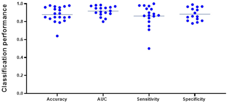
Glioma and brain metastasis can be difficult to distinguish on conventional magnetic resonance imaging (MRI) due to the similarity of imaging features in specific clinical circumstances. Multiple studies have investigated the use of machine learning (ML) models for non-invasive differentiation of glioma from brain metastasis. Many of the studies report promising classification results, however, to date, none have been implemented into clinical practice. After a screening of 12,470 studies, we included 29 eligible studies in our systematic review. From each study, we aggregated data on model design, development, and best classifiers, as well as quality of reporting according to the TRIPOD statement. In a subset of eligible studies, we conducted a meta-analysis of the reported AUC. It was found that data predominantly originated from single-center institutions (n = 25/29) and only two studies performed external validation. The median TRIPOD adherence was 0.48, indicating insufficient quality of reporting among surveyed studies. Our findings illustrate that despite promising classification results, reliable model assessment is limited by poor reporting of study design and lack of algorithm validation and generalizability. Therefore, adherence to quality guidelines and validation on outside datasets is critical for the clinical translation of ML for the differentiation of glioma and brain metastasis.
由于在特定临床情况下成像特征相似,胶质瘤和脑转移瘤在传统磁共振成像(MRI)上可能难以区分。多项研究探讨了使用机器学习(ML)模型对胶质瘤和脑转移瘤进行无创鉴别。许多研究报告了有前景的分类结果,然而,迄今为止,尚无研究应用于临床实践。在筛选了12470项研究后,我们在系统评价中纳入了29项符合条件的研究。我们从每项研究中汇总了关于模型设计、开发和最佳分类器的数据,以及根据TRIPOD声明的报告质量。在一部分符合条件的研究中,我们对报告的AUC进行了荟萃分析。结果发现,数据主要来自单中心机构(n = 25/29),只有两项研究进行了外部验证。TRIPOD依从性中位数为0.48,表明所调查研究的报告质量不足。我们的研究结果表明,尽管分类结果很有前景,但可靠的模型评估受到研究设计报告不佳以及缺乏算法验证和可推广性的限制。因此,遵守质量指南并在外部数据集上进行验证对于将ML用于胶质瘤和脑转移瘤鉴别的临床转化至关重要。