Ye Haotian, Ji Muhuo, Wang Chaoyan, Wang Cong, Li Ying, Chen Yuan, Cheng Lisha, Li Yanfei, Yang Jian-Jun
Department of Anesthesiology, Pain and Perioperative Medicine, The First Affiliated Hospital of Zhengzhou University, Zhengzhou, China.
Department of Anesthesiology, The Second Affiliated Hospital, Nanjing Medical University, Nanjing, China.
Front Psychiatry. 2022 Mar 22;13:848709. doi: 10.3389/fpsyt.2022.848709. eCollection 2022.
Intensive care unit (ICU) medical staffs undergoing sleep deprivation with perennial night shift work were usually at high risk of depression. However, shift work on depression-related resting-state functional magnetic resonance imaging was still not fully understood. The objective of this study was to explore the effects of sleep deprivation in ICU medical staffs after one night of shift work on brain functional connectivity density (FCD) and Hamilton Depression Rating Scale (HAMD) scores. Also, serum neurotransmitter concentrations of serotonin (5-HT) and norepinephrine (NE) were obtained simultaneously.
A total of 21 ICU medical staffs without psychiatric history were recruited. All participants received HAMD score assessment and resting-state functional magnetic resonance imaging scans at two time points: one at rested wakefulness and the other after sleep deprivation (SD) accompanied with one night of shift work. Global FCD, local FCD, and long-range FCD (lrFCD) were used to evaluate spontaneous brain activity in the whole brain. In the meantime, peripheral blood samples were collected for measurement of serum 5-HT and NE levels. All these data were acquired between 7:00 and 8:00 am to limit the influence of biological rhythms. The correlations between the FCD values and HAMD scores and serum levels of neurotransmitters were analyzed concurrently.
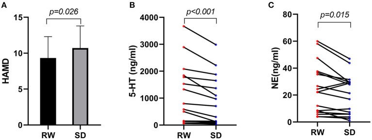
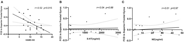
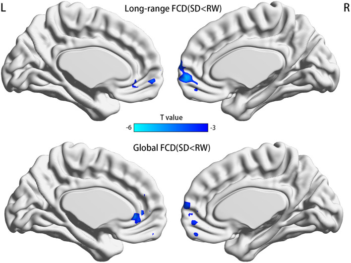
Functional connectivity density mapping manifested that global FCD was decreased in the right medial frontal gyrus and the anterior cingulate gyrus, whereas lrFCD was decreased mainly in the right medial frontal gyrus. Most of these brain areas with FCD differences were components of the default mode network and overlapped with the medial prefrontal cortex. The lrFCD in the medial frontal gyrus showed a negative correlation with HAMD scores after SD. Compared with rested wakefulness, serum levels of 5-HT and NE decreased significantly, whereas HAMD scores were higher after SD within subjects.
Our study suggested that sleep deprivation after night shift work can induce depressive tendency in ICU medical staffs, which might be related to alterative medial prefrontal cortex, raised HAMD scores, and varying monoamine neurotransmitters.
长期值夜班导致睡眠剥夺的重症监护病房(ICU)医护人员通常有较高的抑郁风险。然而,轮班工作对与抑郁相关的静息态功能磁共振成像的影响仍未完全明确。本研究的目的是探讨ICU医护人员在一夜轮班工作后的睡眠剥夺对脑功能连接密度(FCD)和汉密尔顿抑郁量表(HAMD)评分的影响。同时,还获取了血清血清素(5-羟色胺,5-HT)和去甲肾上腺素(NE)的浓度。
共招募了21名无精神病史的ICU医护人员。所有参与者在两个时间点接受HAMD评分评估和静息态功能磁共振成像扫描:一个是在清醒休息时,另一个是在经历一夜轮班工作后的睡眠剥夺(SD)后。采用全脑FCD、局部FCD和长程FCD(lrFCD)来评估全脑的自发脑活动。同时,采集外周血样本以测量血清5-HT和NE水平。所有这些数据均在上午7:00至8:00之间采集,以限制生物节律的影响。同时分析FCD值与HAMD评分及神经递质血清水平之间的相关性。
功能连接密度图谱显示,右侧额内侧回和前扣带回的全脑FCD降低,而lrFCD主要在右侧额内侧回降低。这些FCD存在差异的脑区大多是默认模式网络的组成部分,并与内侧前额叶皮质重叠。内侧前额叶回的lrFCD在SD后与HAMD评分呈负相关。与清醒休息相比,受试者体内血清5-HT和NE水平显著降低,而SD后的HAMD评分更高。
我们的研究表明,夜班工作后的睡眠剥夺可导致ICU医护人员出现抑郁倾向,这可能与内侧前额叶皮质改变、HAMD评分升高以及单胺类神经递质变化有关。