Othman Alaa K, El Kurdi Riham, Badran Adnan, Mesmar Joelle, Baydoun Elias, Patra Digambara
Department of Chemistry, American University of Beirut Beirut Lebanon
Department of Basic Sciences, University of Petra P.O. Box 961343 Amman Jordan.
RSC Adv. 2022 Apr 11;12(18):11282-11292. doi: 10.1039/d2ra00071g. eCollection 2022 Apr 7.
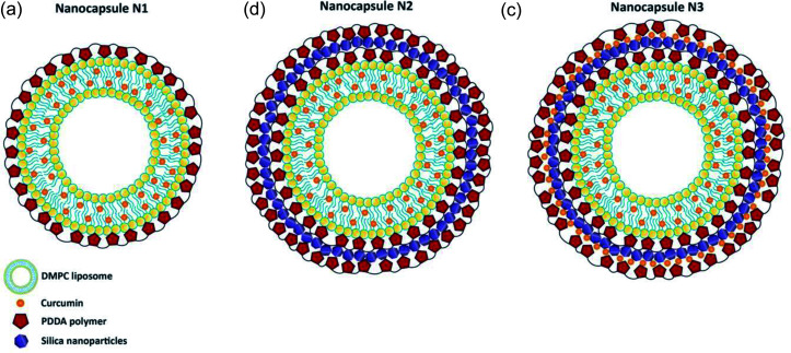
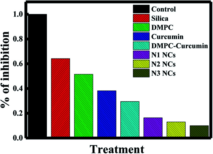
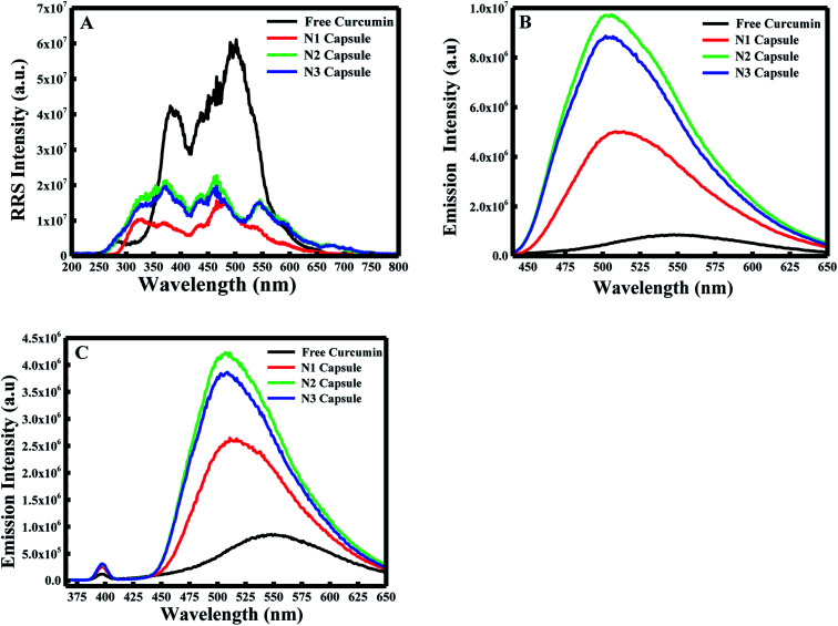
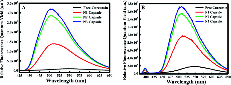
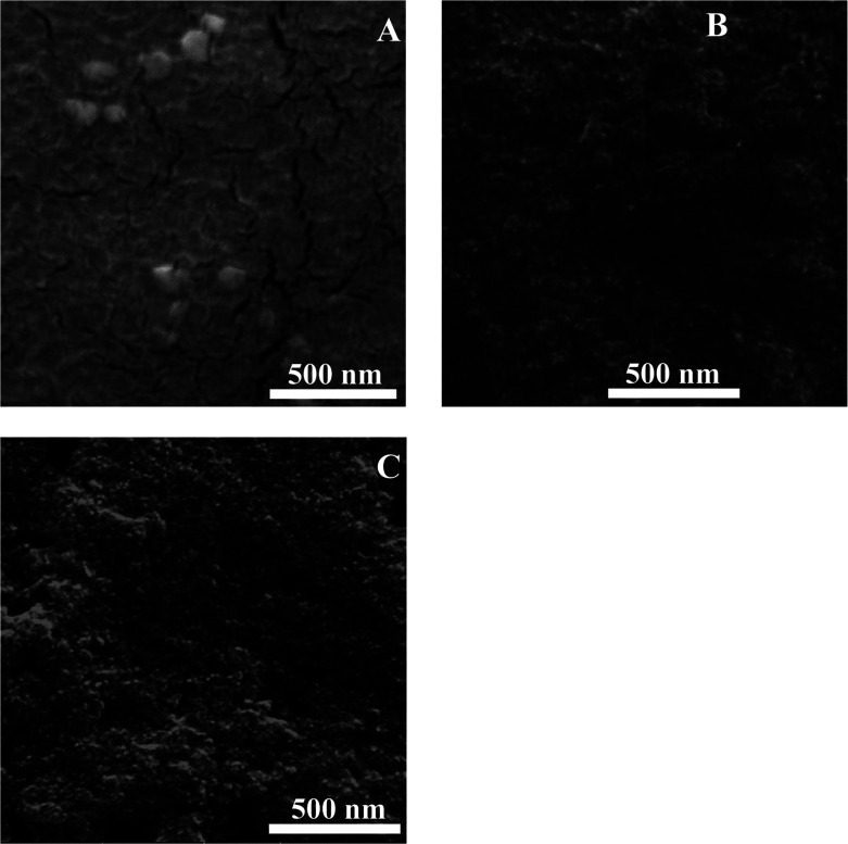
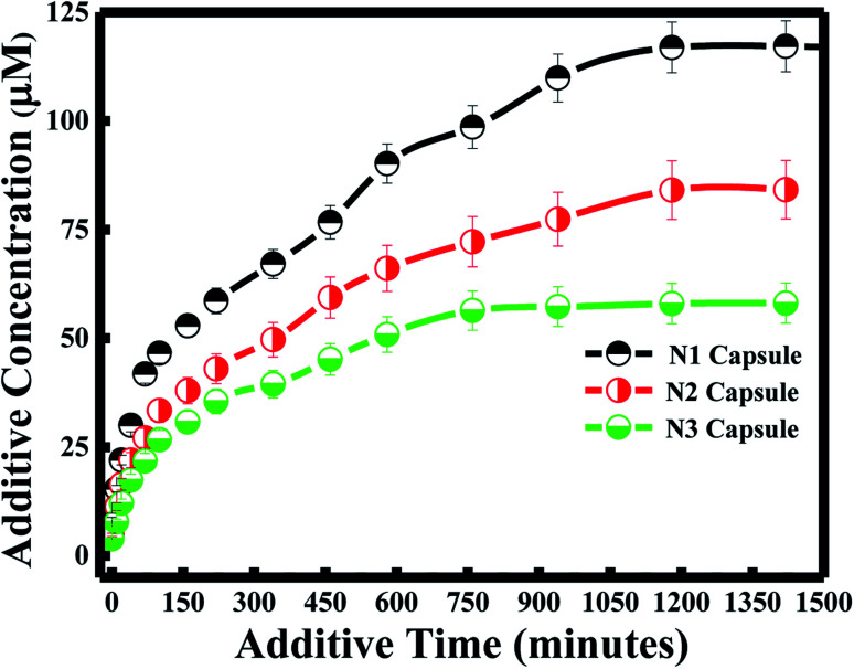
Nanosystems with various compositions and biological properties are being extensively investigated for drug and gene delivery applications. Many nanotechnology methods use novel nanocarriers, such as liposomes, in therapeutically targeted drug delivery systems. However, liposome matrices suffer from several limitations, including drug leakage and instability. Therefore, the surface modification of liposomes by coating them or adding polymers has advanced their application in drug delivery. Hence, the prevention of drug release from the liposome bilayers was the main focus of this work. For this purpose, liposomes were synthesized according to a thin film hydration method by applying various surface modifications. Three different nanocapsules, N1, N2, and N3, were prepared using 1,2-dimyristoyl--3-phosphocholine (DMPC), poly(diallyldimethylammonium)chloride (PDAA) polymer, and silica nanoparticles. PDDA and silica nanoparticles were coated on the surface of liposomes using a layer-by-layer assembly method, completely encapsulating curcumin into the core of the liposome. Fluorescence spectroscopy, TGA, DLS, XRD, SEM, and zeta potential methods were used to characterize the prepared nanocapsules. Interestingly, the fluorescence of curcumin showed a blue shift and the fluorescence efficiency was extraordinarily enhanced ∼25-, ∼54-, and ∼62-fold in the N1, N2, and N3 nanocapsules, respectively. Similarly, encapsulation efficiency, drug loading, and the anticancer activity of dietary curcumin were investigated for the different types of DMPC nanocapsules. The drug efficiencies of the liposomes were established according to the release of curcumin from the liposomes. The results showed that the release of curcumin from the nanocapsules decreased as the number of layers at the surface of the liposomes increased. The release of curcumin follows the Higuchi model; thus, a slow rate of diffusion is observed when a number of layers is added. The better encapsulation and higher anti-cancer activity of curcumin were also observed when more layers were added, which is due to electrostatic interactions inhibiting curcumin from being released.
具有各种组成和生物学特性的纳米系统正在被广泛研究用于药物和基因递送应用。许多纳米技术方法在治疗靶向药物递送系统中使用新型纳米载体,如脂质体。然而,脂质体基质存在一些局限性,包括药物泄漏和不稳定性。因此,通过包被脂质体或添加聚合物对其进行表面修饰推进了它们在药物递送中的应用。因此,防止药物从脂质体双层中释放是这项工作的主要重点。为此,通过应用各种表面修饰,根据薄膜水化法合成脂质体。使用1,2 - 二肉豆蔻酰 - 3 - 磷酸胆碱(DMPC)、聚(二烯丙基二甲基氯化铵)(PDAA)聚合物和二氧化硅纳米颗粒制备了三种不同的纳米胶囊,即N1、N2和N3。使用层层组装法将PDDA和二氧化硅纳米颗粒包被在脂质体表面,将姜黄素完全包裹在脂质体核心中。使用荧光光谱、热重分析(TGA)、动态光散射(DLS)、X射线衍射(XRD)、扫描电子显微镜(SEM)和zeta电位方法对制备的纳米胶囊进行表征。有趣的是,姜黄素的荧光出现蓝移,并且在N1、N2和N3纳米胶囊中荧光效率分别异常增强了约25倍、约54倍和约62倍。同样,针对不同类型的DMPC纳米胶囊研究了膳食姜黄素的包封效率、载药量和抗癌活性。根据姜黄素从脂质体中的释放情况确定脂质体的药物效率。结果表明,随着脂质体表面层数的增加,姜黄素从纳米胶囊中的释放减少。姜黄素的释放遵循Higuchi模型;因此,当添加多层时观察到扩散速率较慢。当添加更多层时,还观察到姜黄素具有更好的包封和更高的抗癌活性,这是由于静电相互作用抑制了姜黄素的释放。